Herrero de la Parte Borja, Irazola Mireia, Pérez-Muñoz Jorge, Rodrigo Irati, Iturrizaga Correcher Sira, Mar Medina Carmen, Castro Kepa, Etxebarria Nestor, Plazaola Fernando, García Jose Ángel, García-Alonso Ignacio, Echevarría-Uraga Jose Javier
Department of Surgery and Radiology and Physical Medicine, University of The Basque Country, ES48940 Leioa, Biscay, Spain.
Biocruces Bizkaia Health Research Institute, ES48903 Barakaldo, Biscay, Spain.
Nanomaterials (Basel). 2021 May 17;11(5):1318. doi: 10.3390/nano11051318.
Hyperthermia (HT) therapy still remains relatively unknown, in terms of both its biological and therapeutic effects. This work aims to analyze the effects of exposure to HT, such as that required in anti-tumor magnetic hyperthermia therapies, using metabolomic and serum parameters routinely analyzed in clinical practice.
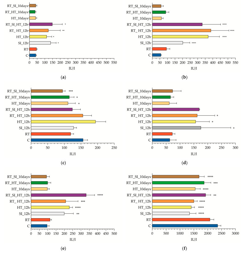
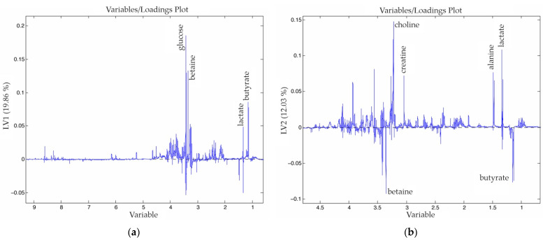
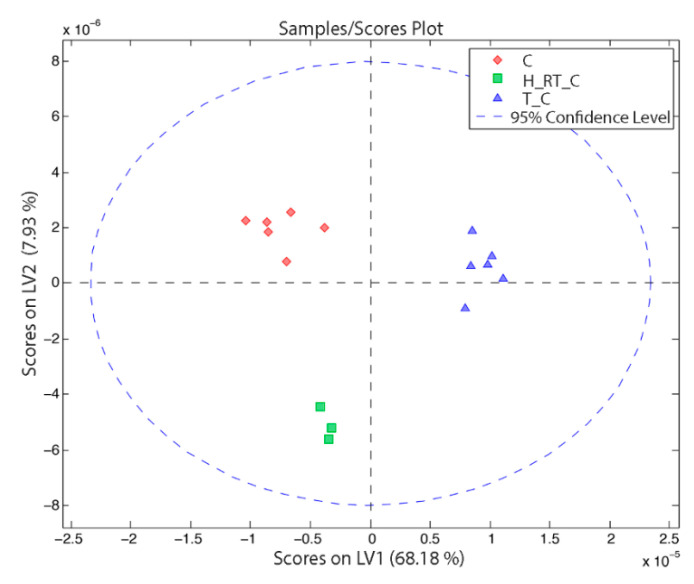
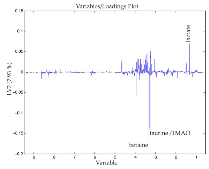
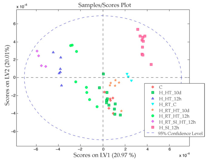
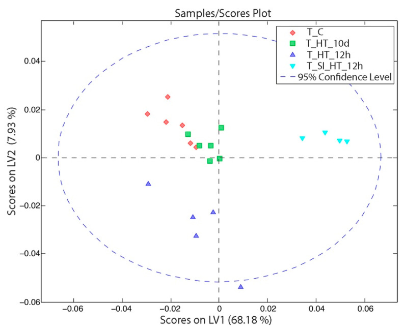
WAG/RigHsd rats were assigned to the different experimental groups needed to emulate all of the procedures involved in the treatment of liver metastases by HT. Twelve hours or ten days after the electromagnetic HT (606 kHz and 14 kA/m during 21 min), blood samples were retrieved and liver samples were obtained. 1H-nuclear-magnetic-resonance spectroscopy (1H-NMR) was used to search for possible diagnostic biomarkers of HT effects on the rat liver tissue. All of the data obtained from the hydrophilic fraction of the tissues were analyzed and modeled using chemometric tools.
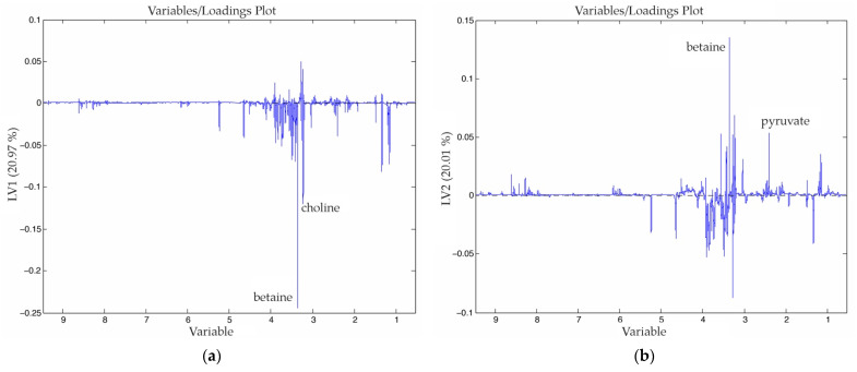
Hepatic enzyme levels were significantly increased in animals that underwent hyperthermia after 12 h, but 10 d later they could not be detected anymore. The metabolomic profile (main metabolic differences were found in phosphatidylcholine, taurine, glucose, lactate and pyruvate, among others) also showed that the therapy significantly altered metabolism in the liver within 12 h (with two different patterns); however, those changes reverted to a control-profile pattern after 10 days.
Magnetic hyperthermia could be considered as a safe therapy to treat liver metastases, since it does not induce irreversible physiological changes after application.
就其生物学和治疗效果而言,热疗(HT)疗法仍然相对鲜为人知。这项工作旨在使用临床实践中常规分析的代谢组学和血清参数,分析暴露于热疗(如抗肿瘤磁热疗疗法中所需的热疗)的效果。
将WAG/RigHsd大鼠分配到不同的实验组,以模拟热疗治疗肝转移瘤所涉及的所有程序。在电磁热疗(606kHz和14kA/m,持续21分钟)后12小时或10天,采集血样并获取肝脏样本。使用1H-核磁共振波谱(1H-NMR)来寻找热疗对大鼠肝脏组织影响的可能诊断生物标志物。使用化学计量工具对从组织的亲水性部分获得的所有数据进行分析和建模。
热疗后12小时,动物的肝酶水平显著升高,但10天后无法再检测到。代谢组学谱(主要代谢差异存在于磷脂酰胆碱、牛磺酸、葡萄糖、乳酸和丙酮酸等之中)也表明,该疗法在12小时内显著改变了肝脏的代谢(有两种不同模式);然而,这些变化在10天后恢复到对照谱模式。
磁热疗可被视为一种治疗肝转移瘤的安全疗法,因为应用后它不会引起不可逆的生理变化。