Chrzan Robert, Bociąga-Jasik Monika, Bryll Amira, Grochowska Anna, Popiela Tadeusz
Department of Radiology, Jagiellonian University Medical College, Kopernika 19, 31-501 Krakow, Poland.
Department of Infectious Diseases, Jagiellonian University Medical College, Jakubowskiego 2, 30-688 Krakow, Poland.
J Pers Med. 2021 May 10;11(5):391. doi: 10.3390/jpm11050391.
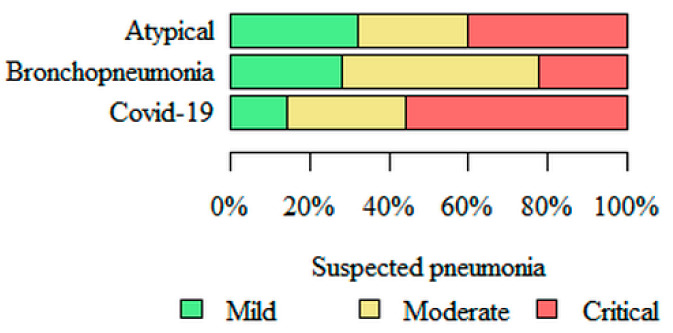
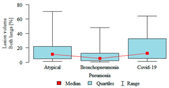
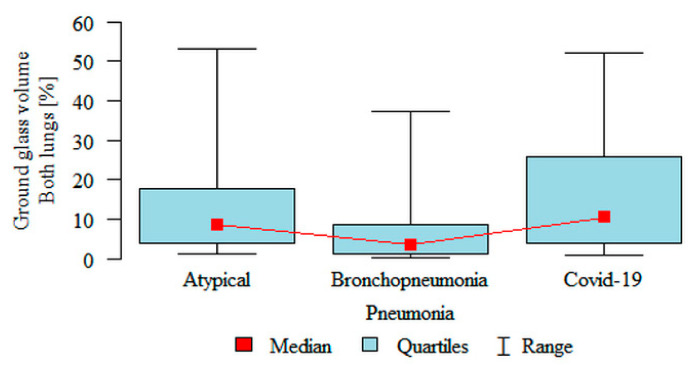
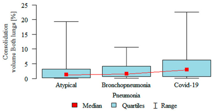
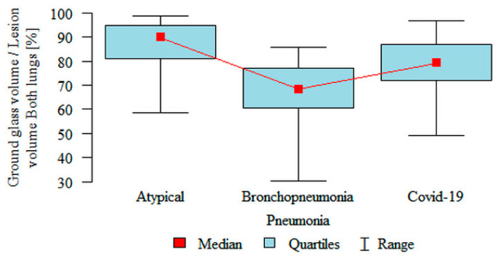
The aim of this study was to compare the results of automatic assessment of high resolution computed tomography (HRCT) by artificial intelligence (AI) in 150 patients from three subgroups: pneumonia in the course of COVID-19, bronchopneumonia and atypical pneumonia. The volume percentage of inflammation and the volume percentage of "ground glass" were significantly higher in the atypical (respectively, 11.04%, 8.61%) and the COVID-19 (12.41%, 10.41%) subgroups compared to the bronchopneumonia (5.12%, 3.42%) subgroup. The volume percentage of consolidation was significantly higher in the COVID-19 (2.95%) subgroup compared to the atypical (1.26%) subgroup. The percentage of "ground glass" in the volume of inflammation was significantly higher in the atypical (89.85%) subgroup compared to the COVID-19 (79.06%) subgroup, which in turn was significantly higher compared to the bronchopneumonia (68.26%) subgroup. HRCT chest images, analyzed automatically by artificial intelligence software, taking into account the structure including "ground glass" and consolidation, significantly differ in three subgroups: COVID-19 pneumonia, bronchopneumonia and atypical pneumonia. However, the partial overlap, particularly between COVID-19 pneumonia and atypical pneumonia, may limit the usefulness of automatic analysis in differentiating the etiology. In our future research, we plan to use artificial intelligence for objective assessment of the dynamics of pulmonary lesions during COVID-19 pneumonia.
本研究的目的是比较人工智能(AI)对150例患者高分辨率计算机断层扫描(HRCT)的自动评估结果,这些患者分为三个亚组:新冠病毒病(COVID-19)病程中的肺炎、支气管肺炎和非典型肺炎。与支气管肺炎亚组(分别为5.12%、3.42%)相比,非典型肺炎亚组(分别为11.04%、8.61%)和COVID-19亚组(12.41%、10.41%)的炎症体积百分比和“磨玻璃”体积百分比显著更高。与非典型肺炎亚组(1.26%)相比,COVID-19亚组(2.95%)的实变体积百分比显著更高。与COVID-19亚组(79.06%)相比,非典型肺炎亚组(89.85%)炎症体积中的“磨玻璃”百分比显著更高,而COVID-19亚组又显著高于支气管肺炎亚组(68.26%)。通过人工智能软件自动分析的胸部HRCT图像,考虑到包括“磨玻璃”和实变在内的结构,在COVID-19肺炎、支气管肺炎和非典型肺炎这三个亚组中存在显著差异。然而,部分重叠,尤其是COVID-19肺炎和非典型肺炎之间的重叠,可能会限制自动分析在鉴别病因方面的实用性。在我们未来的研究中,我们计划使用人工智能对COVID-19肺炎期间肺部病变的动态进行客观评估。