Godoy Eduardo Pires, Alccayhuaman Karol Alí Apaza, Botticelli Daniele, Amaroli Andrea, Balan Vitor Ferreira, Silva Erick Ricardo, Xavier Samuel Porfirio
Department of Oral Biology, Faculty of Dentistry of Ribeirão Preto, University of São Paulo, São Paulo 14040-904, Brazil.
Department of Oral Biology, Medical University of Vienna, 1090 Vienna, Austria.
Dent J (Basel). 2021 May 31;9(6):61. doi: 10.3390/dj9060061.
Due to the lack of data on bone-to-graft contact (BGC) over time in the various regions within the subantral space of the augmented sinus floor, the present study aimed to evaluate the osteoconductivity of deproteinized bovine bone mineral (DBBM) with granules of different sizes applied in maxillary sinus floor elevation.
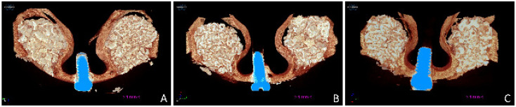
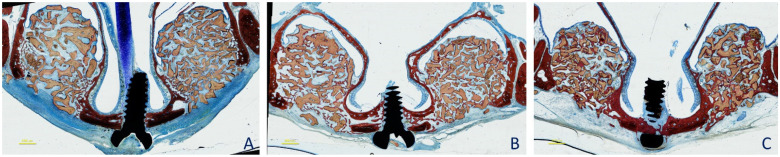
A maxillary sinus augmentation was performed bilaterally in 18 rabbits using DBBM with particle dimensions of either 0.125-1.0 mm or 1-2 mm. The antrostomy was covered using a collagen barrier. The animals were euthanized in groups of six after 2, 4, and 8 weeks of healing. MicroCT and histological analyses were performed.
After 2 weeks of healing, BGC was 10.9% and 11.9% for the small and large granule sites, respectively. After 8 weeks of healing, the BGC increased to 65% and 62% at the small and large granule sites, respectively. The highest values were located close to the bony walls and the bony window. New bone content developed between 2 and 8 weeks from 7.0% to 27.6% and from 6.1% to 27.6% at the small and large granule sites, respectively.
Similar outcomes in osteoconductivity and bone formation were found at both small and large DBBM granule sites.
由于缺乏关于上颌窦底提升术后窦下间隙不同区域骨与移植物接触(BGC)随时间变化的数据,本研究旨在评估不同尺寸颗粒的脱蛋白牛骨矿物质(DBBM)在上颌窦底提升术中的骨传导性。
对18只兔子双侧进行上颌窦提升术,使用粒径为0.125 - 1.0毫米或1 - 2毫米的DBBM。使用胶原屏障覆盖造瘘口。在愈合2、4和8周后,将动物按每组6只进行安乐死。进行了MicroCT和组织学分析。
愈合2周后,小颗粒位点和大颗粒位点的BGC分别为10.9%和11.9%。愈合8周后,小颗粒位点和大颗粒位点的BGC分别增加到65%和62%。最高值位于靠近骨壁和骨窗处。在2至8周之间,小颗粒位点和大颗粒位点的新骨含量分别从7.0%增加到27.6%和从6.1%增加到27.6%。
在DBBM小颗粒位点和大颗粒位点发现了相似的骨传导性和骨形成结果。