Leavesley Silas J, Deal Joshua, Hill Shante, Martin Will A, Lall Malvika, Lopez Carmen, Rider Paul F, Rich Thomas C, Boudreaux Carole W
Department of Chemical and Biomolecular Engineering, University of South Alabama.
Department of Pharmacology, University of South Alabama.
Proc SPIE Int Soc Opt Eng. 2018 Jan-Feb;10489. doi: 10.1117/12.2290696. Epub 2018 Feb 19.
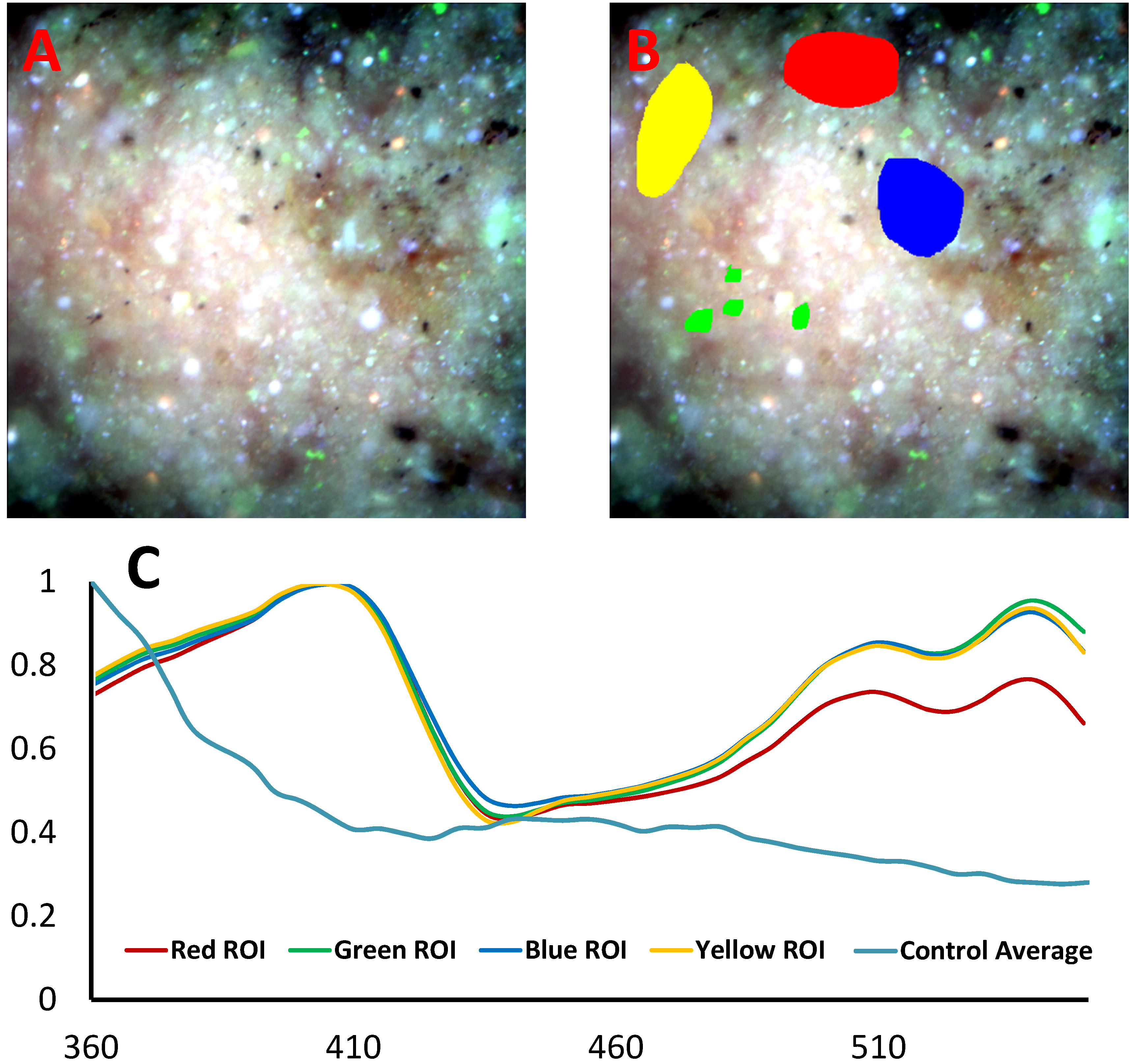
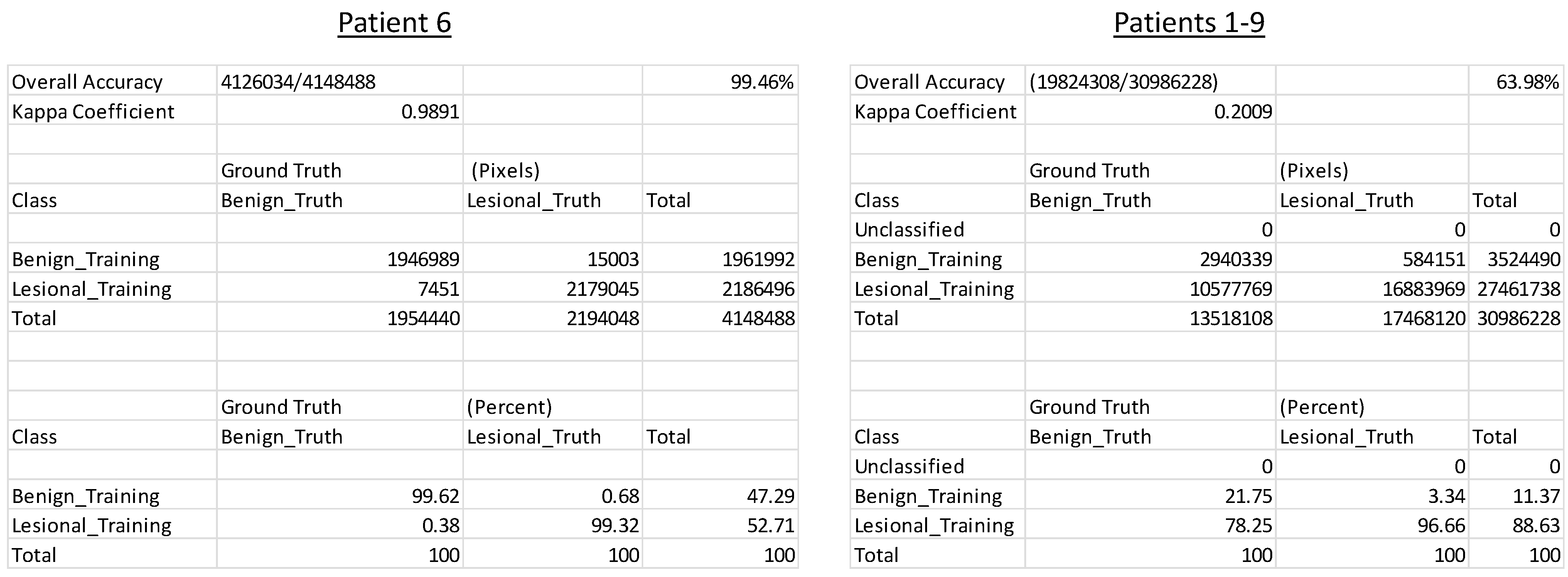
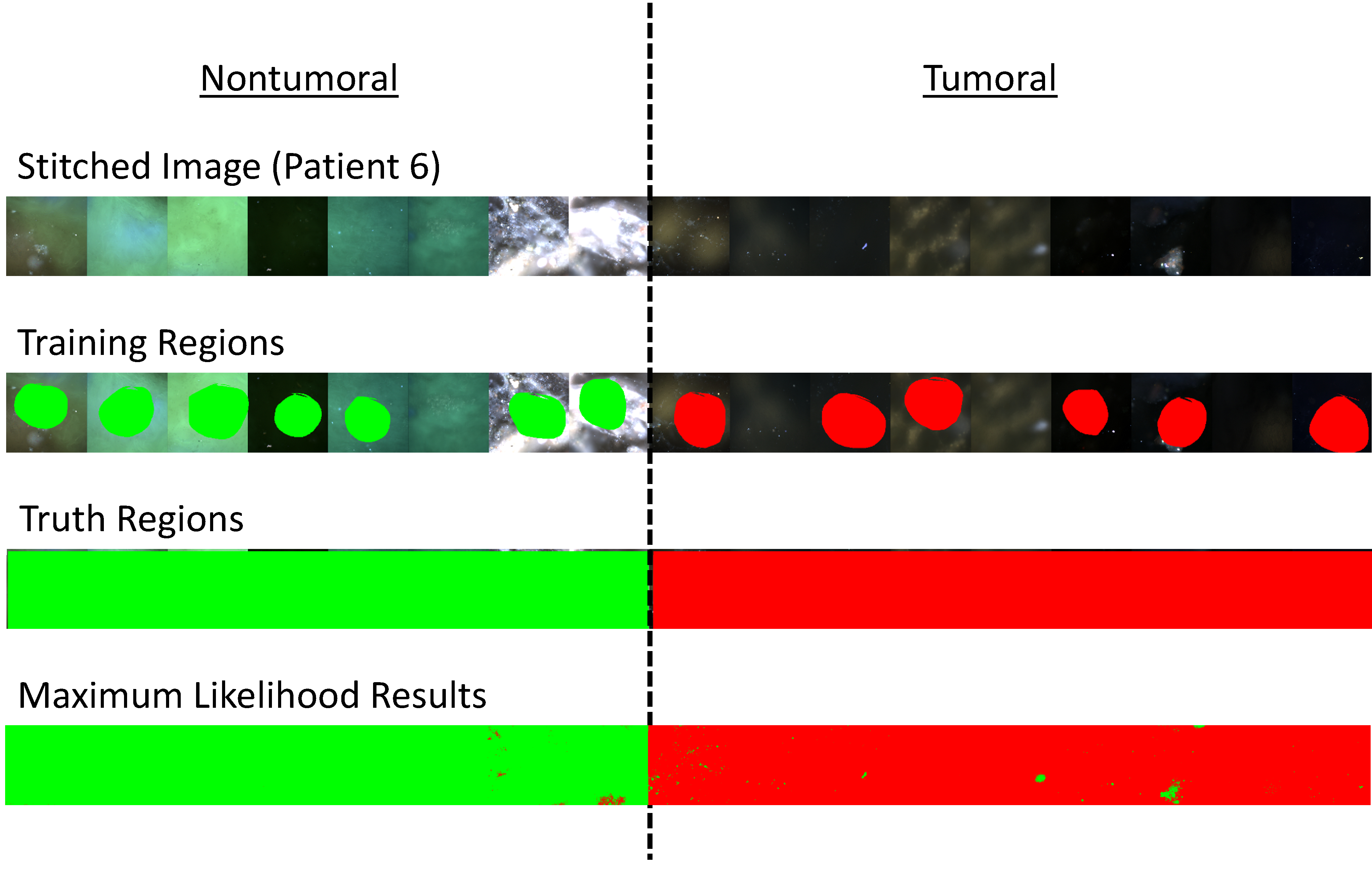
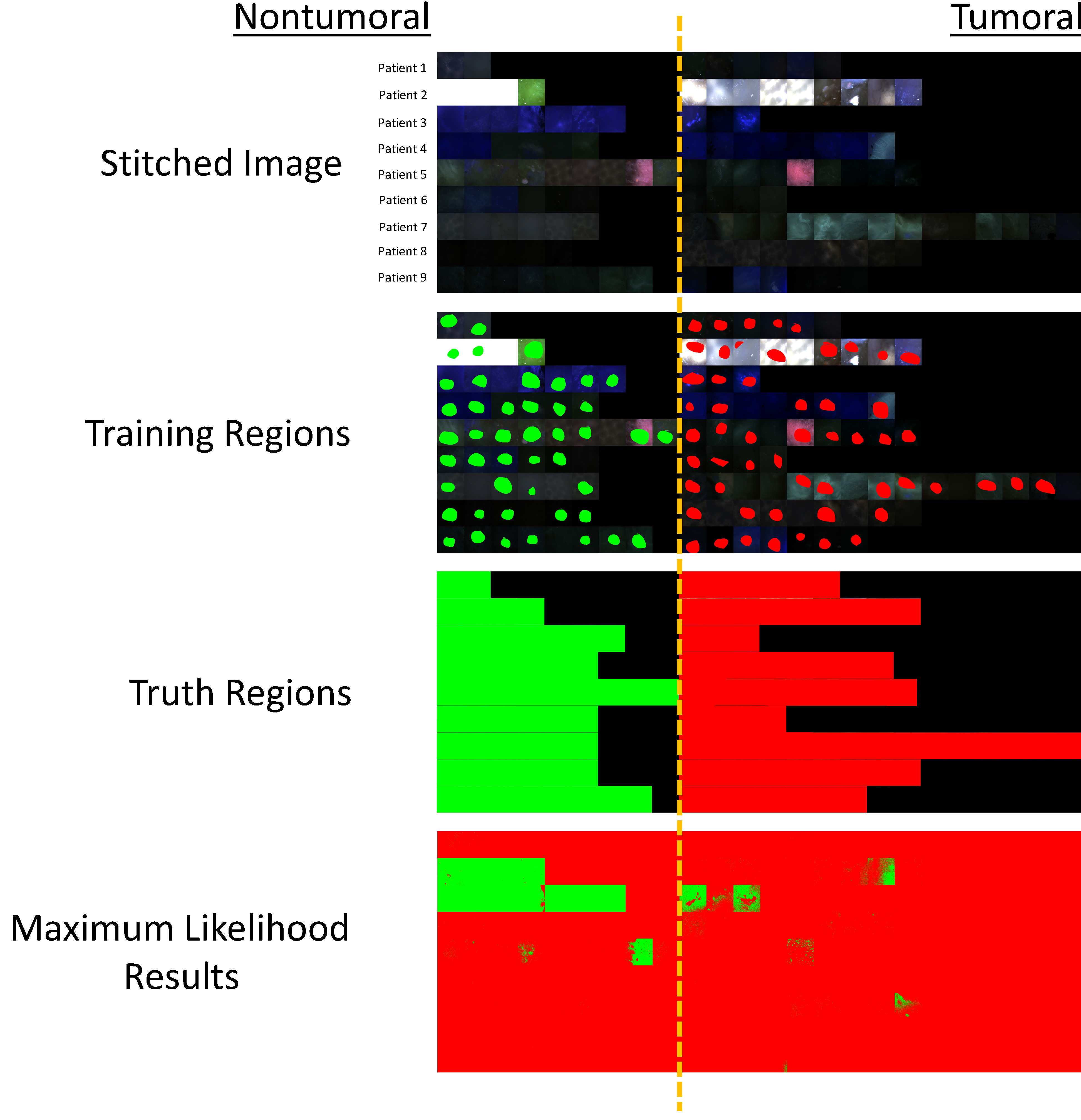
Hyperspectral imaging technologies have shown great promise for biomedical applications. These techniques have been especially useful for detection of molecular events and characterization of cell, tissue, and biomaterial composition. Unfortunately, hyperspectral imaging technologies have been slow to translate to clinical devices - likely due to increased cost and complexity of the technology as well as long acquisition times often required to sample a spectral image. We have demonstrated that hyperspectral imaging approaches which scan the fluorescence excitation spectrum can provide increased signal strength and faster imaging, compared to traditional emission-scanning approaches. We have also demonstrated that excitation-scanning approaches may be able to detect spectral differences between colonic adenomas and adenocarcinomas and normal mucosa in flash-frozen tissues. Here, we report feasibility results from using excitation-scanning hyperspectral imaging to screen pairs of fresh tumoral and nontumoral colorectal tissues. Tissues were imaged using a novel hyperspectral imaging fluorescence excitation scanning microscope, sampling a wavelength range of 360-550 nm, at 5 nm increments. Image data were corrected to achieve a NIST-traceable flat spectral response. Image data were then analyzed using a range of supervised and unsupervised classification approaches within ENVI software (Harris Geospatial Solutions). Supervised classification resulted in >99% accuracy for single-patient image data, but only 64% accuracy for multi-patient classification (n=9 to date), with the drop in accuracy due to increased false-positive detection rates. Hence, initial data indicate that this approach may be a viable detection approach, but that larger patient sample sizes need to be evaluated and the effects of inter-patient variability studied.
高光谱成像技术在生物医学应用方面展现出了巨大潜力。这些技术对于检测分子事件以及表征细胞、组织和生物材料的组成特别有用。不幸的是,高光谱成像技术在转化为临床设备方面进展缓慢,这可能是由于该技术成本增加、复杂性提高,以及采集光谱图像通常需要较长的采集时间。我们已经证明,与传统的发射扫描方法相比,扫描荧光激发光谱的高光谱成像方法能够提供更高的信号强度和更快的成像速度。我们还证明,激发扫描方法或许能够检测冷冻组织中结肠腺瘤、腺癌和正常黏膜之间的光谱差异。在此,我们报告使用激发扫描高光谱成像技术筛选新鲜肿瘤和非肿瘤结直肠组织对的可行性结果。使用一种新型高光谱成像荧光激发扫描显微镜对组织进行成像,采样波长范围为360 - 550 nm,以5 nm为增量。对图像数据进行校正以实现可溯源至美国国家标准与技术研究院(NIST)的平坦光谱响应。然后在ENVI软件(Harris Geospatial Solutions)中使用一系列监督和非监督分类方法对图像数据进行分析。监督分类对于单患者图像数据的准确率>99%,但对于多患者分类(截至目前n = 9)的准确率仅为64%,准确率下降是由于假阳性检测率增加所致。因此,初步数据表明该方法可能是一种可行的检测方法,但需要评估更大的患者样本量,并研究患者间变异性的影响。