Hu Wenxue, Li Guanglan, Lin Jieshan, Dong Wei, Yu Feng, Liu Wei, Wu Yanhua, Hao Wenke, Liang Xinling
Department of Nephrology, Guangdong Provincial People's Hospital, Guangdong Academy of Medical Sciences, Guangdong Provincial Geriatrics Institute, Guangzhou, China.
Shantou University Medical College, Shantou, China.
Front Med (Lausanne). 2021 May 21;8:657232. doi: 10.3389/fmed.2021.657232. eCollection 2021.
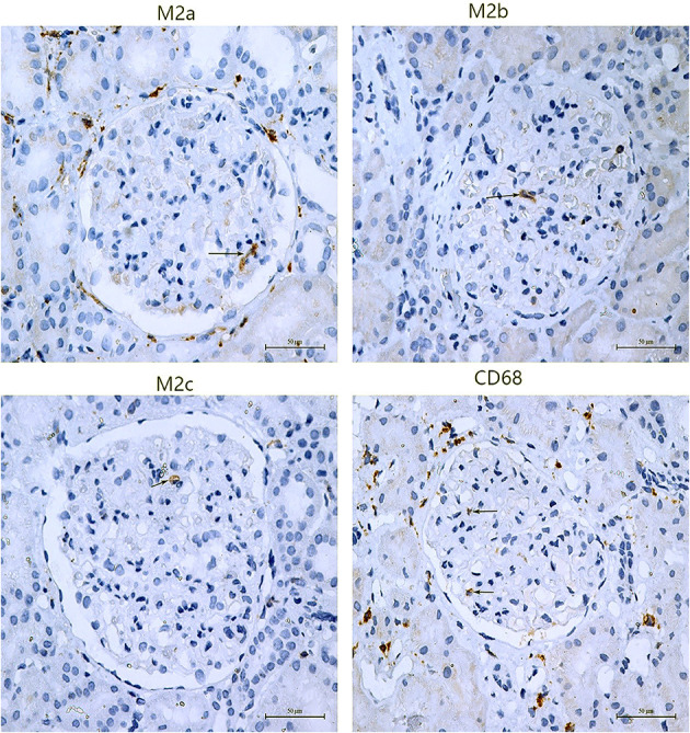
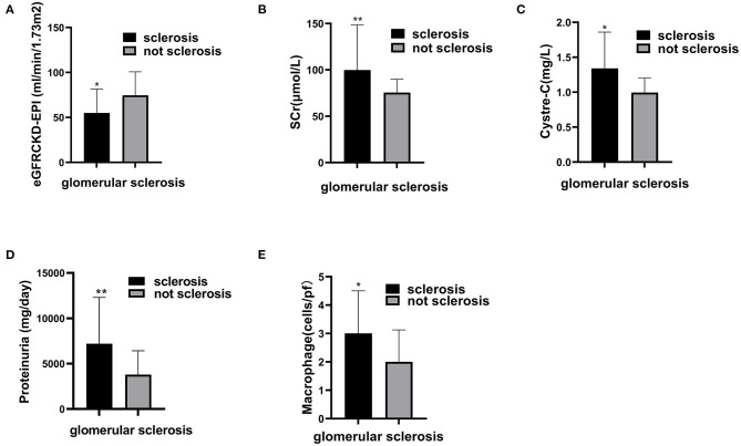
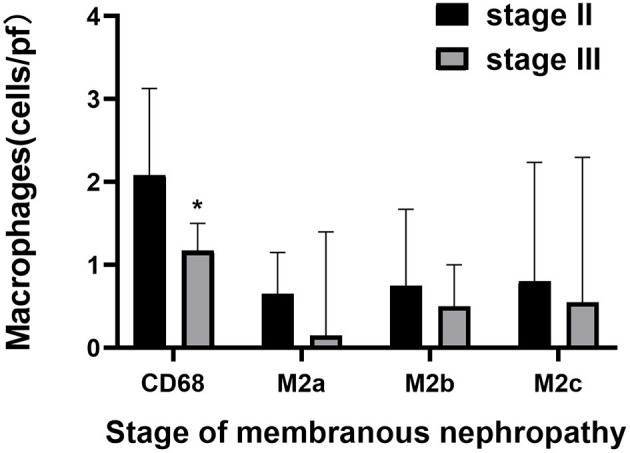
The role of M2 macrophages in the pathogenesis and progression of primary membranous nephropathy (PMN) remains unknown. In this study, we aimed to investigate the relationship between M2 subsets and clinicopathological features of patients with PMN. A total of 55 patients with PMN confirmed by biopsy were recruited. The clinical and pathological data were recorded, respectively. Immunohistochemistry was used to detect the markers of M2 macrophages, including total macrophages (CD68+), M2a (CD206+), M2b (CD86+) and M2c (CD163+). The numbers of glomerular macrophages, M2a, M2b, and M2c macrophages were 1.83 (1.00, 2.67), 0.65 (0.15, 1.15), 0.67 (0.33, 1.50), and 0.80 (0.05, 2.30) per glomerulus, respectively. Higher number of glomerular macrophages was found in stage II compared with stage III (2.08 vs. 1.16, = 0.008). These macrophages also were negatively correlated with serum albumin level ( = -0.331, = 0.014), while positively associated with complement 3 (C3) deposition ( = 0.300, = 0.026) and the severity of glomerulosclerosis ( = 0.276, = 0.041). Moreover, glomerular M2a macrophages were significantly correlated with the deposition of C3 ( = 0.300, = 0.026), immunoglobulin G1 (IgG1) ( = 0.339, = 0.011), immunoglobulin G2 (IgG2) ( = 0.270, = 0.046) and immunoglobulin G3 (IgG3) ( = 0.330, = 0.014) in glomerular basement membrane (GBM). In addition, M2b macrophages were positively associated with IgG1 ( = 0.295, = 0.029) and IgG2 ( = 0.393, = 0.003), while M2c macrophages were negatively correlated with complement 4d (C4d) ( = -0.347, = 0.009) in GBM. Our results showed that M2 macrophage subpopulations in glomeruli are associated with the deposition of IgG subclasses and complements in renal tissue of PMN, which indicate that M2 macrophages may be involved in the pathogenesis and progression of PMN. Moreover, M2a and M2c macrophages might show different tendencies in the pathogenesis of PMN.
M2巨噬细胞在原发性膜性肾病(PMN)发病机制及疾病进展中的作用尚不清楚。在本研究中,我们旨在探究M2亚群与PMN患者临床病理特征之间的关系。共纳入55例经活检确诊的PMN患者。分别记录其临床和病理数据。采用免疫组织化学法检测M2巨噬细胞标志物,包括总巨噬细胞(CD68+)、M2a(CD206+)、M2b(CD86+)和M2c(CD163+)。每个肾小球中肾小球巨噬细胞、M2a、M2b和M2c巨噬细胞的数量分别为1.83(1.00,2.67)、0.65(0.15,1.15)、0.67(0.33,1.50)和0.80(0.05,2.30)。与Ⅲ期相比,Ⅱ期患者肾小球巨噬细胞数量更多(2.08对1.16,P = 0.008)。这些巨噬细胞还与血清白蛋白水平呈负相关(r = -0.331,P = 0.014),而与补体3(C3)沉积呈正相关(r = 0.300,P = 0.026)以及与肾小球硬化严重程度呈正相关(r = 0.276,P = 0.041)。此外,肾小球M2a巨噬细胞与肾小球基底膜(GBM)中C3沉积(r = 0.300,P = 0.026)、免疫球蛋白G1(IgG1)(r = 0.339,P = 0.011)、免疫球蛋白G2(IgG2)(r = 0.270,P = 0.046)和免疫球蛋白G3(IgG3)(r = 0.330,P = 0.014)显著相关。另外,M2b巨噬细胞与GBM中IgG1(r = 0.295,P = 0.029)和IgG2(r = 0.393,P = 0.003)呈正相关,而M2c巨噬细胞与GBM中补体4d(C4d)呈负相关(r = -0.347,P = 0.009)。我们的结果表明,肾小球中的M2巨噬细胞亚群与PMN肾组织中IgG亚类及补体的沉积相关,这表明M2巨噬细胞可能参与PMN的发病机制及疾病进展。此外,M2a和M2c巨噬细胞在PMN发病机制中可能表现出不同趋势。