Department of Rheumatology and Clinical Immunology, Medical Center - University of Freiburg, University of Freiburg, Freiburg, Germany.
Front Immunol. 2021 May 24;12:673912. doi: 10.3389/fimmu.2021.673912. eCollection 2021.
ANCA-associated vasculitides (AAV) affect small- and medium-sized blood vessels. In active disease, vessel wall infiltrates are mainly composed of monocytes and macrophages. Immune checkpoint molecules are crucial for the maintenance of self-tolerance and the prevention of autoimmune diseases. After checkpoint inhibitor therapy, the development of autoimmune vasculitis has been observed. However, defects of immune checkpoint molecules in AAV patients have not been identified yet.
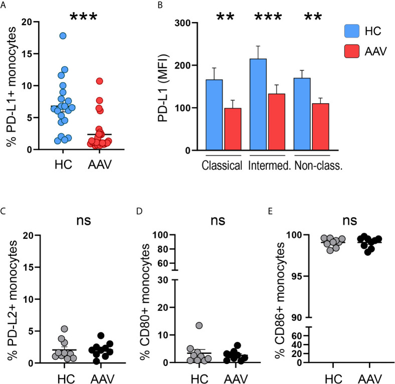
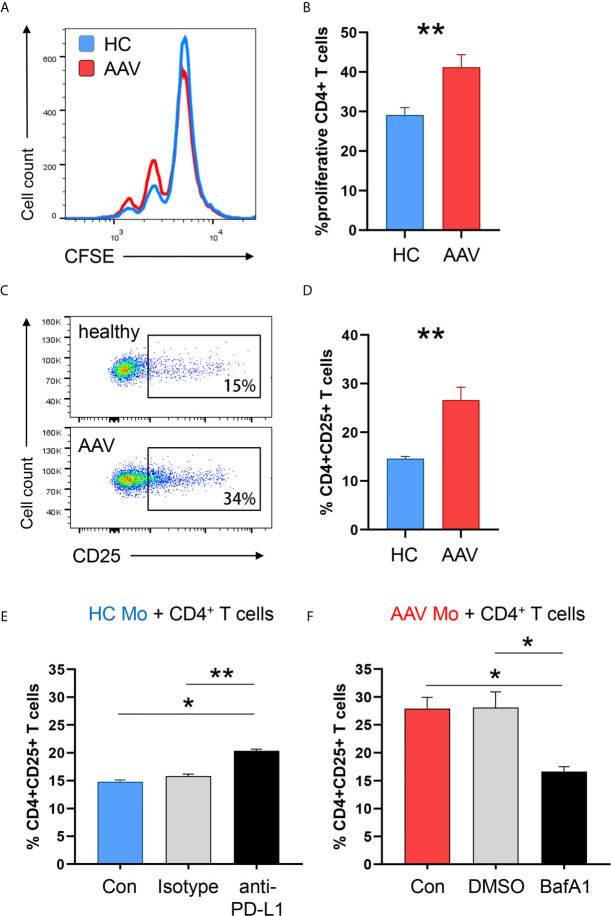
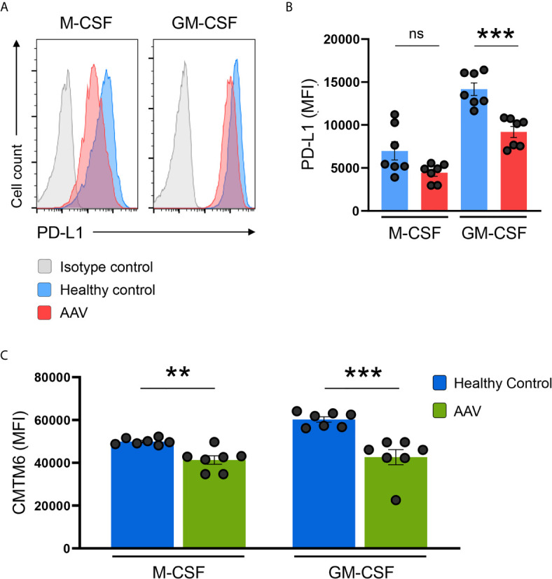
Monocytes and monocyte-derived macrophages from AAV patients and healthy age-matched controls were tested for surface expression of immunoinhibitory checkpoint programmed cell death ligand-1 (PD-L1). Using co-culture approaches, the effect of monocyte PD-L1 expression on CD4 T cell activation and proliferation was tested.
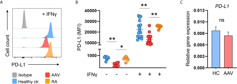
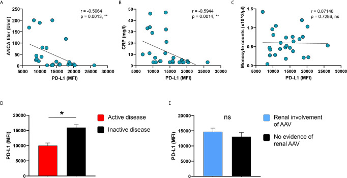
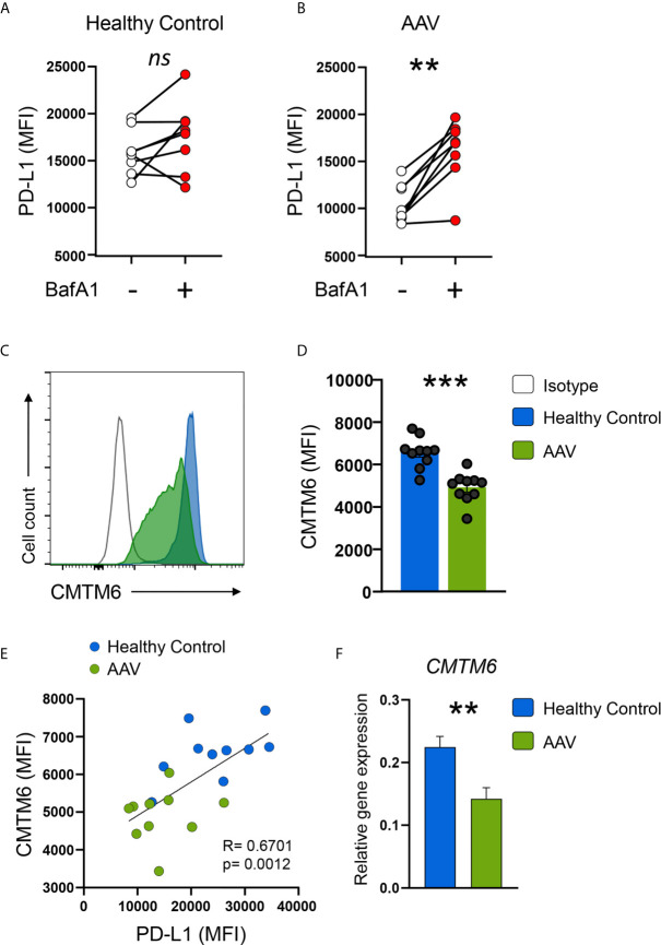
Monocytes from AAV patients displayed lower PD-L1 expression and a defective PD-L1 presentation upon activation, an effect that was correlated with disease activity. Lower PD-L1 expression was due to increased lysosomal degradation of PD-L1 in AAV monocytes. We identified a reduced expression of CMTM6, a protein protecting PD-L1 from lysosomal breakdown, as the underlying molecular defect. PD-L1 AAV monocytes showed increased stimulatory capacity and induced T cell activation and proliferation. Inhibiting lysosomal function corrected this phenotype by increasing PD-L1, thus normalizing the pro-stimulatory behavior of AAV monocytes.
This study identifies a defect of the immunoinhibitory checkpoint PD-L1 in monocytes from patients with AAV. Low expression of CMTM6 results in enhanced lysosomal degradation of PD-L1, thus providing insufficient negative signaling to T cells. Correcting this defect by targeting lysosomal function may represent a novel strategy to treat AAV.
抗中性粒细胞胞浆抗体相关性血管炎(AAV)影响小血管和中等大小的血管。在活动期疾病中,血管壁浸润主要由单核细胞和巨噬细胞组成。免疫检查点分子对于维持自身耐受和预防自身免疫性疾病至关重要。在接受免疫检查点抑制剂治疗后,已观察到自身免疫性血管炎的发展。然而,AAV 患者的免疫检查点分子缺陷尚未确定。
检测 AAV 患者和年龄匹配的健康对照者的单核细胞和单核细胞衍生的巨噬细胞表面表达免疫抑制性检查点程序性细胞死亡配体-1(PD-L1)。采用共培养方法,检测单核细胞 PD-L1 表达对 CD4 T 细胞活化和增殖的影响。
AAV 患者的单核细胞表达 PD-L1 水平较低,活化后 PD-L1 呈递缺陷,这种效应与疾病活动度相关。PD-L1 表达降低是由于 AAV 单核细胞中 PD-L1 的溶酶体降解增加所致。我们发现,CMTM6 的表达减少是导致这一现象的根本分子缺陷,CMTM6 是一种保护 PD-L1 免受溶酶体破坏的蛋白。PD-L1 AAV 单核细胞表现出增强的刺激能力,并诱导 T 细胞活化和增殖。抑制溶酶体功能可通过增加 PD-L1 来纠正这种表型,从而使 AAV 单核细胞的促刺激行为恢复正常。
本研究确定了 AAV 患者单核细胞中免疫抑制性检查点 PD-L1 的缺陷。CMTM6 表达减少导致 PD-L1 的溶酶体降解增加,从而向 T 细胞提供的负信号不足。通过靶向溶酶体功能纠正这种缺陷可能代表治疗 AAV 的一种新策略。