• 文献检索
  • 文档翻译
  • 深度研究
  • 学术资讯
  • Suppr Zotero 插件Zotero 插件
  • 邀请有礼
  • 套餐&价格
  • 历史记录
应用&插件
Suppr Zotero 插件Zotero 插件浏览器插件Mac 客户端Windows 客户端微信小程序
定价
高级版会员购买积分包购买API积分包
服务
文献检索文档翻译深度研究API 文档MCP 服务
关于我们
关于 Suppr公司介绍联系我们用户协议隐私条款
关注我们

Suppr 超能文献

核心技术专利:CN118964589B侵权必究
粤ICP备2023148730 号-1Suppr @ 2026

文献检索

告别复杂PubMed语法,用中文像聊天一样搜索,搜遍4000万医学文献。AI智能推荐,让科研检索更轻松。

立即免费搜索

文件翻译

保留排版,准确专业,支持PDF/Word/PPT等文件格式,支持 12+语言互译。

免费翻译文档

深度研究

AI帮你快速写综述,25分钟生成高质量综述,智能提取关键信息,辅助科研写作。

立即免费体验

症状性单侧颈动脉闭塞患者的软脑膜侧支激活表明其脑血管储备能力严重受损。

Leptomeningeal collateral activation indicates severely impaired cerebrovascular reserve capacity in patients with symptomatic unilateral carotid artery occlusion.

机构信息

Department of Neurosurgery, University Hospital Zurich, University of Zurich, Zurich, Switzerland.

Clinical Neuroscience Center, University Hospital Zurich, University of Zurich, Zurich, Switzerland.

出版信息

J Cereb Blood Flow Metab. 2021 Nov;41(11):3039-3051. doi: 10.1177/0271678X211024373. Epub 2021 Jun 10.

DOI:10.1177/0271678X211024373
PMID:34112002
原文链接:https://pmc.ncbi.nlm.nih.gov/articles/PMC8545056/
Abstract

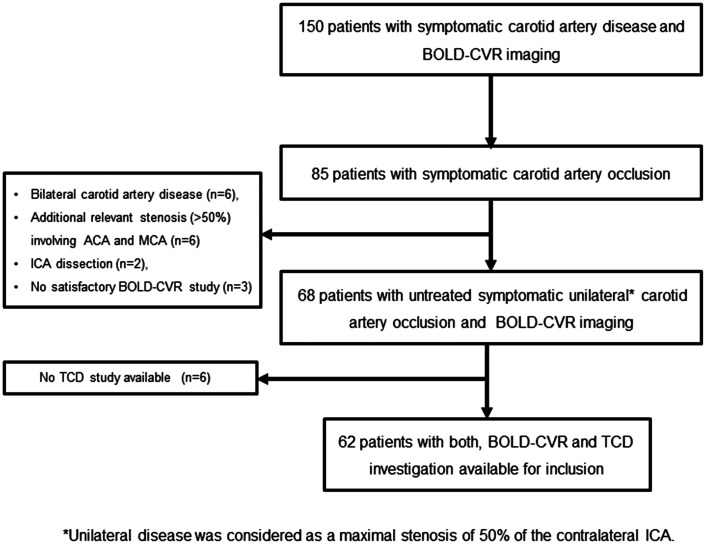
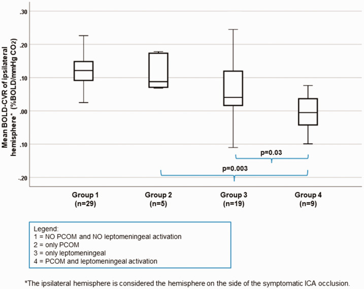
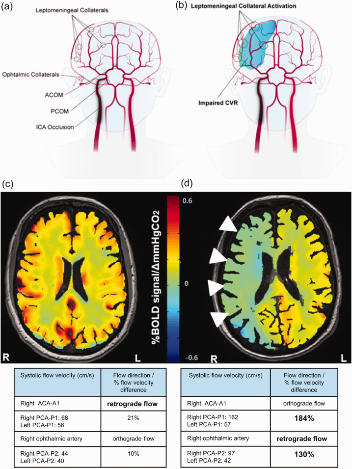
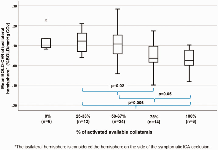
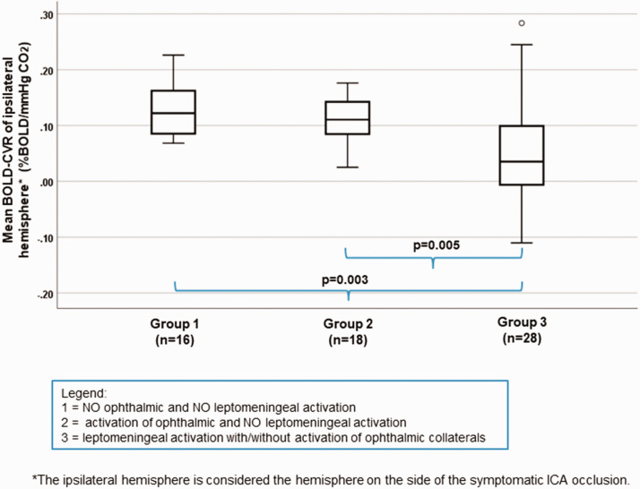
For patients with symptomatic unilateral internal carotid artery (ICA) occlusion, impaired cerebrovascular reactivity (CVR) indicates increased stroke risk. Here, the role of collateral activation remains a matter of debate, whereas angio-anatomical collateral abundancy does not necessarily imply sufficient compensatory flow provided. We aimed to further elucidate the role of collateral activation in the presence of impaired CVR. From a prospective database, 62 patients with symptomatic unilateral ICA occlusion underwent blood oxygenation-level dependent (BOLD) fMRI CVR imaging and a transcranial Doppler (TCD) investigation for primary and secondary collateral activation. Descriptive statistic and multivariate analysis were used to evaluate the relationship between BOLD-CVR values and collateral activation. Patients with activated secondary collaterals exhibited more impaired BOLD-CVR values of the ipsilateral hemisphere (p = 0.02). Specifically, activation of leptomeningeal collaterals showed severely impaired ipsilateral hemisphere BOLD-CVR values when compared to activation of ophthalmic collaterals (0.05 ± 0.09 vs. 0.12 ± 0.04, p = 0.005). Moreover, the prediction analysis showed leptomeningeal collateral activation as a strong independent predictor for ipsilateral hemispheric BOLD-CVR. In our study, ipsilateral leptomeningeal collateral activation is the sole collateral pathway associated with severely impaired BOLD-CVR in patients with symptomatic unilateral ICA occlusion.

摘要

对于有症状的单侧颈内动脉(ICA)闭塞的患者,脑血管反应性(CVR)受损表明中风风险增加。在这里,侧支激活的作用仍然存在争议,而血管解剖学侧支丰富并不一定意味着提供了足够的代偿性血流。我们旨在进一步阐明在 CVR 受损的情况下侧支激活的作用。从一个前瞻性数据库中,我们选择了 62 名有症状的单侧 ICA 闭塞患者,他们接受了血氧水平依赖(BOLD)功能磁共振成像 CVR 成像和经颅多普勒(TCD)检查,以评估原发性和继发性侧支激活。我们使用描述性统计和多元分析来评估 BOLD-CVR 值与侧支激活之间的关系。表现出继发性侧支激活的患者表现出同侧半球更严重受损的 BOLD-CVR 值(p=0.02)。具体来说,与眼动脉侧支相比,软脑膜侧支的激活显示出严重受损的同侧半球 BOLD-CVR 值(0.05±0.09 与 0.12±0.04,p=0.005)。此外,预测分析显示软脑膜侧支激活是同侧半球 BOLD-CVR 的一个强烈独立预测因子。在我们的研究中,同侧软脑膜侧支激活是唯一与症状性单侧 ICA 闭塞患者严重受损的 BOLD-CVR 相关的侧支途径。

https://cdn.ncbi.nlm.nih.gov/pmc/blobs/f97b/8756473/b75509ba7fa0/10.1177_0271678X211024373-fig5.jpg
https://cdn.ncbi.nlm.nih.gov/pmc/blobs/f97b/8756473/2df0b2402056/10.1177_0271678X211024373-fig1.jpg
https://cdn.ncbi.nlm.nih.gov/pmc/blobs/f97b/8756473/7874fffbfe32/10.1177_0271678X211024373-fig2.jpg
https://cdn.ncbi.nlm.nih.gov/pmc/blobs/f97b/8756473/3de53c69dc6f/10.1177_0271678X211024373-fig3.jpg
https://cdn.ncbi.nlm.nih.gov/pmc/blobs/f97b/8756473/be8fc52812a0/10.1177_0271678X211024373-fig4.jpg
https://cdn.ncbi.nlm.nih.gov/pmc/blobs/f97b/8756473/b75509ba7fa0/10.1177_0271678X211024373-fig5.jpg

相似文献

1
Leptomeningeal collateral activation indicates severely impaired cerebrovascular reserve capacity in patients with symptomatic unilateral carotid artery occlusion.症状性单侧颈动脉闭塞患者的软脑膜侧支激活表明其脑血管储备能力严重受损。
J Cereb Blood Flow Metab. 2021 Nov;41(11):3039-3051. doi: 10.1177/0271678X211024373. Epub 2021 Jun 10.
2
Reduced contralateral cerebrovascular reserve in patients with unilateral steno-occlusive disease.单侧狭窄闭塞性疾病患者对侧脑血管储备减少。
Cerebrovasc Dis. 2014;38(2):94-100. doi: 10.1159/000362084. Epub 2014 Oct 2.
3
Mapping Cerebrovascular Reactivity Impairment in Patients With Symptomatic Unilateral Carotid Artery Disease.绘制症状性单侧颈动脉疾病患者脑血管反应性损害图。
J Am Heart Assoc. 2021 Jun 15;10(12):e020792. doi: 10.1161/JAHA.121.020792. Epub 2021 Jun 9.
4
Predictors of poor cerebral collaterals and cerebrovascular reserve in patients with chronic total carotid occlusion.慢性颈总动脉闭塞患者脑侧支循环不良和脑血管储备的预测因素
Int J Neurosci. 2019 May;129(5):455-460. doi: 10.1080/00207454.2018.1538990. Epub 2018 Nov 27.
5
Carotid endarterectomy improves cerebrovascular reserve capacity preferentially in patients with preoperative impairment as indicated by asymmetric BOLD response to hypercapnia.如高碳酸血症时非对称BOLD反应所示,颈动脉内膜切除术优先改善术前有损伤患者的脑血管储备能力。
Eur J Vasc Endovasc Surg. 2009 Nov;38(5):546-51. doi: 10.1016/j.ejvs.2009.06.010. Epub 2009 Sep 9.
6
Bilateral severe carotid artery stenosis or occlusion - cerebral autoregulation dynamics and collateral flow patterns.双侧严重颈动脉狭窄或闭塞——脑自动调节动力学及侧支血流模式
Acta Neurochir (Wien). 2003 Dec;145(12):1053-9; discussion 1059-60. doi: 10.1007/s00701-003-0137-8. Epub 2003 Nov 3.
7
Flow-augmentation STA-MCA bypass for acute and subacute ischemic stroke due to internal carotid artery occlusion and the role of advanced neuroimaging with hemodynamic and flow-measurement in the decision-making: preliminary data.用于因颈内动脉闭塞所致急性和亚急性缺血性卒中的血流增强颞浅动脉-大脑中动脉搭桥术以及先进的血流动力学和血流测量神经影像学在决策中的作用:初步数据
Quant Imaging Med Surg. 2024 Jan 3;14(1):777-788. doi: 10.21037/qims-23-876. Epub 2024 Jan 2.
8
A longitudinal study of collateral flow patterns in the circle of Willis and the ophthalmic artery in patients with a symptomatic internal carotid artery occlusion.一项针对有症状的颈内动脉闭塞患者 Willis 环和眼动脉侧支血流模式的纵向研究。
Stroke. 2000 Aug;31(8):1913-20. doi: 10.1161/01.str.31.8.1913.
9
[The supratrochlear artery as an indicator of cerebral hemodynamics in carotid occlusion].[滑车上动脉作为颈动脉闭塞时脑血流动力学的指标]
Ultraschall Med. 1998 Jun;19(3):114-9. doi: 10.1055/s-2007-1000473.
10
Pattern of Activated Pathways and Quality of Collateral Status in Patients with Symptomatic Internal Carotid Artery Occlusion.症状性颈内动脉闭塞患者的激活通路模式和侧支循环状态的质量。
Cerebrovasc Dis. 2019;48(3-6):244-250. doi: 10.1159/000504663. Epub 2019 Dec 17.

引用本文的文献

1
Reperfusion failure after successful thrombectomy of large vessel occlusion stroke: clinical and imaging evidence.大血管闭塞性卒中成功取栓术后的再灌注失败:临床及影像学证据
Front Neurol. 2025 Aug 22;16:1639880. doi: 10.3389/fneur.2025.1639880. eCollection 2025.
2
Flow-Augmentation Bypass Surgery: Indications and Decision-Making.流量增加旁路手术:适应症与决策制定
Acta Neurochir Suppl. 2025;136:113-118. doi: 10.1007/978-3-031-89844-0_14.
3
The Value of Non-Invasive Optimal Vessel Analysis Quantitative Magnetic Resonance Angiography for Studying Flow and Collateral Patterns in Patients with Bilateral Carotid Steno-Occlusive Disease.

本文引用的文献

1
Carotid occlusion: Impact of cerebral hemodynamic impairment on cognitive performances.颈动脉闭塞:脑血流动力学障碍对认知功能的影响。
Int J Geriatr Psychiatry. 2021 Jan;36(1):197-206. doi: 10.1002/gps.5414. Epub 2020 Sep 11.
2
Crossed Cerebellar Diaschisis Indicates Hemodynamic Compromise in Ischemic Stroke Patients.交叉性小脑 diaschisis 提示缺血性脑卒中患者存在血液动力学障碍。
Transl Stroke Res. 2021 Feb;12(1):39-48. doi: 10.1007/s12975-020-00821-0. Epub 2020 Jun 6.
3
Impaired cerebrovascular reactivity is associated with recurrent stroke in patients with severe intracranial arterial stenosis: A C02 BOLD fMRI study.
无创性最佳血管分析定量磁共振血管造影在研究双侧颈动脉狭窄闭塞性疾病患者血流和侧支循环模式中的价值
Brain Sci. 2025 Feb 19;15(2):211. doi: 10.3390/brainsci15020211.
4
Recruitment of pial collaterals and carotid occlusive disease in large-vessel occlusion ischemic stroke.大脑中动脉闭塞性缺血性卒中软脑膜侧支循环的募集与颈动脉闭塞性疾病
Front Neurol. 2024 Oct 28;15:1423967. doi: 10.3389/fneur.2024.1423967. eCollection 2024.
5
Early Cerebral Microvasculature Impairment and Increased Body Mass Index in Patients with Psoriasis.银屑病患者早期脑微血管损伤与体重指数增加
Biomedicines. 2024 Jul 23;12(8):1627. doi: 10.3390/biomedicines12081627.
6
BOLD Cerebrovascular Reactivity and NOVA Quantitative MR Angiography in Adult Patients with Moyamoya Vasculopathy Undergoing Cerebral Bypass Surgery.大脑中动脉狭窄或闭塞性脑血管病成年患者行脑搭桥手术时的血氧水平依赖性功能磁共振成像脑血管反应性及NOVA定量磁共振血管造影
Brain Sci. 2024 Jul 29;14(8):762. doi: 10.3390/brainsci14080762.
7
Exploring ischemic core growth rate and endovascular therapy benefit in large core patients.探索大梗死核心患者的缺血核心生长速率及血管内治疗的益处。
J Cereb Blood Flow Metab. 2024 Dec;44(12):1593-1604. doi: 10.1177/0271678X241242911. Epub 2024 Jul 26.
8
Association of Carotid Artery Disease with Collateralization and Infarct Growth in Patients with Acute Middle Cerebral Artery Occlusion.颈动脉疾病与急性大脑中动脉闭塞患者的侧支循环和梗死灶生长的关系。
AJNR Am J Neuroradiol. 2024 May 9;45(5):574-580. doi: 10.3174/ajnr.A8180.
9
Flow-augmentation STA-MCA bypass for acute and subacute ischemic stroke due to internal carotid artery occlusion and the role of advanced neuroimaging with hemodynamic and flow-measurement in the decision-making: preliminary data.用于因颈内动脉闭塞所致急性和亚急性缺血性卒中的血流增强颞浅动脉-大脑中动脉搭桥术以及先进的血流动力学和血流测量神经影像学在决策中的作用:初步数据
Quant Imaging Med Surg. 2024 Jan 3;14(1):777-788. doi: 10.21037/qims-23-876. Epub 2024 Jan 2.
10
Hemodynamic Failure Staging With Blood Oxygenation Level-Dependent Cerebrovascular Reactivity and Acetazolamide-Challenged (O-)HO-Positron Emission Tomography Across Individual Cerebrovascular Territories.个体脑血管域内血氧水平依赖脑血管反应性和乙酰唑胺挑战 (O-)HO-正电子发射断层扫描的血液动力学衰竭分期。
J Am Heart Assoc. 2023 Dec 19;12(24):e029491. doi: 10.1161/JAHA.123.029491. Epub 2023 Dec 12.
脑血流反应性受损与颅内动脉重度狭窄患者的复发性卒中相关:一项 CO2 BOLD fMRI 研究。
J Neuroradiol. 2021 Sep;48(5):339-345. doi: 10.1016/j.neurad.2020.04.005. Epub 2020 May 17.
4
DSC perfusion-based collateral imaging and quantitative T2 mapping to assess regional recruitment of leptomeningeal collaterals and microstructural cortical tissue damage in unilateral steno-occlusive vasculopathy.基于 DSC 灌注的侧支循环成像和定量 T2 映射评估单侧狭窄性血管病变中的软脑膜侧支循环的区域性募集和皮质组织的微观结构损伤。
J Cereb Blood Flow Metab. 2021 Jan;41(1):67-81. doi: 10.1177/0271678X19898148. Epub 2020 Jan 27.
5
Pattern of Activated Pathways and Quality of Collateral Status in Patients with Symptomatic Internal Carotid Artery Occlusion.症状性颈内动脉闭塞患者的激活通路模式和侧支循环状态的质量。
Cerebrovasc Dis. 2019;48(3-6):244-250. doi: 10.1159/000504663. Epub 2019 Dec 17.
6
Transcranial Doppler ultrasonography in neurological surgery and neurocritical care.经颅多普勒超声在神经外科学和神经危重症中的应用。
Neurosurg Focus. 2019 Dec 1;47(6):E2. doi: 10.3171/2019.9.FOCUS19611.
7
Collateral grade of the Willis' circle predicts outcomes of acute intracranial internal carotid artery occlusion before thrombectomy.Willis 环侧支循环分级可预测急性颈内动脉颅内段闭塞取栓前的结局。
Brain Behav. 2019 Dec;9(12):e01452. doi: 10.1002/brb3.1452. Epub 2019 Nov 7.
8
Cognition and Quality of Life in Symptomatic Carotid Occlusion.有症状性颈动脉闭塞患者的认知与生活质量
J Stroke Cerebrovasc Dis. 2019 Aug;28(8):2250-2254. doi: 10.1016/j.jstrokecerebrovasdis.2019.05.007. Epub 2019 Jun 4.
9
Combination of Plaque Characteristics, Pial Collaterals, and Hypertension Contributes to Misery Perfusion in Patients With Symptomatic Middle Cerebral Artery Stenosis.斑块特征、软脑膜侧支循环和高血压的组合导致有症状的大脑中动脉狭窄患者出现灌注不良。
J Magn Reson Imaging. 2020 Jan;51(1):195-204. doi: 10.1002/jmri.26778. Epub 2019 May 8.
10
BOLD cerebrovascular reactivity as a novel marker for crossed cerebellar diaschisis.脑血容量反应性作为交叉性小脑失联络的新型标志物。
Neurology. 2018 Oct 2;91(14):e1328-e1337. doi: 10.1212/WNL.0000000000006287. Epub 2018 Sep 5.