Department of Pathology, Faculty of Veterinary Medicine, Cairo University, Giza, 12211, Egypt.
Department of Biochemistry and Molecular Biology, Cairo University, Giza, Egypt.
Stem Cell Res Ther. 2021 Jun 10;12(1):336. doi: 10.1186/s13287-021-02413-7.
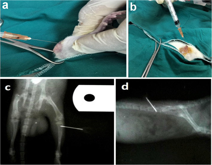
Intra-bone marrow injection (IBMI) in rats is adopted in many studies for stem cell and hematopoietic cell transplantation. IBMI in the tibia or the femur results in severe distress to the animal. Therefore, this study aims to evaluate intra-iliac injections as an alternative approach for IBMI.
Twenty-seven Sprague Dawley rats were assigned into 3 groups, 9 rats each, for 4 weeks. The control group rats were not injected. Tibia group rats were injected intra-tibial and the iliac group rats were injected intra-iliac with saline. Behavioral, radiological, histopathological, and stress evaluation was performed. Total bilirubin, cortisol, and insulin-like growth factor-1 (IGF1) were measured.
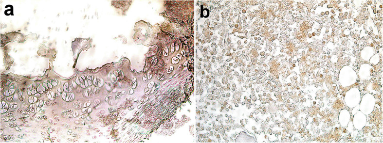
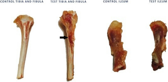
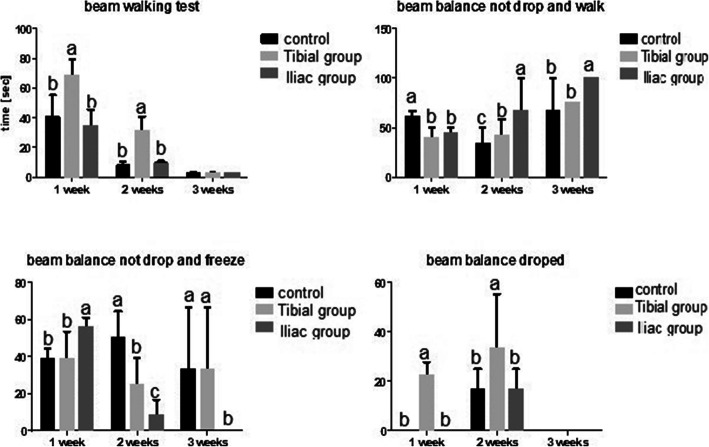
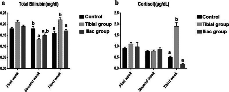
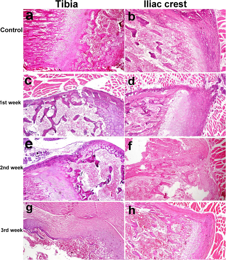
Behavioral measurements revealed deviation compared to control, in both injected groups, on the 1st and 2nd week. By the 3rd week, it was equivalent to control in the iliac group only. Bilirubin and cortisol levels were increased by intra-tibial injection compared to intra-iliac injection. The IGF-1 gene expression increased compared to control at 1st and 2nd weeks in intra-iliac injection and decreased by intra-tibial injection at 2nd week. The thickness of the iliac crest was not different from the control group, whereas there were significant differences between the control and tibia groups. Healing of the iliac crest was faster compared to the tibia. In the 3rd week, the tibia showed fibrosis at the site of injection whereas the iliac crest showed complete bone reconstruction.
Intra-iliac injections exert less distress on animals, and by 3 weeks, they regained their normal activity in comparison to intra-tibial injections.
在许多干细胞和造血细胞移植研究中,大鼠采用骨髓内注射(IBMI)。胫骨或股骨内的 IBMI 会给动物带来严重的不适。因此,本研究旨在评估髂内注射作为 IBMI 的替代方法。
将 27 只 Sprague Dawley 大鼠分为 3 组,每组 9 只,持续 4 周。对照组大鼠未注射。胫骨组大鼠进行胫骨内注射,髂骨组大鼠进行髂内注射生理盐水。进行行为、影像学、组织病理学和应激评估。测量总胆红素、皮质醇和胰岛素样生长因子-1(IGF1)。
行为测量显示,在注射后第 1 周和第 2 周,与对照组相比,两组注射组均出现偏差。到第 3 周,仅髂骨组与对照组相当。与髂内注射相比,胫骨内注射会导致胆红素和皮质醇水平升高。与对照组相比,IGF-1 基因表达在髂内注射后第 1 周和第 2 周增加,而在胫骨内注射后第 2 周下降。髂嵴的厚度与对照组无差异,而对照组与胫骨组之间存在显著差异。髂嵴的愈合速度比胫骨快。在第 3 周,胫骨显示注射部位纤维化,而髂嵴显示完全骨重建。
与胫骨内注射相比,髂内注射对动物的不适程度较小,到第 3 周,与胫骨内注射相比,它们恢复了正常活动。