Division of Cardiovascular Medicine, University of Cambridge, Box 110, ACCI, Addenbrooke's Hospital, Cambridge CB2 2QQ, UK.
Nuclear Dynamics Programme, Babraham Institute, Cambridge, UK.
Cardiovasc Res. 2022 Jun 22;118(7):1713-1727. doi: 10.1093/cvr/cvab208.
Traditional markers of cell senescence including p16, Lamin B1, and senescence-associated beta galactosidase (SAβG) suggest very high frequencies of senescent cells in atherosclerosis, while their removal via 'senolysis' has been reported to reduce atherogenesis. However, selective killing of a variety of different cell types can exacerbate atherosclerosis. We therefore examined the specificity of senescence markers in vascular smooth muscle cells (VSMCs) and the effects of genetic or pharmacological senolysis in atherosclerosis.
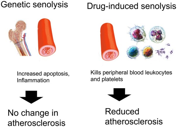
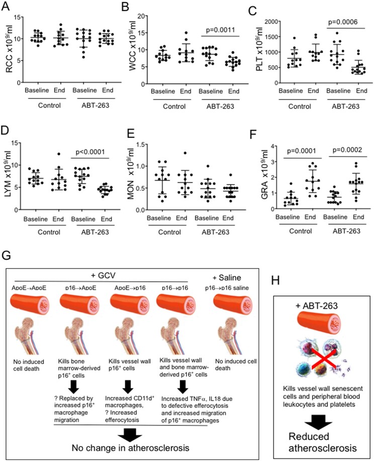
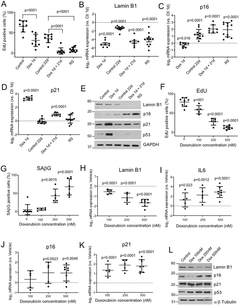
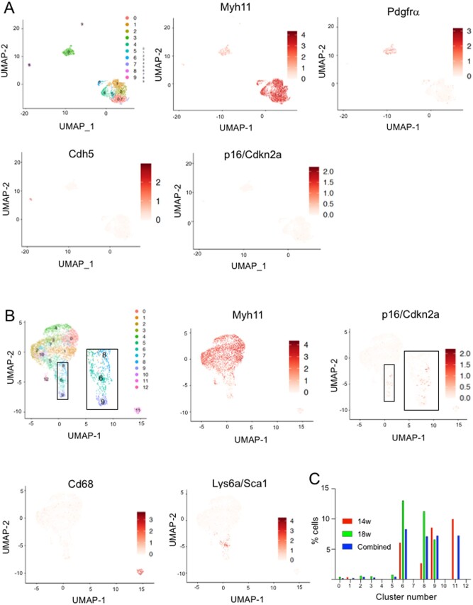
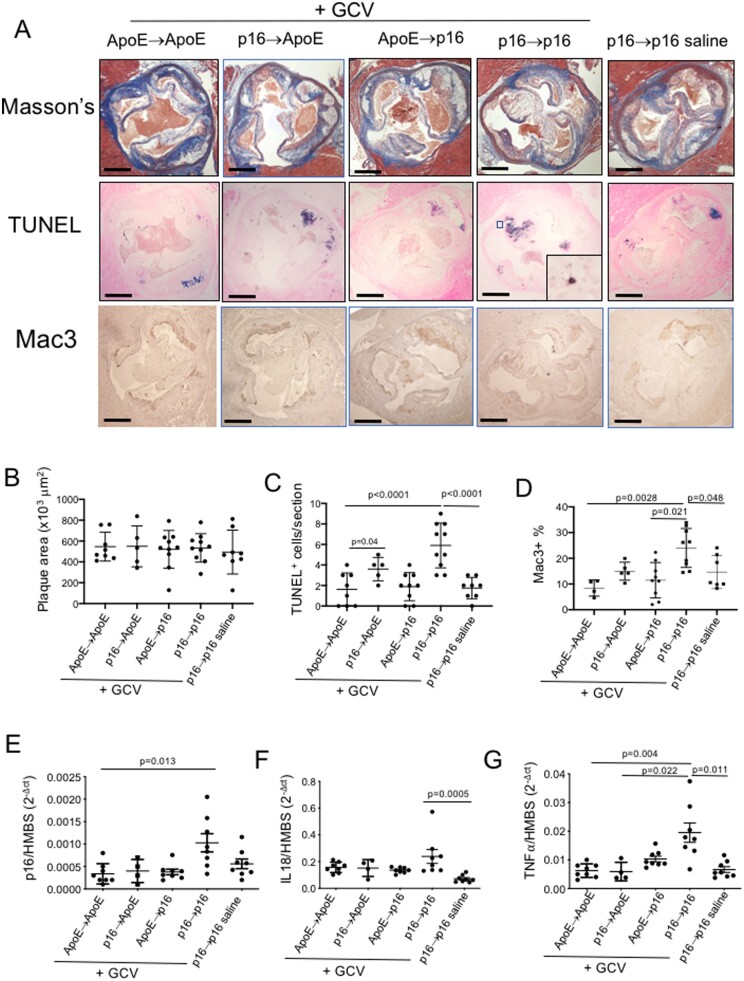
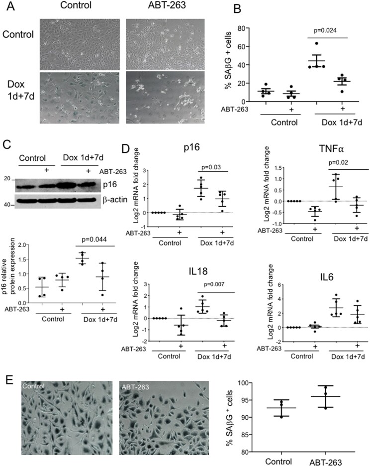
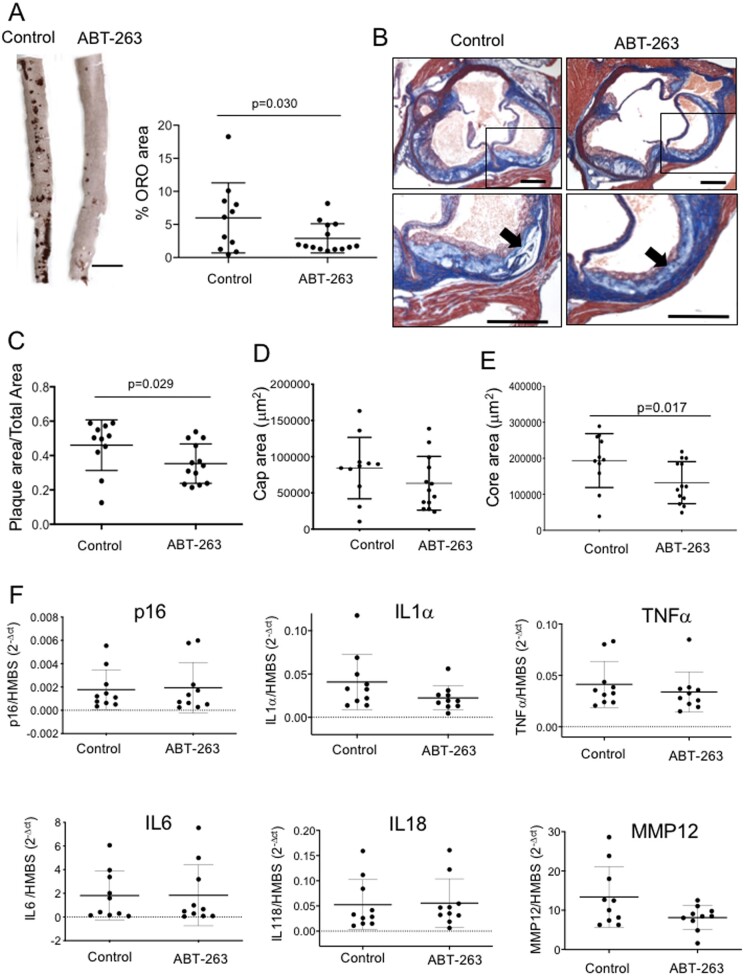
We examined traditional senescence markers in human and mouse VSMCs in vitro, and in mouse atherosclerosis. p16 and SAβG increased and Lamin B1 decreased in replicative senescence and stress-induced premature senescence (SIPS) of cultured human VSMCs. In contrast, mouse VSMCs undergoing SIPS showed only modest p16 up-regulation, and proliferating mouse monocyte/macrophages also expressed p16 and SAβG. Single cell RNA-sequencing (scRNA-seq) of lineage-traced mice showed increased p16 expression in VSMC-derived cells in plaques vs. normal arteries, but p16 localized to Stem cell antigen-1 (Sca1)+ or macrophage-like populations. Activation of a p16-driven suicide gene to remove p16+ vessel wall- and/or bone marrow-derived cells increased apoptotic cells, but also induced inflammation and did not change plaque size or composition. In contrast, the senolytic ABT-263 selectively reduced senescent VSMCs in culture, and markedly reduced atherogenesis. However, ABT-263 did not reduce senescence markers in vivo, and significantly reduced monocyte and platelet counts and interleukin 6 as a marker of systemic inflammation.
We show that genetic and pharmacological senolysis have variable effects on atherosclerosis, and may promote inflammation and non-specific effects respectively. In addition, traditional markers of cell senescence such as p16 have significant limitations to identify and remove senescent cells in atherosclerosis, suggesting that senescence studies in atherosclerosis and new senolytic drugs require more specific and lineage-restricted markers before ascribing their effects entirely to senolysis.
传统的细胞衰老标志物,如 p16、Lamin B1 和衰老相关的β半乳糖苷酶(SAβG),表明动脉粥样硬化中存在非常高频率的衰老细胞,而通过“衰老细胞消除”去除这些标志物已被报道可以减少动脉粥样硬化的发生。然而,选择性杀死各种不同的细胞类型可能会加剧动脉粥样硬化。因此,我们研究了血管平滑肌细胞(VSMCs)中衰老标志物的特异性,以及遗传或药理学衰老细胞消除在动脉粥样硬化中的作用。
我们在体外培养的人和鼠 VSMCs 中以及在鼠动脉粥样硬化模型中研究了传统的衰老标志物。在人 VSMCs 的复制性衰老和应激诱导的早衰(SIPS)中,p16 和 SAβG 增加,而 Lamin B1 减少。相比之下,发生 SIPS 的鼠 VSMCs 仅表现出适度的 p16 上调,增殖的鼠单核/巨噬细胞也表达 p16 和 SAβG。对谱系追踪的鼠进行单细胞 RNA 测序(scRNA-seq)显示,斑块中 VSMC 衍生细胞的 p16 表达增加,但 p16 定位于干细胞抗原-1(Sca1)+或巨噬细胞样群体。激活 p16 驱动的自杀基因以去除 p16+血管壁和/或骨髓来源的细胞会增加凋亡细胞,但也会诱导炎症,并且不会改变斑块大小或组成。相比之下,选择性衰老细胞消除剂 ABT-263 在体外显著减少了衰老的 VSMCs,并显著减少了动脉粥样硬化的发生。然而,ABT-263 并没有减少体内的衰老标志物,并且显著降低了单核细胞和血小板计数以及白细胞介素 6 作为全身炎症的标志物。
我们表明,遗传和药理学衰老细胞消除对动脉粥样硬化有不同的影响,并且可能分别促进炎症和非特异性效应。此外,传统的细胞衰老标志物,如 p16,在识别和去除动脉粥样硬化中的衰老细胞方面存在显著的局限性,这表明在将衰老研究和新型衰老细胞消除药物的作用完全归因于衰老细胞消除之前,需要更特异和谱系限制的标志物。