Pierrakos Charalampos, Smit Marry R, Hagens Laura A, Heijnen Nanon F L, Hollmann Markus W, Schultz Marcus J, Paulus Frederique, Bos Lieuwe D J
Department of Intensive Care, Amsterdam UMC, University of Amsterdam, Amsterdam, Netherlands.
Department of Intensive Care, Brugmann University Hospital, Université Libre de Bruxelles, Brussels, Belgium.
Front Physiol. 2021 Jun 4;12:666941. doi: 10.3389/fphys.2021.666941. eCollection 2021.
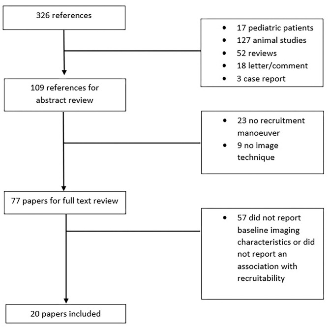
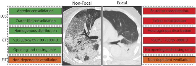
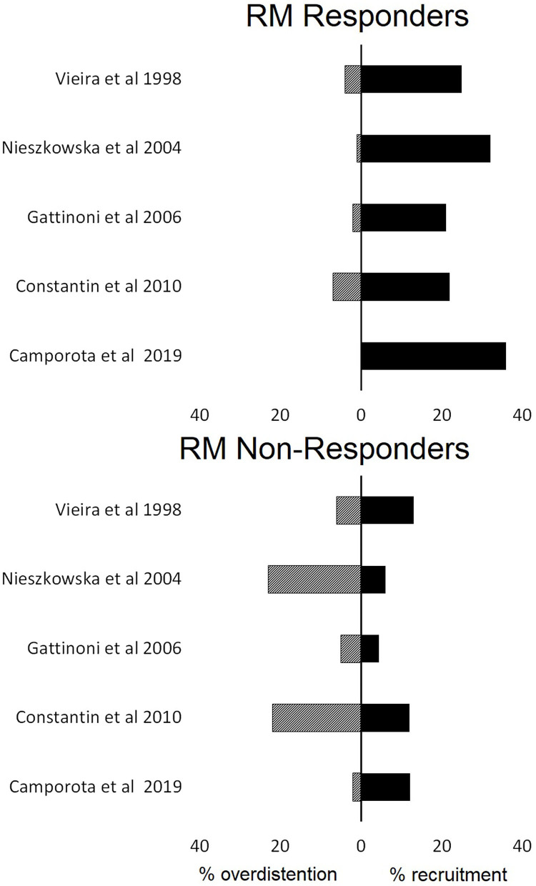
Recruitment maneuvers (RMs) have heterogeneous effects on lung aeration and have adverse side effects. We aimed to identify morphological, anatomical, and functional imaging characteristics that might be used to predict the RMs on lung aeration in invasively ventilated patients. We performed a systemic review. Studies included invasively ventilated patients who received an RM and in whom re-aeration was examined with chest computed tomography (CT), electrical impedance tomography (EIT), and lung ultrasound (LUS) were included. Twenty studies were identified. Different types of RMs were applied. The amount of re-aerated lung tissue after an RM was highly variable between patients in all studies, irrespective of the used imaging technique and the type of patients (ARDS or non-ARDS). Imaging findings suggesting a non-focal morphology (i.e., radiologic findings consistent with attenuations with diffuse or patchy loss of aeration) were associated with higher likelihood of recruitment and lower chance of overdistention than a focal morphology (i.e., radiological findings suggestive of lobar or segmental loss of aeration). This was independent of the used imaging technique but only observed in patients with ARDS. In patients without ARDS, the results were inconclusive. ARDS patients with imaging findings suggestive of non-focal morphology show most re-aeration of previously consolidated lung tissue after RMs. The role of imaging techniques in predicting the effect of RMs on re-aeration in patients without ARDS remains uncertain.
肺复张手法(RMs)对肺通气有不同的影响且存在不良副作用。我们旨在确定可能用于预测有创通气患者肺复张手法对肺通气影响的形态学、解剖学和功能成像特征。我们进行了一项系统评价。纳入的研究包括接受肺复张手法且使用胸部计算机断层扫描(CT)、电阻抗断层扫描(EIT)和肺部超声(LUS)检查再通气情况的有创通气患者。共确定了20项研究。应用了不同类型的肺复张手法。在所有研究中,无论使用何种成像技术及患者类型(急性呼吸窘迫综合征[ARDS]或非ARDS),肺复张手法后再通气的肺组织量在患者之间差异很大。与局灶性形态(即提示肺叶或肺段通气丧失的影像学表现)相比,提示非局灶性形态(即与弥漫性或斑片状通气丧失导致的肺组织衰减一致 的影像学表现)的影像学结果与更高的肺复张可能性及更低的过度扩张几率相关。这与所使用的成像技术无关,但仅在ARDS患者中观察到。在非ARDS患者中,结果尚无定论。影像学表现提示非局灶性形态的ARDS患者在肺复张手法后,先前实变的肺组织再通气最为明显。成像技术在预测非ARDS患者肺复张手法对再通气的影响方面的作用仍不确定。