Division of Biomedical Sciences, School of Medicine, University of California, Riverside, Riverside, CA, 92521, USA.
Present address: Division of Mathematics and Sciences, Delta State University, Cleveland, MS, 38733, USA.
J Neuroinflammation. 2021 Jun 21;18(1):140. doi: 10.1186/s12974-021-02183-2.
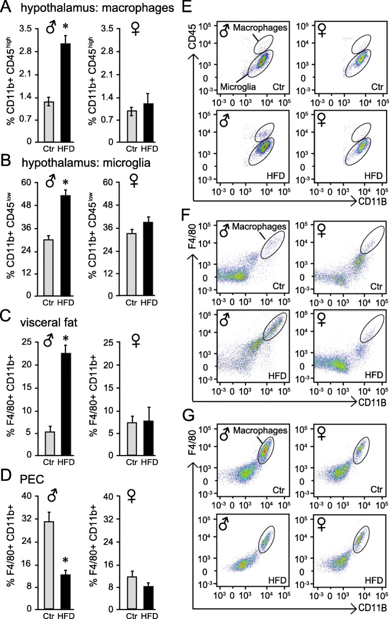
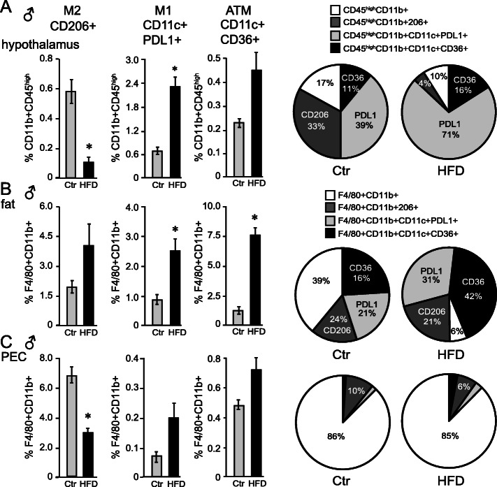
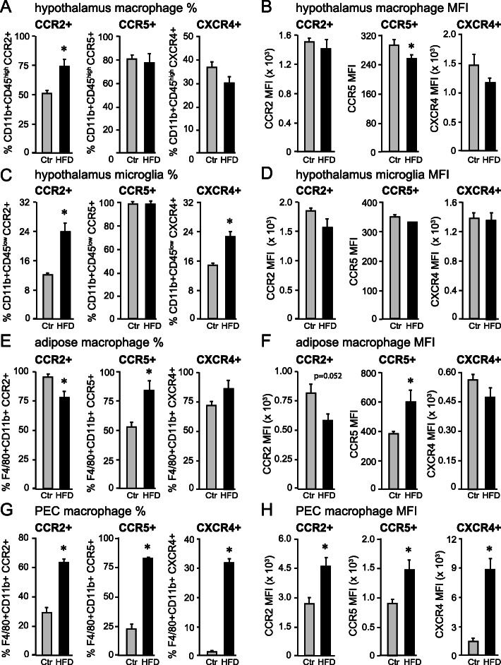
Obesity is characterized by a systemic inflammation and hypothalamic neuroinflammation. Systemic inflammation is caused by macrophages that infiltrate obese adipose tissues. We previously demonstrated that high-fat diet (HFD)-fed male mice exhibited peripheral macrophage infiltration into the hypothalamus, in addition to activation of resident microglia. Since this infiltration contributes to neuroinflammation and neuronal impairment, herein we characterize the phenotype and origin of these hypothalamic macrophages in HFD mice.
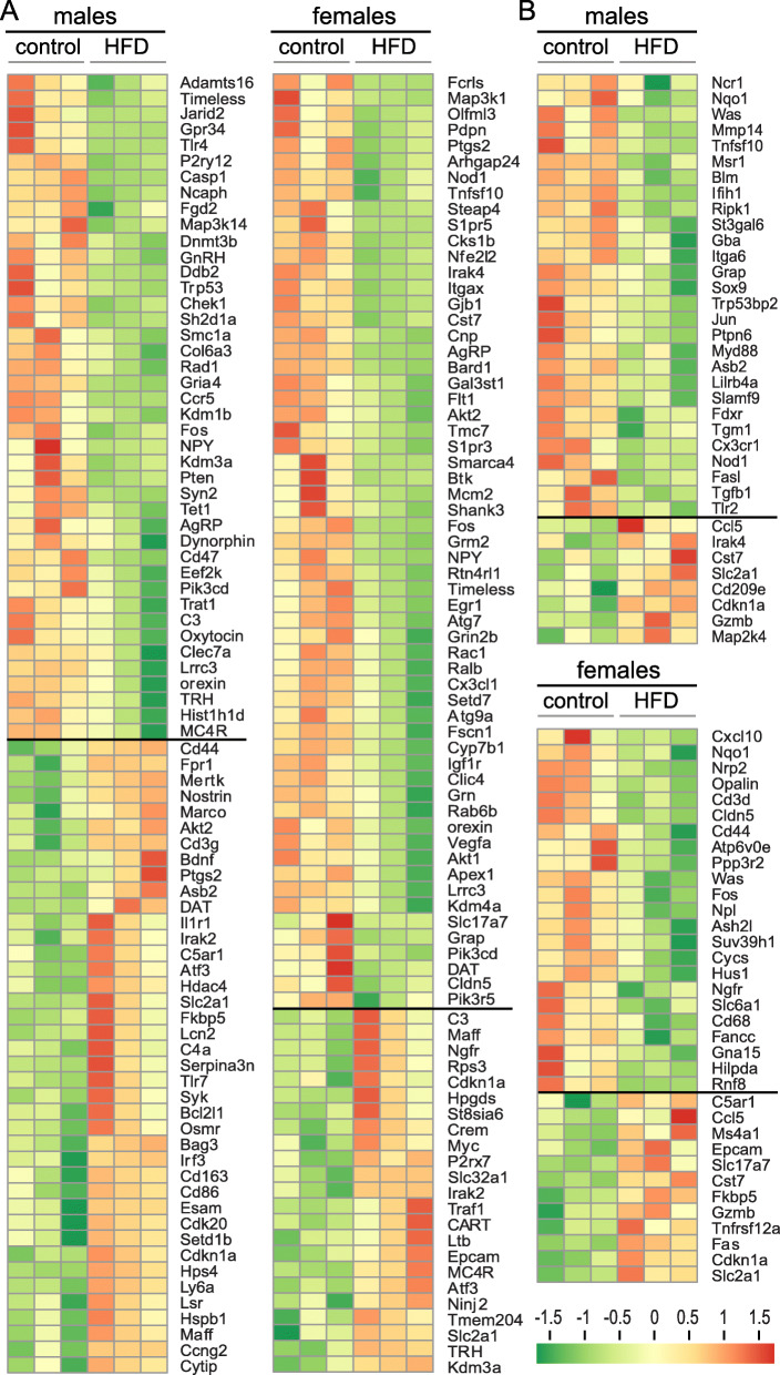
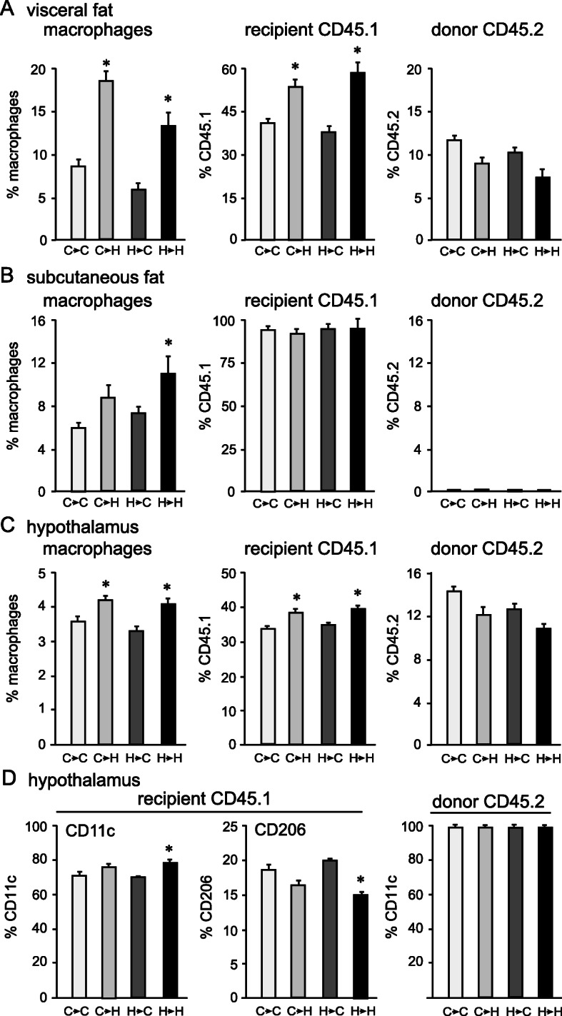
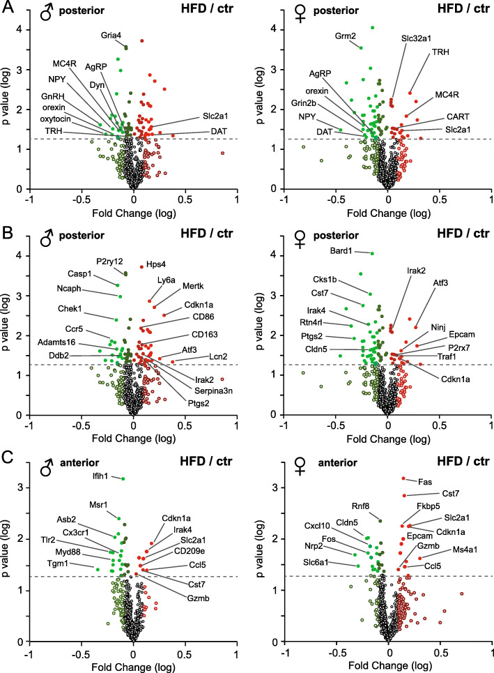
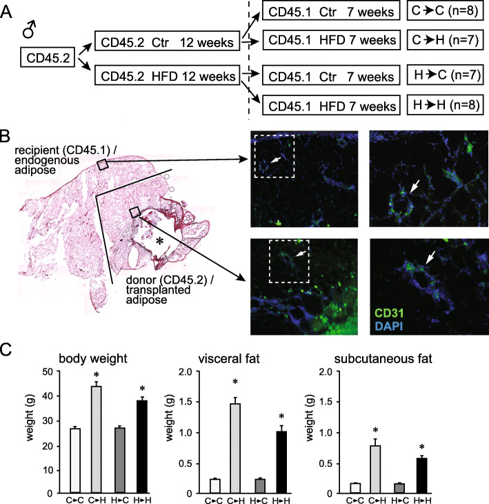
C57BL/6J mice were fed HFD (60% kcal from fat) or control diet with matching sucrose levels, for 12-16 weeks. Males and females were analyzed separately to determine sex-specific responses to HFD. Differences in hypothalamic gene expression in HFD-fed male and female mice, compared to their lean controls, in two different areas of the hypothalamus, were determined using the NanoString neuroinflammation panel. Phenotypic changes in macrophages that infiltrated the hypothalamus in HFD-fed mice were determined by analyzing cell surface markers using flow cytometry and compared to changes in macrophages from the adipose tissue and peritoneal cavity. Adipose tissue transplantation was performed to determine the source of hypothalamic macrophages.
We determined that hypothalamic gene expression profiles demonstrate sex-specific and region-specific diet-induced changes. Sex-specific changes included larger changes in males, while region-specific changes included larger changes in the area surrounding the median eminence. Several genes were identified that may provide partial protection to female mice. We also identified diet-induced changes in macrophage migration into the hypothalamus, adipose tissue, and peritoneal cavity, specifically in males. Further, we determined that hypothalamus-infiltrating macrophages express pro-inflammatory markers and markers of metabolically activated macrophages that were identical to markers of adipose tissue macrophages in HFD-fed mice. Employing adipose tissue transplant, we demonstrate that hypothalamic macrophages can originate from the visceral adipose tissue.
HFD-fed males experience higher neuroinflammation than females, likely because they accumulate more visceral fat, which provides a source of pro-inflammatory macrophages that migrate to other tissues, including the hypothalamus. Our findings may explain the male bias for neuroinflammation and the metabolic syndrome. Together, our results demonstrate a new connection between the adipose tissue and the hypothalamus in obesity that contributes to neuroinflammation and hypothalamic pathologies.
肥胖的特征是全身炎症和下丘脑神经炎症。全身炎症是由浸润肥胖脂肪组织的巨噬细胞引起的。我们之前的研究表明,高脂肪饮食(HFD)喂养的雄性小鼠除了激活常驻小胶质细胞外,还表现出外周巨噬细胞浸润到下丘脑。由于这种浸润有助于神经炎症和神经元损伤,因此本文我们描述了 HFD 小鼠下丘脑巨噬细胞的表型和来源。
C57BL/6J 小鼠分别用 HFD(60%热量来自脂肪)或含有匹配蔗糖水平的对照饮食喂养 12-16 周。雄性和雌性小鼠分别进行分析,以确定 HFD 对雄性和雌性小鼠的性别特异性反应。通过使用 NanoString 神经炎症面板,确定了与瘦对照组相比,HFD 喂养的雄性和雌性小鼠下丘脑两个不同区域的下丘脑基因表达差异。通过流式细胞术分析浸润到 HFD 喂养小鼠下丘脑的巨噬细胞的表型变化,并与脂肪组织和腹腔巨噬细胞的变化进行比较。进行脂肪组织移植以确定下丘脑巨噬细胞的来源。
我们确定了下丘脑基因表达谱显示出性别特异性和区域特异性的饮食诱导变化。性别特异性变化包括雄性变化更大,而区域特异性变化包括围绕正中隆起的区域变化更大。鉴定出一些可能为雌性小鼠提供部分保护的基因。我们还发现,饮食诱导的巨噬细胞向下丘脑、脂肪组织和腹腔的迁移发生了变化,特别是在雄性中。此外,我们确定,浸润到下丘脑的巨噬细胞表达促炎标志物和代谢激活巨噬细胞的标志物,与 HFD 喂养小鼠的脂肪组织巨噬细胞的标志物相同。通过脂肪组织移植,我们证明下丘脑巨噬细胞可以源自内脏脂肪组织。
与女性相比,HFD 喂养的雄性经历更高的神经炎症,这可能是因为他们积累了更多的内脏脂肪,这为迁移到其他组织(包括下丘脑)的促炎巨噬细胞提供了来源。我们的发现可能解释了男性对神经炎症和代谢综合征的偏向。总之,我们的研究结果表明,肥胖症中脂肪组织和下丘脑之间存在新的联系,这有助于神经炎症和下丘脑病变。