Asan Institute for Life Sciences, University of Ulsan College of Medicine, Seoul, 05505, Republic of Korea.
Department of Biomedical Science, Asan Medical Institute of Convergence Science and Technology, Asan Medical Center and University of Ulsan College of Medicine, Seoul, 05505, Republic of Korea.
J Neuroinflammation. 2019 Nov 14;16(1):221. doi: 10.1186/s12974-019-1607-0.
Obese mice on a high-fat diet (HFD) display signs of inflammation in the hypothalamic arcuate nucleus (ARC), a critical area for controlling systemic energy metabolism. This has been suggested as a key mechanism of obesity-associated hypothalamic dysfunction. We reported earlier that bone marrow-derived macrophages accumulate in the ARC to sustain hypothalamic inflammation upon chronic exposure to an HFD. However, the mechanism underlying hypothalamic macrophage accumulation has remained unclear.
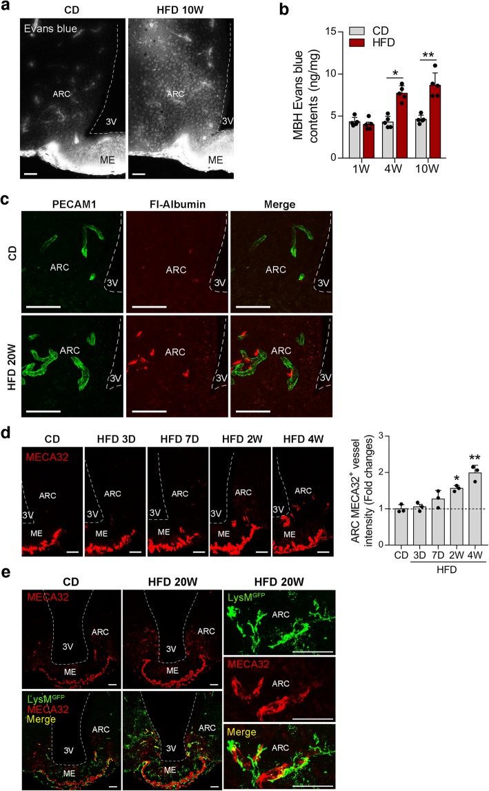
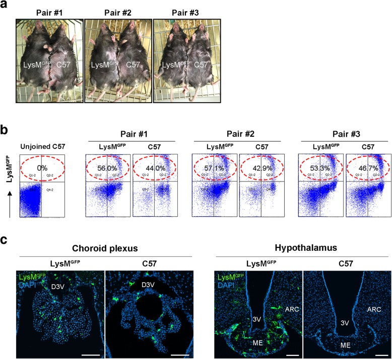
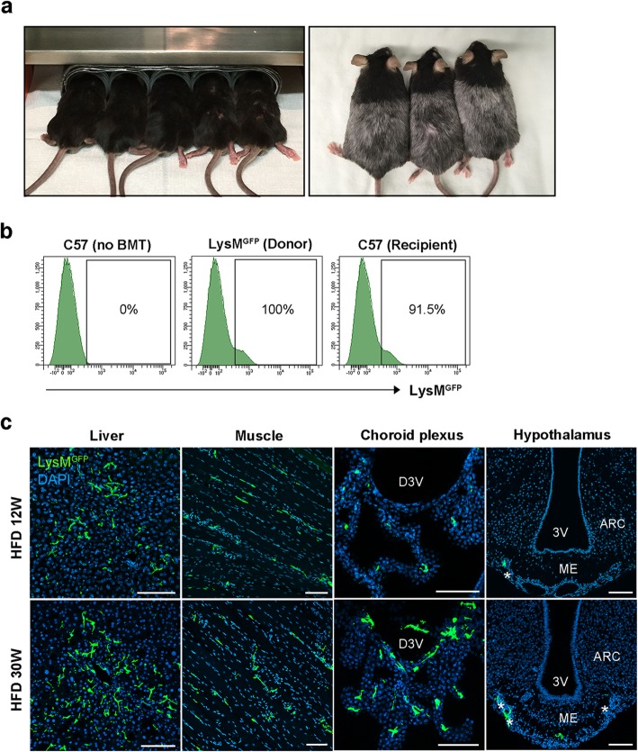
We investigated whether circulating monocytes or myeloid precursors contribute to hypothalamic macrophage expansion during chronic HFD feeding. To trace circulating myeloid cells, we generated mice that express green fluorescent protein (GFP) in their lysozyme M-expressing myeloid cells (LysM mice). We conducted parabiosis and bone marrow transplantation experiments using these animals. Mice received an HFD for 12 or 30 weeks and were then sacrificed to analyze LysM cells in the hypothalamus. Hypothalamic vascular permeability in the HFD-fed obese mice was also tested by examining the extravascular leakage of Evans blue and fluorescence-labeled albumin. The timing of LysM cell entry to the hypothalamus during development was also evaluated.
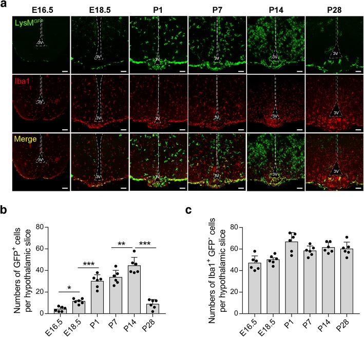
Our parabiosis and bone marrow transplantation experiments revealed a significant infiltration of circulating LysM cells into the liver, skeletal muscle, choroid plexus, and leptomeninges but not in the hypothalamic ARC during chronic HFD feeding, despite increased hypothalamic vascular permeability. These results suggested that the recruitment of circulating monocytes is not a major mechanism for maintaining and expanding the hypothalamic macrophage population in diet-induced obesity. We demonstrated instead that LysM cells infiltrate the hypothalamus during its development. LysM cells appeared in the hypothalamic area from the late embryonic period. This cellular pool suddenly increased immediately after birth, peaked at the postnatal second week, and adopted an adult pattern of distribution after weaning.
Bone marrow-derived macrophages mostly populate the hypothalamus in early postnatal life and may maintain their pool without significant recruitment of circulating monocytes throughout life, even under conditions of chronic HFD feeding.
高脂肪饮食(HFD)喂养的肥胖小鼠在弓状核(ARC)表现出炎症迹象,ARC 是控制全身能量代谢的关键区域。这被认为是肥胖相关下丘脑功能障碍的关键机制。我们之前报道过,骨髓来源的巨噬细胞在慢性暴露于 HFD 时会积累在 ARC 中,以维持下丘脑炎症。然而,下丘脑巨噬细胞积累的机制仍不清楚。
我们研究了循环单核细胞或髓样前体是否有助于慢性 HFD 喂养期间下丘脑巨噬细胞的扩增。为了追踪循环髓样细胞,我们生成了在溶酶体相关膜蛋白(LysM)表达的髓样细胞中表达绿色荧光蛋白(GFP)的小鼠(LysM 小鼠)。我们使用这些动物进行了联体共生和骨髓移植实验。小鼠接受 HFD 喂养 12 或 30 周,然后处死以分析下丘脑的 LysM 细胞。还通过检查伊文思蓝和荧光标记白蛋白的血管外渗漏来测试肥胖 HFD 喂养小鼠的下丘脑血管通透性。还评估了 LysM 细胞在发育过程中进入下丘脑的时间。
我们的联体共生和骨髓移植实验显示,尽管下丘脑血管通透性增加,但在慢性 HFD 喂养期间,循环 LysM 细胞会大量浸润肝脏、骨骼肌、脉络丛和软脑膜,但不会浸润下丘脑 ARC。这些结果表明,循环单核细胞的募集不是维持和扩大饮食诱导肥胖症中下丘脑巨噬细胞群体的主要机制。相反,我们证明 LysM 细胞在其发育过程中会浸润下丘脑。LysM 细胞从胚胎后期开始出现在下丘脑区域。这个细胞池在出生后立即突然增加,在出生后第二周达到峰值,在断奶后采用成人分布模式。
骨髓来源的巨噬细胞主要在生命早期定植于下丘脑,并且即使在慢性 HFD 喂养条件下,也可能在没有循环单核细胞大量募集的情况下维持其细胞池。