Pain Research Center and Department of Physiology, Zhongshan School of Medicine, Sun Yat-Sen University, Guangzhou, China.
Departments of Physiology and.
Pain. 2021 Dec 1;162(12):2865-2880. doi: 10.1097/j.pain.0000000000002279.
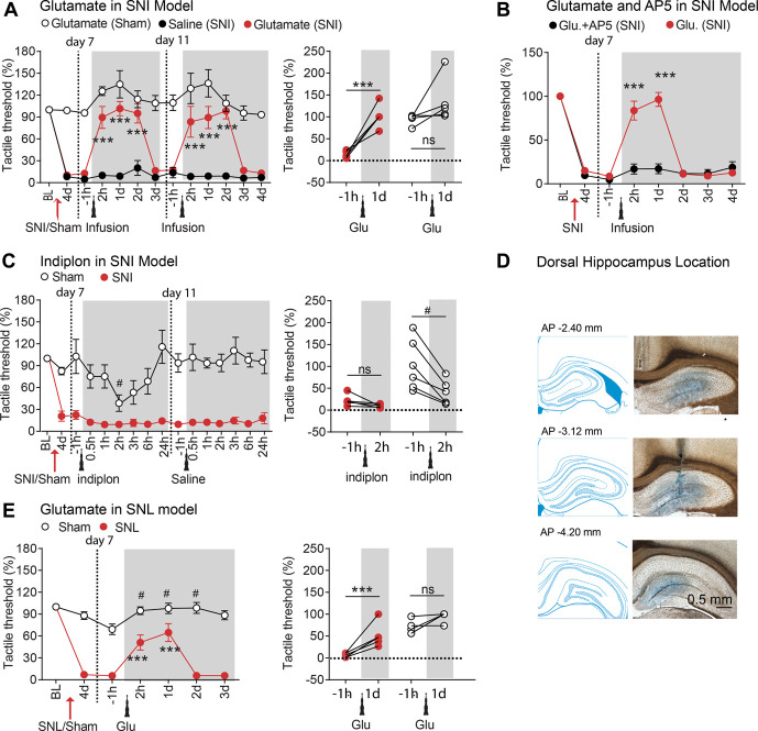
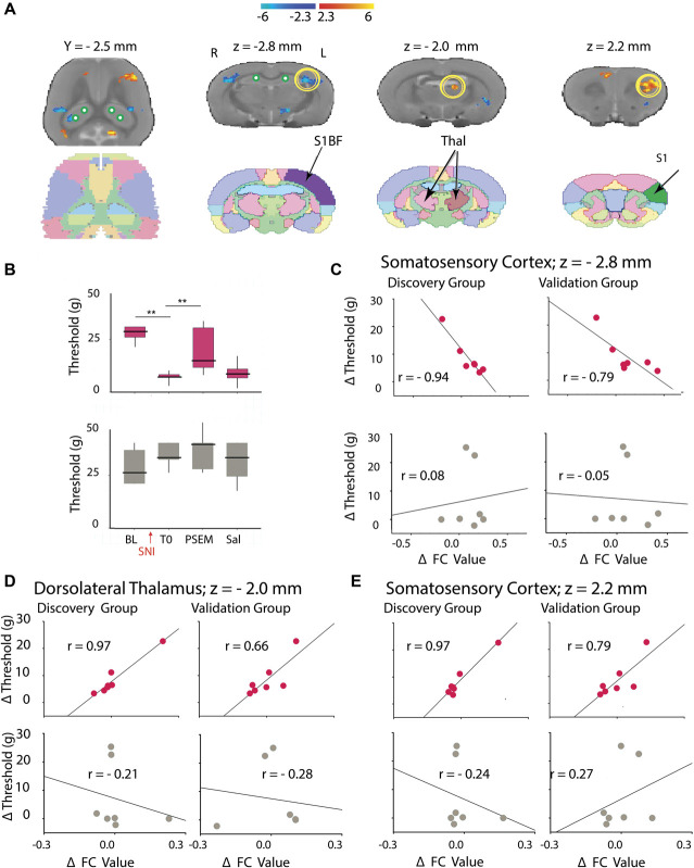
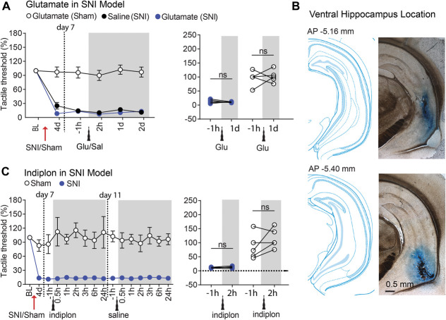
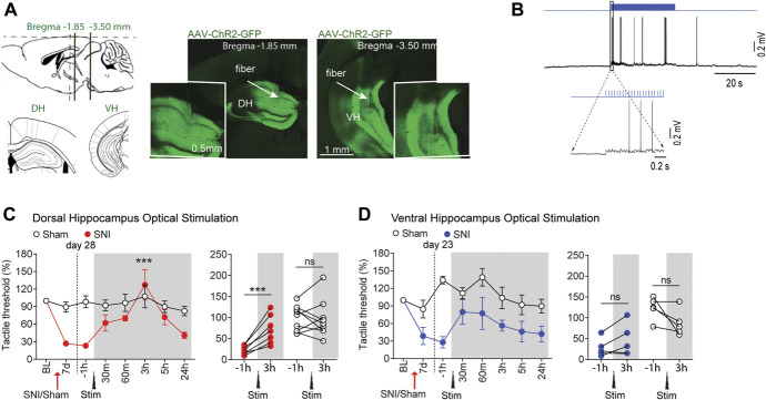
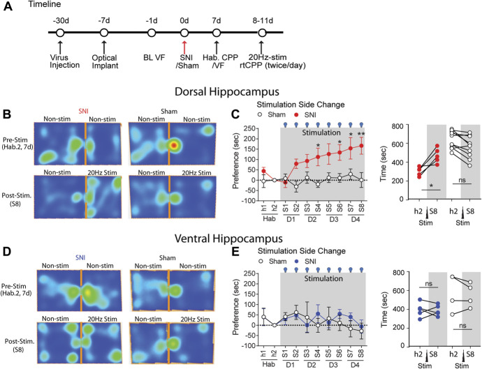
Accumulating evidence suggests hippocampal impairment under the chronic pain phenotype. However, it is unknown whether neuropathic behaviors are related to dysfunction of the hippocampal circuitry. Here, we enhanced hippocampal activity by pharmacological, optogenetic, and chemogenetic techniques to determine hippocampal influence on neuropathic pain behaviors. We found that excitation of the dorsal (DH), but not the ventral (VH) hippocampus induces analgesia in 2 rodent models of neuropathic pain (SNI and SNL) and in rats and mice. Optogenetic and pharmacological manipulations of DH neurons demonstrated that DH-induced analgesia was mediated by N-Methyl-D-aspartate and μ-opioid receptors. In addition to analgesia, optogenetic stimulation of the DH in SNI mice also resulted in enhanced real-time conditioned place preference for the chamber where the DH was activated, a finding consistent with pain relief. Similar manipulations in the VH were ineffective. Using chemo-functional magnetic resonance imaging (fMRI), where awake resting-state fMRI was combined with viral vector-mediated chemogenetic activation (PSAM/PSEM89s) of DH neurons, we demonstrated changes of functional connectivity between the DH and thalamus and somatosensory regions that tracked the extent of relief from tactile allodynia. Moreover, we examined hippocampal functional connectivity in humans and observe differential reorganization of its anterior and posterior subdivisions between subacute and chronic back pain. Altogether, these results imply that downregulation of the DH circuitry during chronic neuropathic pain aggravates pain-related behaviors. Conversely, activation of the DH reverses pain-related behaviors through local excitatory and opioidergic mechanisms affecting DH functional connectivity. Thus, this study exhibits a novel causal role for the DH but not the VH in controlling neuropathic pain-related behaviors.
越来越多的证据表明,慢性疼痛表型下的海马损伤。然而,尚不清楚神经病理性行为是否与海马回路功能障碍有关。在这里,我们通过药理学、光遗传学和化学遗传学技术增强海马活性,以确定海马对神经病理性疼痛行为的影响。我们发现,背侧(DH)而不是腹侧(VH)海马的兴奋在 2 种神经病理性疼痛模型(SNI 和 SNL)以及大鼠和小鼠中引起镇痛。DH 神经元的光遗传学和药理学操作表明,DH 诱导的镇痛是由 N-甲基-D-天冬氨酸和 μ 阿片受体介导的。除了镇痛之外,SNI 小鼠 DH 的光遗传学刺激还导致对 DH 被激活的腔室的实时条件性位置偏好增强,这一发现与疼痛缓解一致。在 VH 中的类似操作则无效。使用化学功能磁共振成像(fMRI),将清醒静息状态 fMRI 与病毒载体介导的 DH 神经元化学遗传激活(PSAM/PSEM89s)相结合,我们证明了 DH 与丘脑和体感区域之间的功能连接发生变化,这些变化与触觉过敏缓解的程度相吻合。此外,我们还观察了人类海马的功能连接,并观察到亚急性和慢性背痛之间其前后部分的差异重组。总之,这些结果表明,慢性神经病理性疼痛期间 DH 回路的下调会加重与疼痛相关的行为。相反,DH 的激活通过影响 DH 功能连接的局部兴奋性和阿片能机制来逆转与疼痛相关的行为。因此,这项研究表明,DH 而不是 VH 在控制神经病理性疼痛相关行为方面具有新的因果作用。