Department of Behavioral Physiology and Sociobiology, Biocenter, University of Würzburg, 97074, Würzburg, Germany.
Department of Life Sciences, Imperial College London, Silwood Park, Buckhurst Road, Ascot, Berkshire, SL5 7PY, UK.
Cell Tissue Res. 2021 Oct;386(1):29-45. doi: 10.1007/s00441-021-03482-z. Epub 2021 Jun 28.
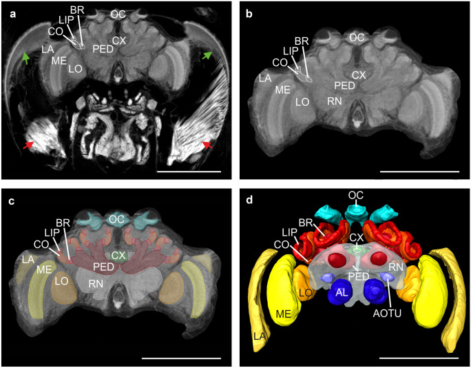
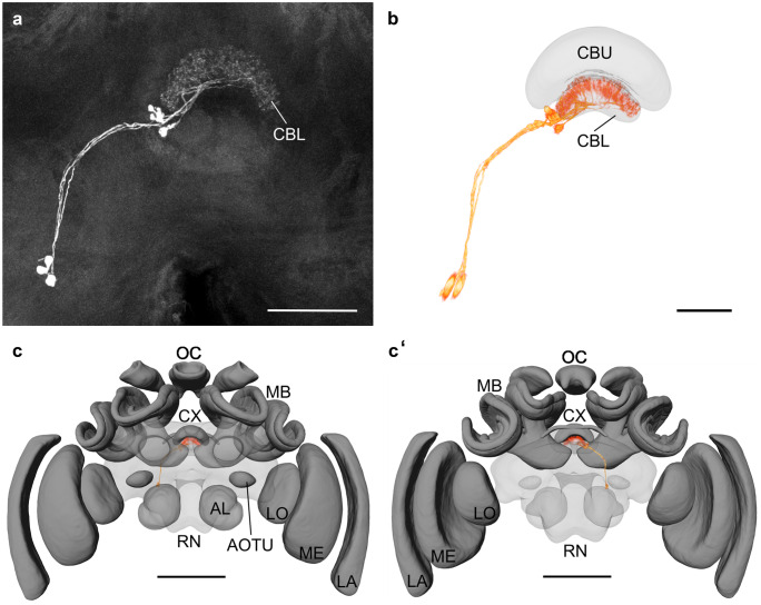
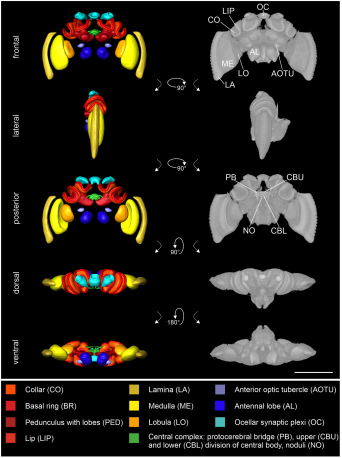
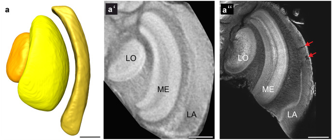
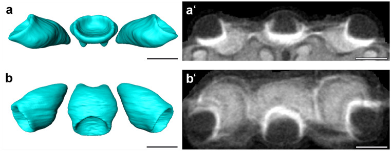
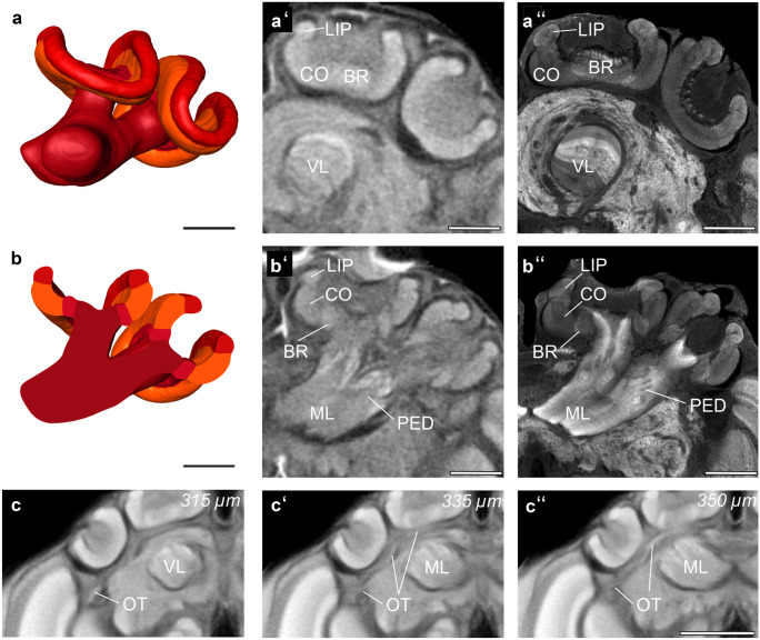
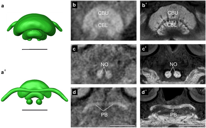
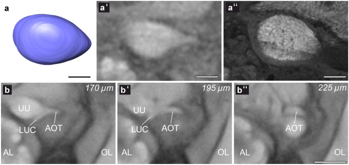
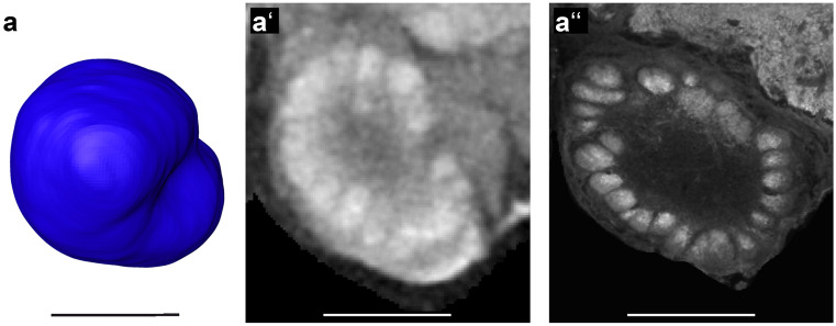
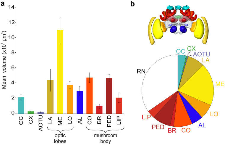
In recent years, bumblebees have become a prominent insect model organism for a variety of biological disciplines, particularly to investigate learning behaviors as well as visual performance. Understanding these behaviors and their underlying neurobiological principles requires a clear understanding of brain anatomy. Furthermore, to be able to compare neuronal branching patterns across individuals, a common framework is required, which has led to the development of 3D standard brain atlases in most of the neurobiological insect model species. Yet, no bumblebee 3D standard brain atlas has been generated. Here we present a brain atlas for the buff-tailed bumblebee Bombus terrestris using micro-computed tomography (micro-CT) scans as a source for the raw data sets, rather than traditional confocal microscopy, to produce the first ever micro-CT-based insect brain atlas. We illustrate the advantages of the micro-CT technique, namely, identical native resolution in the three cardinal planes and 3D structure being better preserved. Our Bombus terrestris brain atlas consists of 30 neuropils reconstructed from ten individual worker bees, with micro-CT allowing us to segment neuropils completely intact, including the lamina, which is a tissue structure often damaged when dissecting for immunolabeling. Our brain atlas can serve as a platform to facilitate future neuroscience studies in bumblebees and illustrates the advantages of micro-CT for specific applications in insect neuroanatomy.
近年来,熊蜂已成为各种生物学学科的重要昆虫模式生物,特别是用于研究学习行为和视觉表现。要理解这些行为及其潜在的神经生物学原理,需要清楚地了解大脑解剖结构。此外,为了能够比较个体之间的神经元分支模式,需要一个通用的框架,这导致了大多数神经生物学昆虫模式物种的 3D 标准脑图谱的发展。然而,目前还没有生成熊蜂的 3D 标准脑图谱。在这里,我们使用微计算机断层扫描(micro-CT)扫描为原始数据集提供来源,而不是传统的共聚焦显微镜,为黄胫熊蜂 Bombus terrestris 生成了第一个基于 micro-CT 的昆虫脑图谱。我们展示了 micro-CT 技术的优势,即在三个主平面上具有相同的固有分辨率,并且 3D 结构得到更好的保留。我们的 Bombus terrestris 脑图谱由十个工蜂个体重建的 30 个神经节组成,micro-CT 允许我们完全完整地分割神经节,包括通常在免疫标记时解剖会损坏的组织结构——神经薄板。我们的脑图谱可以作为一个平台,促进未来在熊蜂中的神经科学研究,并展示了 micro-CT 在昆虫神经解剖学中的特定应用的优势。