Cutaneous Biology Research Center, Massachusetts General Hospital, Harvard Medical School, Boston, MA, USA.
Laboratory for Functional Optical Imaging, Department of Biomedical Engineering, Columbia University, New York, NY, USA.
Sci Rep. 2021 Jun 28;11(1):13411. doi: 10.1038/s41598-021-92712-z.
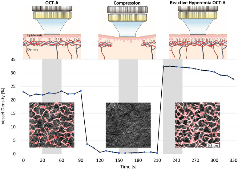
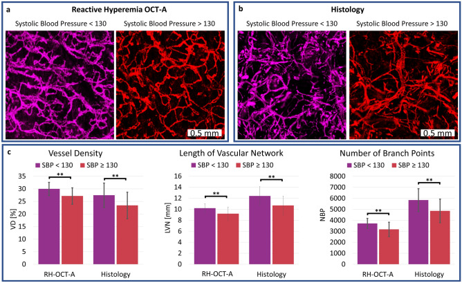
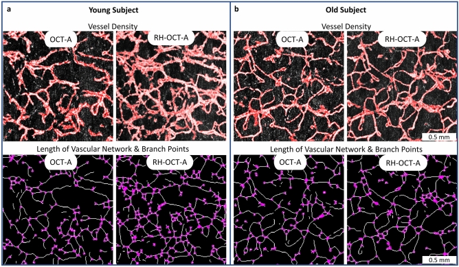
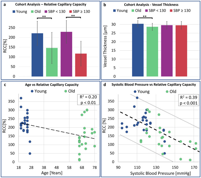
Visualization and quantification of the skin microvasculature are important for studying the health of the human microcirculation. We correlated structural and pathophysiological changes of the dermal capillary-level microvasculature with age and blood pressure by using the reactive hyperemia optical coherence tomography angiography (RH-OCT-A) technique and evaluated both conventional OCT-A and the RH-OCT-A method as non-invasive imaging alternatives to histopathology. This observational pilot study acquired OCT-A and RH-OCT-A images of the dermal microvasculature of 13 young and 12 old healthy Caucasian female subjects. Two skin biopsies were collected per subject for histological analysis. The dermal microvasculature in OCT-A, RH-OCT-A, and histological images were automatically quantified and significant indications of vessel rarefaction in both old subjects and subjects with high blood pressure were observed by RH-OCT-A and histopathology. We showed that an increase in dermal microvasculature perfusion in response to reactive hyperemia was significantly lower in high blood pressure subjects compared to normal blood pressure subjects (117% vs. 229%). These results demonstrate that RH-OCT-A imaging holds functional information of the microvasculature with respect to physiological factors such as age and blood pressure that may help to monitor early disease progression and assess overall vascular health. Additionally, our results suggest that RH-OCT-A images may serve as a non-invasive alternative to histopathology for vascular analysis.
皮肤微血管的可视化和量化对于研究人体微循环的健康状况非常重要。我们使用反应性充血光相干断层扫描血管造影术(RH-OCT-A)技术,将皮肤毛细血管水平微血管的结构和病理生理学变化与年龄和血压相关联,并评估了常规 OCT-A 和 RH-OCT-A 方法作为组织病理学的非侵入性成像替代方法。这项观察性初步研究采集了 13 名年轻和 12 名健康白种女性受试者的皮肤微血管 OCT-A 和 RH-OCT-A 图像。每个受试者采集了两个皮肤活检样本用于组织学分析。在 OCT-A、RH-OCT-A 和组织学图像中,皮肤微血管被自动量化,通过 RH-OCT-A 和组织病理学观察到,在老年受试者和高血压受试者中,血管稀疏的迹象明显。我们表明,与正常血压受试者相比,高血压受试者对反应性充血的皮肤微血管灌注增加明显较低(117%对 229%)。这些结果表明,RH-OCT-A 成像具有与年龄和血压等生理因素有关的微血管功能信息,这可能有助于监测早期疾病进展和评估整体血管健康。此外,我们的结果表明,RH-OCT-A 图像可能作为血管分析的组织病理学的非侵入性替代方法。