Department of Obstetrics and Gynecology, Kobe University Graduate School of Medicine, 7-5-2 Kusunoki-cho, Chuo-ku, Kobe, Hyogo, 650-0017, Japan.
J Ovarian Res. 2021 Jun 29;14(1):87. doi: 10.1186/s13048-021-00835-8.
Serous endometrial intraepithelial carcinoma (SEIC) is now considered to represent an early stage of uterine serous carcinoma (USC). It is an intraepithelial lesion but has been reported to cause extrauterine metastases. We report a case of SEIC with serous ovarian carcinoma and lymph node metastasis.
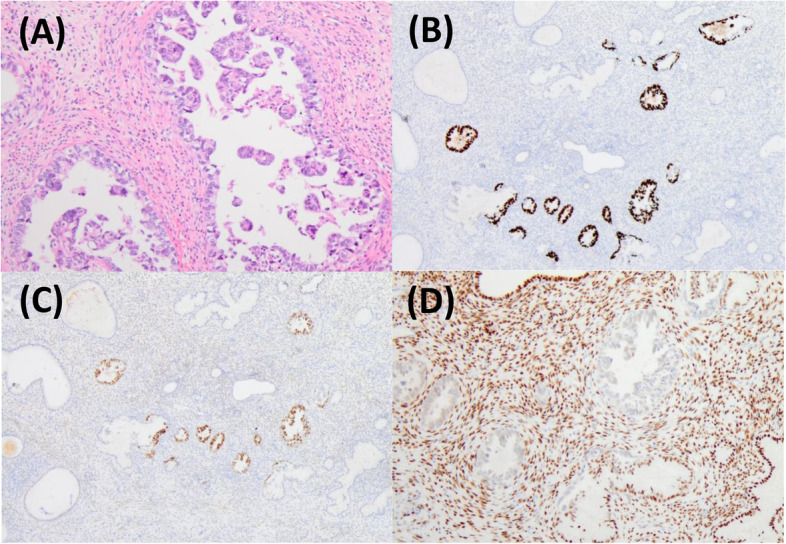
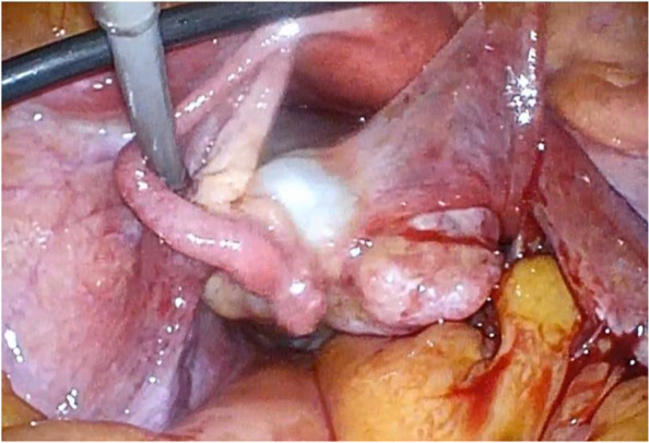
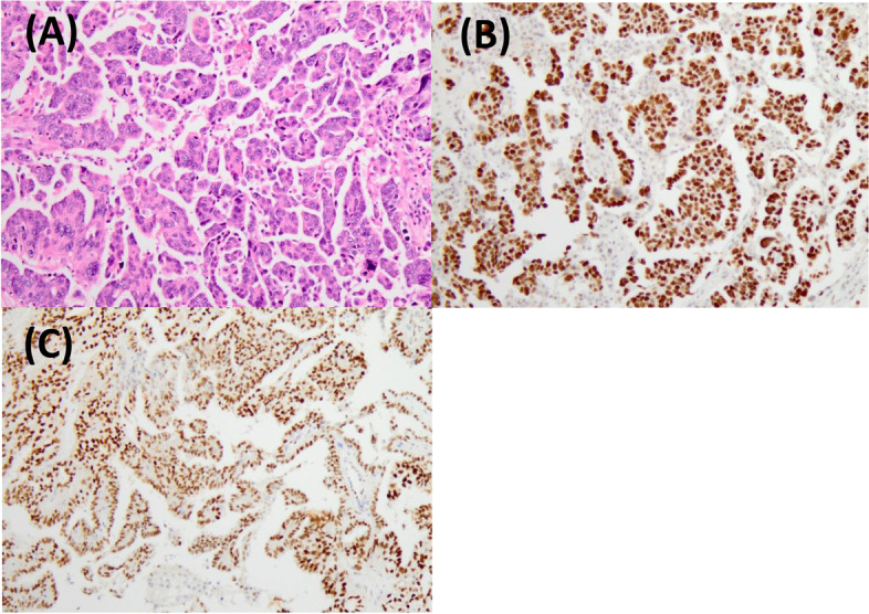
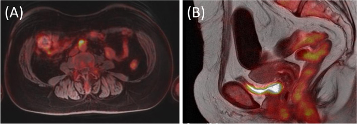
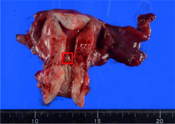
A 57-year-old post-menopausal woman (gravida 3, para 2, SA1) was referred to our hospital with lower abdominal pain. An ultrasound and MRI showed that the ovary had swollen to 8 cm in size and had a solid lesion. The uterus was normal. The patient underwent exploratory laparoscopy on the suspicion of torsion of the ovarian tumor. Intraoperative findings showed a right ovarian tumor, but no ovarian tumor torsion was observed. A small amount of bloody ascites was found in the Douglas fossa, and bleeding was observed from the tumor itself. A right salpingo-oophorectomy was then performed. Histopathological results revealed a high-grade serous carcinoma. Forty days after the first surgery, we performed a staging laparotomy: a total abdominal hysterectomy, left salpingo-oophorectomy, systematic pelvic and paraaortic lymphadenectomy, and a partial omentectomy. A complete cytoreduction was achieved. In the pathological examination, the invasion of the serous carcinoma was observed in the left ovarian ligament, and lymph node metastasis was found in the paraaortic lymph nodes. Atypical columnar cells formed irregular papillary lesions which had proliferated in the endometrium, and this was diagnosed as SEIC. The final diagnosis was serous ovarian cancer, FIGO stage IIIA1(ii), pT2bN1M0, with SEIC.
We report a case of SEIC with synchronous serous carcinoma of the adnexa uteri. Both were serous carcinomas and, thus, it was difficult to identify the primary lesion. The distinction between metastatic cancer and two independent primary tumors is important for an accurate diagnosis and tumor staging. Histological diagnostic criteria remain controversial, and further development of a method for differentiating between both diseases is required.
浆液性子宫内膜上皮内癌(SEIC)现在被认为是子宫浆液性癌(USC)的早期阶段。它是一种上皮内病变,但已有报道称其会引起子宫外转移。我们报告了一例 SEIC 合并浆液性卵巢癌和淋巴结转移。
一名 57 岁绝经后妇女(孕 3 产 2,SA1)因下腹疼痛就诊。超声和 MRI 显示卵巢增大至 8 厘米大小,有实性病变。子宫正常。患者因怀疑卵巢肿瘤扭转而行腹腔镜探查术。术中发现右侧卵巢肿瘤,但未见卵巢肿瘤扭转。Douglas 窝有少量血性腹水,肿瘤本身有出血。随后行右侧附件切除术。组织病理学结果显示高级别浆液性癌。第一次手术后 40 天,我们进行了分期剖腹手术:全子宫切除术、左侧附件切除术、系统盆腔和腹主动脉旁淋巴结切除术以及部分网膜切除术。达到完全肿瘤细胞减灭术。在病理检查中,左侧卵巢韧带可见浆液性癌浸润,腹主动脉旁淋巴结发现淋巴结转移。不典型柱状细胞在子宫内膜中形成不规则的乳头状病变,诊断为 SEIC。最终诊断为浆液性卵巢癌,FIGO 分期 IIIA1(ii),pT2bN1M0,伴 SEIC。
我们报告了一例 SEIC 合并子宫附件的浆液性癌。两者均为浆液性癌,因此很难确定原发灶。鉴别转移性癌症和两个独立的原发性肿瘤对于准确诊断和肿瘤分期很重要。组织学诊断标准仍存在争议,需要进一步开发区分这两种疾病的方法。