Klíma Karel, Ulmann Dan, Bartoš Martin, Španko Michal, Dušková Jaroslava, Vrbová Radka, Pinc Jan, Kubásek Jiří, Ulmannová Tereza, Foltán René, Brizman Eitan, Drahoš Milan, Beňo Michal, Čapek Jaroslav
Department of Stomatology-Maxillofacial Surgery, General Teaching Hospital and First Faculty of Medicine, Charles University, 121 08 Prague, Czech Republic.
Department of Anatomy, First Faculty of Medicine, Charles University, 121 08 Prague, Czech Republic.
Materials (Basel). 2021 Jun 13;14(12):3271. doi: 10.3390/ma14123271.
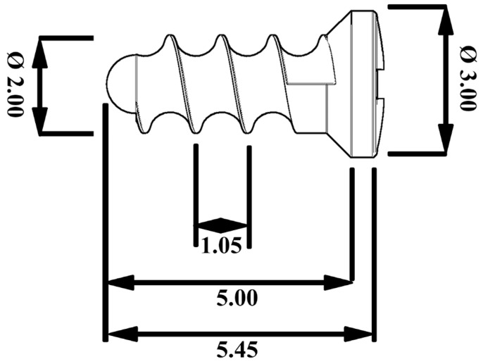
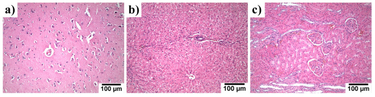
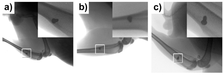
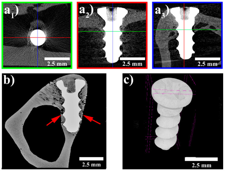
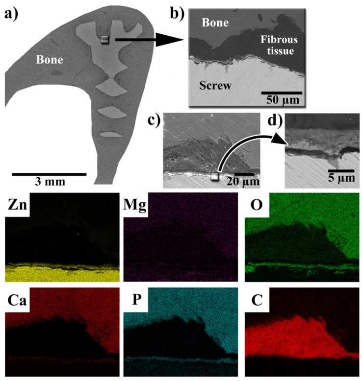
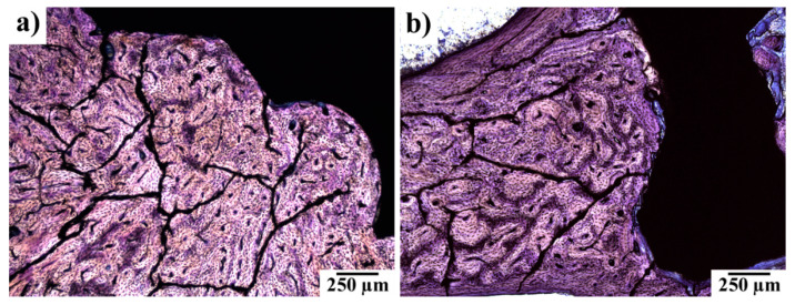
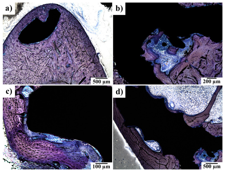
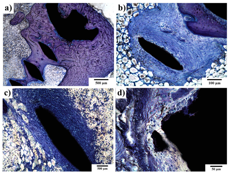
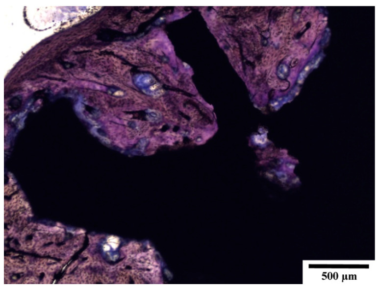
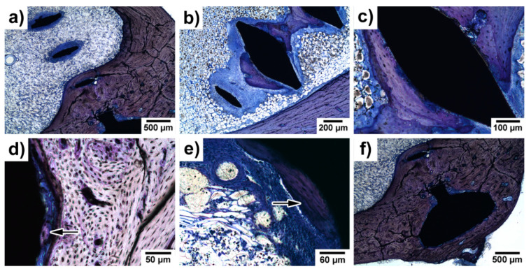
In this pilot study, we investigated the biocompatibility and degradation rate of an extruded Zn-0.8Mg-0.2Sr (wt.%) alloy on a rabbit model. An alloy screw was implanted into one of the tibiae of New Zealand White rabbits. After 120 days, the animals were euthanized. Evaluation included clinical assessment, microCT, histological examination of implants, analyses of the adjacent bone, and assessment of zinc, magnesium, and strontium in vital organs (liver, kidneys, brain). The bone sections with the implanted screw were examined via scanning electron microscopy and energy dispersive spectroscopy (SEM-EDS). This method showed that the implant was covered by a thin layer of phosphate-based solid corrosion products with a thickness ranging between 4 and 5 µm. Only negligible changes of the implant volume and area were observed. The degradation was not connected with gas evolution. The screws were fibrointegrated, partially osseointegrated histologically. We observed no inflammatory reaction or bone resorption. Periosteal apposition and formation of new bone with a regular structure were frequently observed near the implant surface. The histological evaluation of the liver, kidneys, and brain showed no toxic changes. The levels of Zn, Mg, and Sr after 120 days in the liver, kidneys, and brain did not exceed the reference values for these elements. The alloy was safe, biocompatible, and well-tolerated.
在这项初步研究中,我们在兔模型上研究了挤压态Zn-0.8Mg-0.2Sr(重量百分比)合金的生物相容性和降解率。将一枚合金螺钉植入新西兰白兔的一侧胫骨。120天后,对动物实施安乐死。评估内容包括临床评估、微型计算机断层扫描(microCT)、植入物的组织学检查、相邻骨的分析以及重要器官(肝脏、肾脏、脑)中锌、镁和锶的评估。通过扫描电子显微镜和能谱分析(SEM-EDS)对带有植入螺钉的骨切片进行检查。该方法显示,植入物被一层厚度在4至5μm之间的磷酸盐基固体腐蚀产物覆盖。仅观察到植入物体积和面积的微小变化。降解过程未伴随气体释放。螺钉在组织学上呈纤维整合,部分呈骨整合。我们未观察到炎症反应或骨吸收。在植入物表面附近经常观察到骨膜附着和结构规则的新骨形成。肝脏、肾脏和脑的组织学评估未显示毒性变化。120天后肝脏、肾脏和脑中锌、镁和锶的水平未超过这些元素的参考值。该合金安全、具有生物相容性且耐受性良好。