Chigoho Dora Mugoli, Lecocq Quentin, Awad Robin Maximilian, Breckpot Karine, Devoogdt Nick, Keyaerts Marleen, Caveliers Vicky, Xavier Catarina, Bridoux Jessica
In Vivo Cellular and Molecular Imaging Laboratory (ICMI), Medical Imaging Department (MIMA), Vrije Universiteit Brussel, 1090 Brussels, Belgium.
Laboratory for Molecular and Cellular Therapy (LMCT), Department of Biomedical Sciences, Vrije Universiteit Brussel, 1090 Brussels, Belgium.
Pharmaceuticals (Basel). 2021 Jun 8;14(6):550. doi: 10.3390/ph14060550.
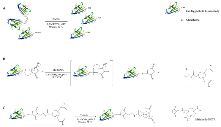
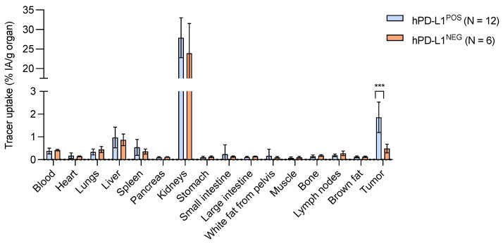
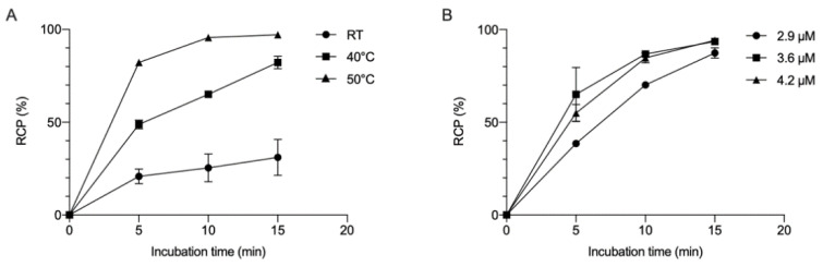
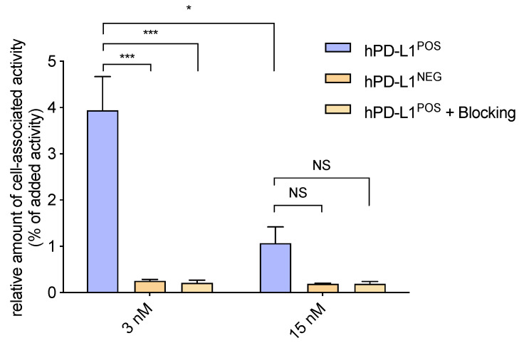
Immune checkpoint inhibitors targeting the programmed cell death-1 (PD-1) and its ligand PD-L1 have proven to be efficient cancer therapies in a subset of patients. From all the patients with various cancer types, only 20% have a positive response. Being able to distinguish patients that do express PD-1/PD-L1 from patients that do not allows patients to benefit from a more personalized and efficient treatment of tumor lesion(s). Expression of PD-1 and PD-L1 is typically assessed via immunohistochemical detection in a tumor biopsy. However, this method does not take in account the expression heterogeneity within the lesion, nor the possible metastasis. To visualize whole-body PD-L1 expression by PET imaging, we developed a nanobody-based radio-immunotracer targeting PD-L1 site-specifically labeled with gallium-68. The cysteine-tagged nanobody was site-specifically conjugated with a maleimide (mal)-NOTA chelator and radiolabeling was tested at different nanobody concentrations and temperatures. Affinity and specificity of the tracer, referred to as [Ga]Ga-NOTA-mal-hPD-L1 Nb, were assayed by surface plasmon resonance and on PD-L1 or PD-L1 624-MEL cells. Xenografted athymic nude mice bearing 624-MEL PD-L1 or PD-L1 tumors were injected with the tracer and ex vivo biodistribution was performed 1 h 20 min post-injection. Ideal Ga-labeling conditions were found at 50 °C for 15 min. [Ga]Ga-NOTA-mal-hPD-L1 Nb was obtained in 80 ± 5% DC-RCY with a RCP > 99%, and was stable in injection buffer and human serum up to 3 h (>99% RCP). The in vitro characterization showed that the NOTA-functionalized Nb retained its affinity and specificity. Ex vivo biodistribution revealed a tracer uptake of 1.86 ± 0.67% IA/g in the positive tumors compared with 0.42 ± 0.04% IA/g in the negative tumors. Low background uptake was measured in the other organs and tissues, except for the kidneys and bladder, due to the expected excretion route of Nbs. The data obtained show that the site-specific Ga-labeled NOTA-mal-hPD-L1 Nb is a promising PET radio-immunotracer due to its ease of production, stability and specificity for PD-L1.
靶向程序性细胞死亡蛋白1(PD-1)及其配体PD-L1的免疫检查点抑制剂已被证明在一部分患者中是有效的癌症治疗方法。在所有患有各种癌症类型的患者中,只有20%有阳性反应。能够区分表达PD-1/PD-L1的患者和不表达的患者,可使患者受益于对肿瘤病灶更个性化、更有效的治疗。PD-1和PD-L1的表达通常通过肿瘤活检中的免疫组织化学检测来评估。然而,这种方法没有考虑到病灶内的表达异质性,也没有考虑到可能的转移情况。为了通过PET成像可视化全身PD-L1表达,我们开发了一种基于纳米抗体的放射性免疫示踪剂,该示踪剂靶向用镓-68特异性标记的PD-L1。将带有半胱氨酸标签的纳米抗体与马来酰亚胺(mal)-NOTA螯合剂进行位点特异性偶联,并在不同的纳米抗体浓度和温度下测试放射性标记。通过表面等离子体共振以及在PD-L1或PD-L1 624-MEL细胞上测定了称为[Ga]Ga-NOTA-mal-hPD-L1 Nb的示踪剂的亲和力和特异性。给携带624-MEL PD-L1或PD-L1肿瘤的异种移植无胸腺裸鼠注射该示踪剂,并在注射后1小时20分钟进行离体生物分布研究。发现50℃下15分钟是理想的镓标记条件。[Ga]Ga-NOTA-mal-hPD-L1 Nb的放射性化学产率为80±5%,放射化学纯度>99%,并且在注射缓冲液和人血清中3小时内稳定(放射化学纯度>99%)。体外表征表明,NOTA功能化的纳米抗体保留了其亲和力和特异性。离体生物分布显示,阳性肿瘤中示踪剂摄取为1.86±0.67%IA/g,而阴性肿瘤中为0.42±0.04%IA/g。除肾脏和膀胱外,由于纳米抗体预期的排泄途径,在其他器官和组织中测得的背景摄取较低。所获得的数据表明,位点特异性镓标记的NOTA-mal-hPD-L1 Nb由于其易于生产、稳定性以及对PD-L1的特异性,是一种有前景的PET放射性免疫示踪剂。