Department of Radiology, UT Southwestern Medical Center, Dallas, Texas.
Advanced Imaging Research Center, UT Southwestern Medical Center, Dallas, Texas.
Clin Cancer Res. 2021 Sep 1;27(17):4794-4806. doi: 10.1158/1078-0432.CCR-21-0706. Epub 2021 Jul 1.
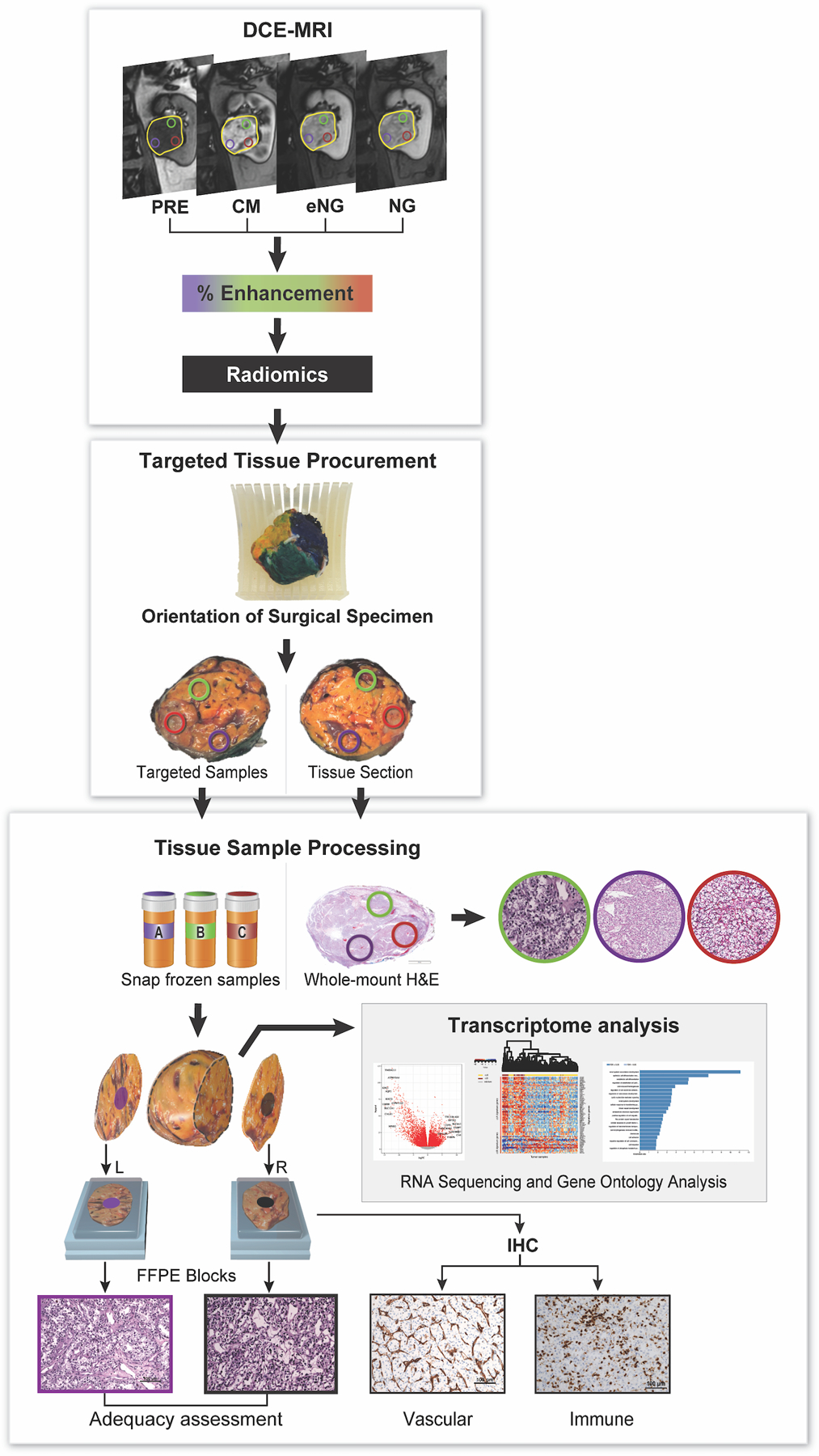
Intratumoral heterogeneity (ITH) challenges the molecular characterization of clear cell renal cell carcinoma (ccRCC) and is a confounding factor for therapy selection. Most approaches to evaluate ITH are limited by two-dimensional tissue analyses. Dynamic contrast-enhanced magnetic resonance imaging (DCE-MRI) can noninvasively assess the spatial landscape of entire tumors in their natural milieu. To assess the potential of DCE-MRI, we developed a vertically integrated radiogenomics colocalization approach for multi-region tissue acquisition and analyses. We investigated the potential of spatial imaging features to predict molecular subtypes using histopathologic and transcriptome correlatives.
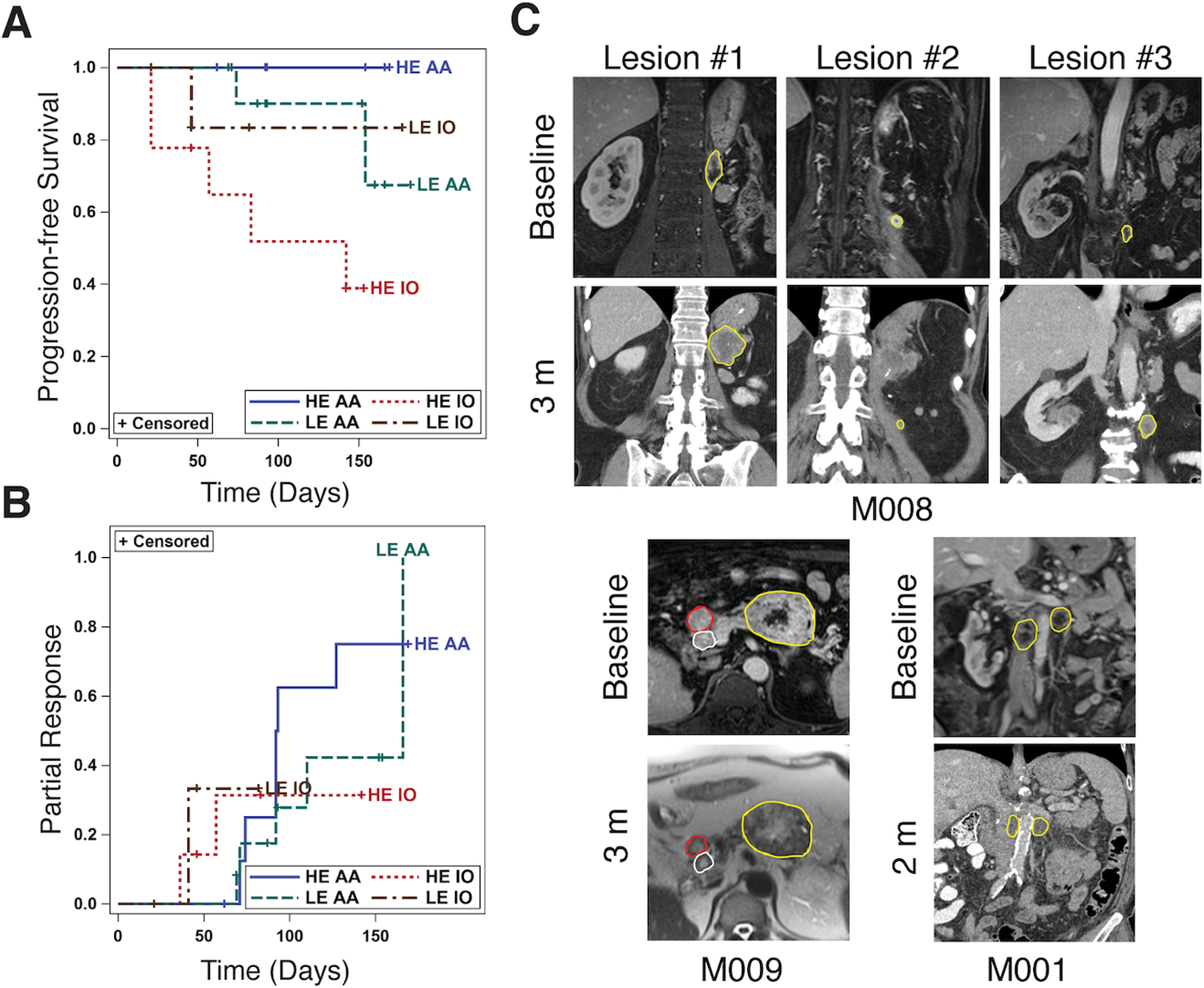
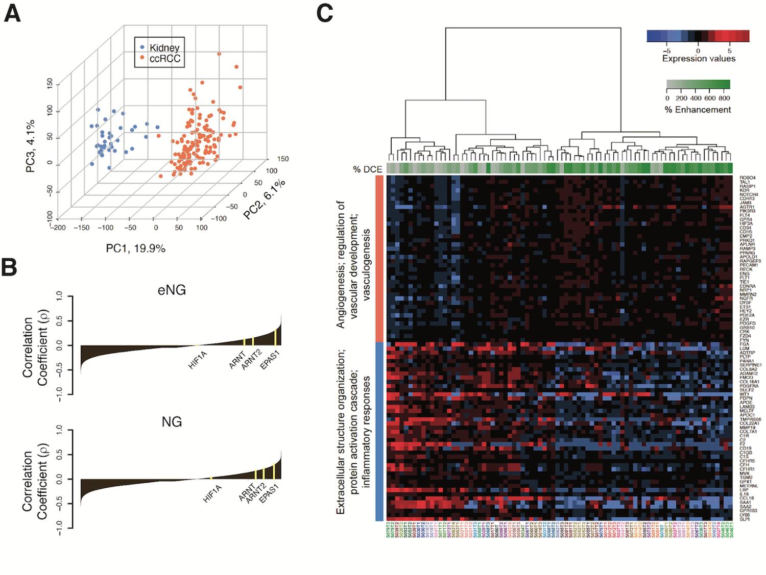
We report the results of a prospective study of 49 patients with ccRCC who underwent DCE-MRI prior to nephrectomy. Surgical specimens were sectioned to match the MRI acquisition plane. RNA sequencing data from multi-region tumor sampling (80 samples) were correlated with percent enhancement on DCE-MRI in spatially colocalized regions of the tumor. Independently, we evaluated clinical applicability of our findings in 19 patients with metastatic RCC (39 metastases) treated with first-line antiangiogenic drugs or checkpoint inhibitors.
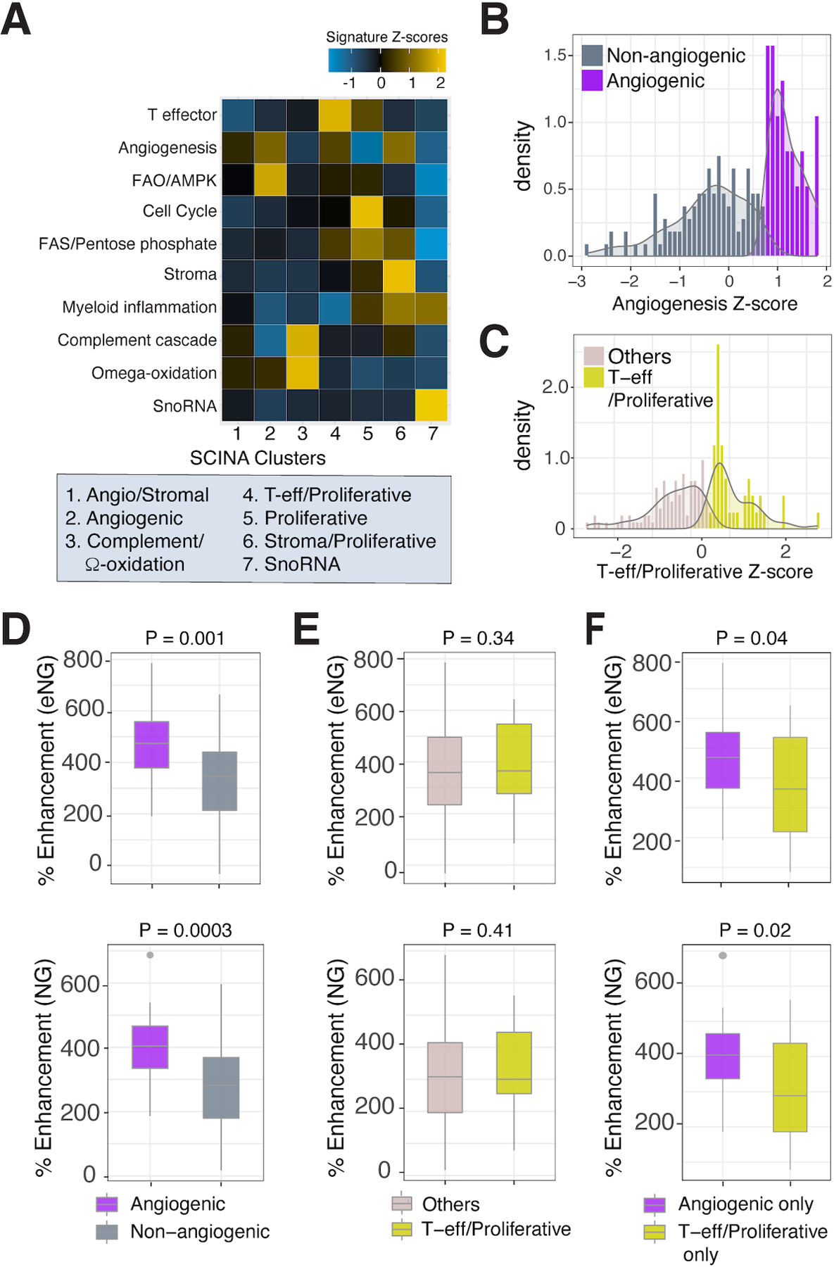
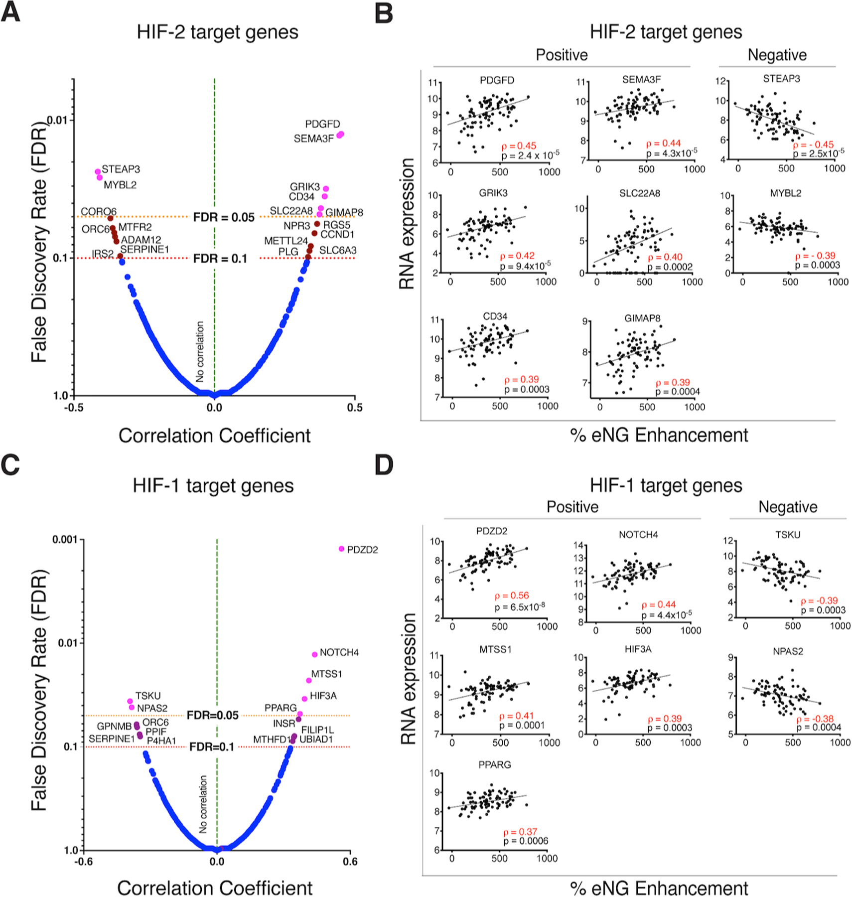
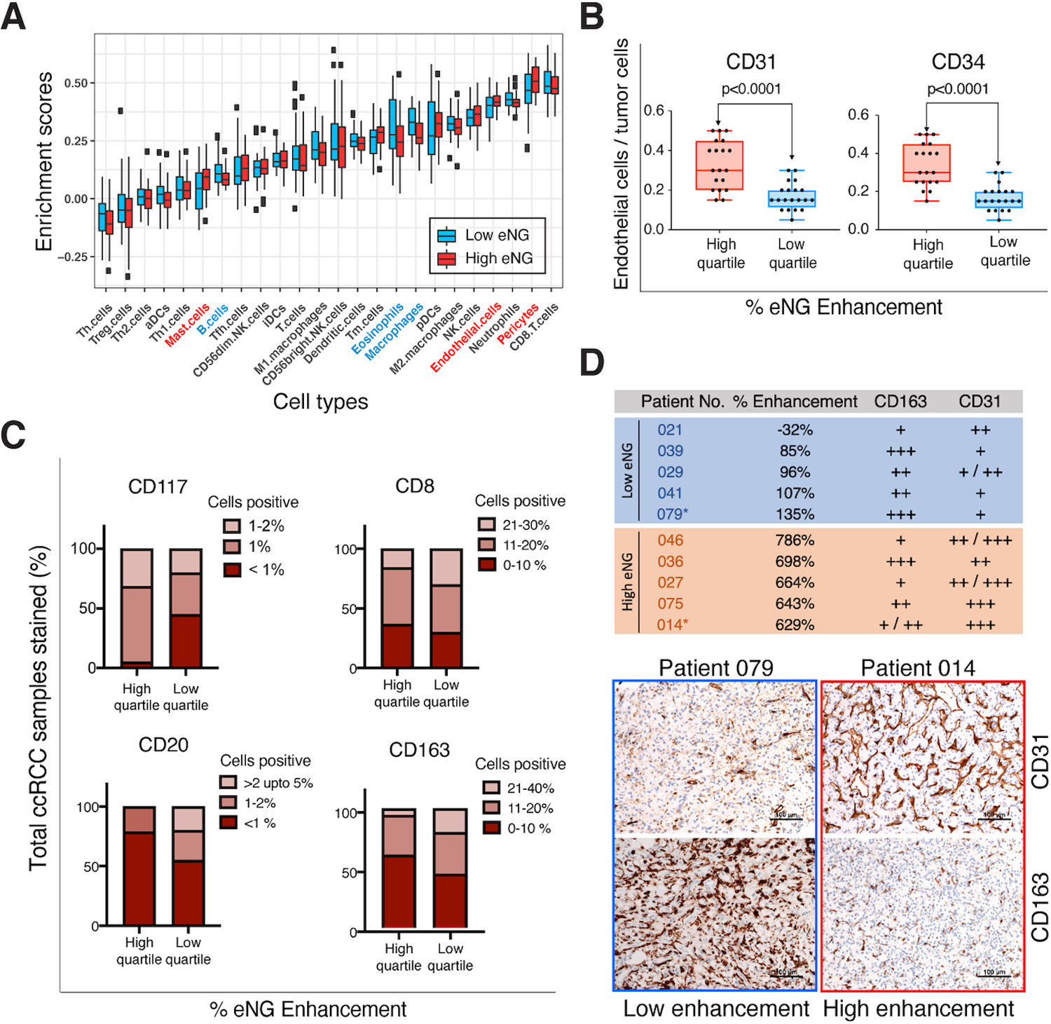
DCE-MRI identified tumor features associated with angiogenesis and inflammation, which differed within and across tumors, and likely contribute to the efficacy of antiangiogenic drugs and immunotherapies. Our vertically integrated analyses show that angiogenesis and inflammation frequently coexist and spatially anti-correlate in the same tumor. Furthermore, MRI contrast enhancement identifies phenotypes with better response to antiangiogenic therapy among patients with metastatic RCC.
These findings have important implications for decision models based on biopsy samples and highlight the potential of more comprehensive imaging-based approaches.
肿瘤内异质性(ITH)挑战了透明细胞肾细胞癌(ccRCC)的分子特征,并且是治疗选择的混杂因素。评估 ITH 的大多数方法都受到二维组织分析的限制。动态对比增强磁共振成像(DCE-MRI)可以非侵入性地评估整个肿瘤在其自然环境中的空间景观。为了评估 DCE-MRI 的潜力,我们开发了一种垂直整合的放射组学共定位方法,用于多区域组织采集和分析。我们研究了空间成像特征预测分子亚型的潜力,使用组织病理学和转录组相关物进行了研究。
我们报告了 49 例接受 DCE-MRI 检查的 ccRCC 患者的前瞻性研究结果,这些患者在肾切除术前接受了检查。手术标本被切成与 MRI 采集平面相匹配的切片。从多区域肿瘤取样(80 个样本)获得的 RNA 测序数据与肿瘤空间共定位区域的 DCE-MRI 上的增强百分比相关。独立地,我们评估了在接受一线抗血管生成药物或检查点抑制剂治疗的 19 例转移性 RCC 患者(39 个转移灶)中发现的临床适用性。
DCE-MRI 确定了与血管生成和炎症相关的肿瘤特征,这些特征在肿瘤内和肿瘤间存在差异,并且可能对抗血管生成药物和免疫疗法的疗效有贡献。我们的垂直整合分析表明,血管生成和炎症经常共存,并且在同一肿瘤中空间上呈负相关。此外,MRI 对比增强可以识别出转移性 RCC 患者中对抗血管生成治疗反应更好的表型。
这些发现对基于活检样本的决策模型具有重要意义,并强调了更全面的基于成像的方法的潜力。