• 文献检索
  • 文档翻译
  • 深度研究
  • 学术资讯
  • Suppr Zotero 插件Zotero 插件
  • 邀请有礼
  • 套餐&价格
  • 历史记录
应用&插件
Suppr Zotero 插件Zotero 插件浏览器插件Mac 客户端Windows 客户端微信小程序
定价
高级版会员购买积分包购买API积分包
服务
文献检索文档翻译深度研究API 文档MCP 服务
关于我们
关于 Suppr公司介绍联系我们用户协议隐私条款
关注我们

Suppr 超能文献

核心技术专利:CN118964589B侵权必究
粤ICP备2023148730 号-1Suppr @ 2026

文献检索

告别复杂PubMed语法,用中文像聊天一样搜索,搜遍4000万医学文献。AI智能推荐,让科研检索更轻松。

立即免费搜索

文件翻译

保留排版,准确专业,支持PDF/Word/PPT等文件格式,支持 12+语言互译。

免费翻译文档

深度研究

AI帮你快速写综述,25分钟生成高质量综述,智能提取关键信息,辅助科研写作。

立即免费体验

未增强磁共振成像与超声在检测极微小肝细胞癌中的比较

Comparison of unenhanced magnetic resonance imaging and ultrasound in detecting very small hepatocellular carcinoma.

作者信息

Tarao Kazuo, Nozaki Akito, Komatsu Hirokazu, Komatsu Tatsuji, Taguri Masataka, Tanaka Katsuaki, Yoshida Testuo, Koyasu Hideki, Chuma Makoto, Numata Kazushi, Maeda Shin

机构信息

Tarao's Gastroenterological Clinic, Yokohama 241-0821, Japan.

Gastroenterological Center, Yokohama City University Medical Center, Yokohama 232-0024, Japan.

出版信息

World J Hepatol. 2021 Jun 27;13(6):699-708. doi: 10.4254/wjh.v13.i6.699.

DOI:10.4254/wjh.v13.i6.699
PMID:34239704
原文链接:https://pmc.ncbi.nlm.nih.gov/articles/PMC8239491/
Abstract

BACKGROUND

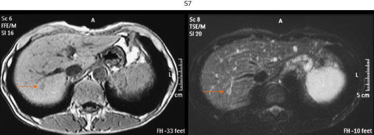
In hepatocellular carcinoma (HCC), detection and treatment prior to growth beyond 2 cm are important as a larger tumor size is more frequently associated with microvascular invasion and/or satellites. In the surveillance of very small HCC nodules (≤ 2 cm in maximum diameter, Barcelona clinical stage 0), we demonstrated that the tumor markers alpha-fetoprotein and PIVKA-Ⅱ are not so useful. Therefore, we must survey with imaging modalities. The superiority of magnetic resonance imaging (MRI) over ultrasound (US) to detect HCC was confirmed in many studies. Although enhanced MRI is now performed to accurately diagnose HCC, in conventional clinical practice for HCC surveillance in liver diseases, unenhanced MRI is widely performed throughout the world. While, MRI has made marked improvements in recent years.

AIM

To make a comparison of unenhanced MRI and US in detecting very small HCC that was examined in the last ten years in patients in whom MRI and US examinations were performed nearly simultaneously.

METHODS

In 394 patients with very small HCC nodules, those who underwent MRI and US at nearly the same time (on the same day whenever possible or at least within 14 days of one another) at the first diagnosis of HCC were selected. The detection rate of HCC with unenhanced MRI was investigated and compared with that of unenhanced US.

RESULTS

The sensitivity of unenhanced MRI for detecting very small HCC was 95.1% (97/102, 95% confidence interval: 90.9-99.3) and that of unenhanced US was 69.6% (71/102, 95% confidence interval: 60.7-78.5). The sensitivity of unenhanced MRI for detecting very small HCC was significantly higher than that of unenhanced US ( < 0.001). Regarding the location of HCC in the liver in patients in whom detection by US was unsuccessful, S was identified in 51.7%.

CONCLUSION

Currently, unenhanced MRI is a very useful tool for the surveillance of very small HCC in conventional clinical follow-up practice.

摘要

背景

在肝细胞癌(HCC)中,在肿瘤生长超过2 cm之前进行检测和治疗很重要,因为更大的肿瘤尺寸更常与微血管侵犯和/或卫星灶相关。在监测非常小的HCC结节(最大直径≤2 cm,巴塞罗那临床分期0期)时,我们发现肿瘤标志物甲胎蛋白和异常凝血酶原不太有用。因此,我们必须采用影像学检查方法。许多研究证实了磁共振成像(MRI)在检测HCC方面优于超声(US)。尽管现在进行增强MRI以准确诊断HCC,但在肝病中HCC监测的传统临床实践中,全世界广泛进行的是平扫MRI。同时,近年来MRI有了显著改进。

目的

比较平扫MRI和超声在检测过去十年中几乎同时进行MRI和超声检查的患者中非常小的HCC方面的差异。

方法

在394例有非常小的HCC结节的患者中,选择首次诊断HCC时几乎同时(尽可能在同一天或至少在彼此14天内)接受MRI和超声检查的患者。研究平扫MRI检测HCC的检出率,并与平扫超声的检出率进行比较。

结果

平扫MRI检测非常小的HCC的敏感性为95.1%(97/102,95%置信区间:90.9 - 99.3),平扫超声的敏感性为69.6%(71/102,95%置信区间:60.7 - 78.5)。平扫MRI检测非常小的HCC的敏感性显著高于平扫超声(<0.001)。对于超声检测未成功的患者,HCC在肝脏中的位置,S在51.7%中被识别。

结论

目前,在传统临床随访实践中,平扫MRI是监测非常小的HCC的非常有用的工具。

https://cdn.ncbi.nlm.nih.gov/pmc/blobs/4524/8239491/47a8336a2f37/WJH-13-699-g005.jpg
https://cdn.ncbi.nlm.nih.gov/pmc/blobs/4524/8239491/885312b97a64/WJH-13-699-g001.jpg
https://cdn.ncbi.nlm.nih.gov/pmc/blobs/4524/8239491/46b39a8c9adb/WJH-13-699-g002.jpg
https://cdn.ncbi.nlm.nih.gov/pmc/blobs/4524/8239491/bc973103700a/WJH-13-699-g003.jpg
https://cdn.ncbi.nlm.nih.gov/pmc/blobs/4524/8239491/26a032e457a4/WJH-13-699-g004.jpg
https://cdn.ncbi.nlm.nih.gov/pmc/blobs/4524/8239491/47a8336a2f37/WJH-13-699-g005.jpg

相似文献

1
Comparison of unenhanced magnetic resonance imaging and ultrasound in detecting very small hepatocellular carcinoma.未增强磁共振成像与超声在检测极微小肝细胞癌中的比较
World J Hepatol. 2021 Jun 27;13(6):699-708. doi: 10.4254/wjh.v13.i6.699.
2
Real impact of tumor marker AFP and PIVKA-II in detecting very small hepatocellular carcinoma (≤ 2 cm, Barcelona stage 0) - assessment with large number of cases.肿瘤标志物甲胎蛋白(AFP)和异常凝血酶原(PIVKA-II)在检测极微小肝细胞癌(≤2cm,巴塞罗那分期0期)中的实际影响——大量病例评估
World J Hepatol. 2020 Nov 27;12(11):1046-1054. doi: 10.4254/wjh.v12.i11.1046.
3
Noncontrast magnetic resonance imaging versus ultrasonography for hepatocellular carcinoma surveillance (MIRACLE-HCC): study protocol for a prospective randomized trial.非对比磁共振成像与超声检查用于肝细胞癌监测的比较(MIRACLE-HCC):一项前瞻性随机试验的研究方案。
BMC Cancer. 2018 Sep 24;18(1):915. doi: 10.1186/s12885-018-4827-2.
4
Gadoxetic acid-enhanced magnetic resonance imaging can predict the pathologic stage of solitary hepatocellular carcinoma.钆塞酸增强磁共振成像可预测单发肝细胞癌的病理分期。
World J Gastroenterol. 2019 Jun 7;25(21):2636-2649. doi: 10.3748/wjg.v25.i21.2636.
5
Comparison of biannual ultrasonography and annual non-contrast liver magnetic resonance imaging as surveillance tools for hepatocellular carcinoma in patients with liver cirrhosis (MAGNUS-HCC): a study protocol.超声造影每半年 1 次与非增强磁共振成像每年 1 次作为肝硬化患者肝癌监测工具的比较(MAGNUS-HCC):研究方案。
BMC Cancer. 2017 Dec 21;17(1):877. doi: 10.1186/s12885-017-3819-y.
6
7
Hepatocellular carcinoma in liver transplantation candidates: detection with gadobenate dimeglumine-enhanced MRI.肝移植候选者中的肝细胞癌:钆贝葡胺增强MRI检测
AJR Am J Roentgenol. 2008 Aug;191(2):529-36. doi: 10.2214/AJR.07.2565.
8
Preoperative evaluation of malignant liver tumors: comparison of unenhanced and SPIO (Resovist)-enhanced MR imaging with biphasic CTAP and intraoperative US.恶性肝肿瘤的术前评估:未增强和超顺磁性氧化铁(Resovist)增强磁共振成像与双期CT动脉门静脉造影及术中超声的比较
Eur Radiol. 2003 Feb;13(2):262-72. doi: 10.1007/s00330-002-1677-7. Epub 2002 Sep 10.
9
Contrast-Enhanced Ultrasound: a Simple and Effective Tool in Defining a Rapid Diagnostic Work-up for Small Nodules Detected in Cirrhotic Patients during Surveillance.超声造影:肝硬化患者监测中发现小结节时快速诊断的有效工具
J Gastrointestin Liver Dis. 2016 Jun;25(2):205-11. doi: 10.15403/jgld.2014.1121.252.chu.
10
Double-contrast MRI (DC-MRI) in the study of the cirrhotic liver: utility of administering Gd-DTPA as a complement to examinations in which SPIO liver uptake and distribution alterations (SPIO-LUDA) are present and in the identification and characterisation of focal lesions.双对比磁共振成像(DC-MRI)在肝硬化肝脏研究中的应用:给予钆喷酸葡胺作为补充检查的效用,用于存在超顺磁性氧化铁肝脏摄取和分布改变(SPIO-LUDA)的情况以及局灶性病变的识别和特征描述。
Radiol Med. 2006 Dec;111(8):1087-102. doi: 10.1007/s11547-006-0107-3. Epub 2006 Dec 20.

引用本文的文献

1
Radiomics analysis based on dynamic contrast-enhanced MRI for predicting early recurrence after hepatectomy in hepatocellular carcinoma patients.基于动态对比增强磁共振成像的影像组学分析预测肝细胞癌患者肝切除术后早期复发
Sci Rep. 2025 Jul 1;15(1):22240. doi: 10.1038/s41598-025-02291-6.
2
ASAP Score GALAD Score for detection of hepatitis C-related hepatocellular carcinoma: A multicenter case-control analysis.用于检测丙型肝炎相关肝细胞癌的ASAP评分与GALAD评分:一项多中心病例对照分析。
Front Oncol. 2022 Sep 30;12:1018396. doi: 10.3389/fonc.2022.1018396. eCollection 2022.

本文引用的文献

1
Real impact of tumor marker AFP and PIVKA-II in detecting very small hepatocellular carcinoma (≤ 2 cm, Barcelona stage 0) - assessment with large number of cases.肿瘤标志物甲胎蛋白(AFP)和异常凝血酶原(PIVKA-II)在检测极微小肝细胞癌(≤2cm,巴塞罗那分期0期)中的实际影响——大量病例评估
World J Hepatol. 2020 Nov 27;12(11):1046-1054. doi: 10.4254/wjh.v12.i11.1046.
2
MRI With Liver-Specific Contrast for Surveillance of Patients With Cirrhosis at High Risk of Hepatocellular Carcinoma.MRI 联合肝脏特异性对比剂用于监测肝硬化高危 HCC 患者
JAMA Oncol. 2017 Apr 1;3(4):456-463. doi: 10.1001/jamaoncol.2016.3147.
3
Validation of diagnostic criteria using gadoxetic acid-enhanced and diffusion-weighted MR imaging for small hepatocellular carcinoma (<= 2.0 cm) in patients with hepatitis-induced liver cirrhosis.
使用钆塞酸二钠增强磁共振成像和扩散加权磁共振成像对肝炎后肝硬化患者小肝细胞癌(≤2.0 cm)诊断标准的验证
Acta Radiol. 2013 Mar 1;54(2):127-36. doi: 10.1258/ar.2012.120262. Epub 2012 Nov 12.
4
Hepatocellular carcinoma.肝细胞癌。
Lancet. 2012 Mar 31;379(9822):1245-55. doi: 10.1016/S0140-6736(11)61347-0. Epub 2012 Feb 20.
5
Detection of pancreatic carcinoma and liver metastases with gadoxetic acid-enhanced MR imaging: comparison with contrast-enhanced multi-detector row CT.钆塞酸增强磁共振成像检测胰腺癌和肝转移瘤:与增强多排 CT 的比较。
Radiology. 2011 Aug;260(2):446-53. doi: 10.1148/radiol.11103548. Epub 2011 Jun 21.
6
Intraindividual comparison of gadoxetate disodium-enhanced MR imaging and 64-section multidetector CT in the Detection of hepatocellular carcinoma in patients with cirrhosis.钆塞酸二钠增强磁共振成像与 64 层多排 CT 检测肝硬化患者肝细胞癌的个体内比较。
Radiology. 2010 Sep;256(3):806-16. doi: 10.1148/radiol.10091334.
7
Detection of hepatic metastases by superparamagnetic iron oxide-enhanced MR imaging: prospective comparison between 1.5-T and 3.0-T images in the same patients.超顺磁性氧化铁增强磁共振成像检测肝转移瘤:同患者 1.5T 与 3.0T 图像的前瞻性对比研究。
Eur Radiol. 2010 Sep;20(9):2265-73. doi: 10.1007/s00330-010-1798-3. Epub 2010 Apr 29.
8
Surveillance for hepatocellular carcinoma in patients with cirrhosis improves outcome.对肝硬化患者进行肝细胞癌监测可改善预后。
Am J Med. 2008 Feb;121(2):119-26. doi: 10.1016/j.amjmed.2007.09.020.
9
Diffusion-weighed MR imaging of pancreatic carcinoma.胰腺癌的扩散加权磁共振成像
Abdom Imaging. 2007 Jul-Aug;32(4):481-3. doi: 10.1007/s00261-007-9192-6.
10
Accuracy of ultrasonography, spiral CT, magnetic resonance, and alpha-fetoprotein in diagnosing hepatocellular carcinoma: a systematic review.超声检查、螺旋CT、磁共振成像及甲胎蛋白诊断肝细胞癌的准确性:一项系统评价
Am J Gastroenterol. 2006 Mar;101(3):513-23. doi: 10.1111/j.1572-0241.2006.00467.x.