• 文献检索
  • 文档翻译
  • 深度研究
  • 学术资讯
  • Suppr Zotero 插件Zotero 插件
  • 邀请有礼
  • 套餐&价格
  • 历史记录
应用&插件
Suppr Zotero 插件Zotero 插件浏览器插件Mac 客户端Windows 客户端微信小程序
定价
高级版会员购买积分包购买API积分包
服务
文献检索文档翻译深度研究API 文档MCP 服务
关于我们
关于 Suppr公司介绍联系我们用户协议隐私条款
关注我们

Suppr 超能文献

核心技术专利:CN118964589B侵权必究
粤ICP备2023148730 号-1Suppr @ 2026

文献检索

告别复杂PubMed语法,用中文像聊天一样搜索,搜遍4000万医学文献。AI智能推荐,让科研检索更轻松。

立即免费搜索

文件翻译

保留排版,准确专业,支持PDF/Word/PPT等文件格式,支持 12+语言互译。

免费翻译文档

深度研究

AI帮你快速写综述,25分钟生成高质量综述,智能提取关键信息,辅助科研写作。

立即免费体验

严重 COVID-19 康复与髓系细胞免疫调节表型的及时获得有关。

Severe COVID-19 Recovery Is Associated with Timely Acquisition of a Myeloid Cell Immune-Regulatory Phenotype.

机构信息

Instituto de Medicina Molecular João Lobo Antunes, Faculdade de Medicina, Universidade de Lisboa, Lisbon, Portugal.

Clinica Universitária de Medicina Intensiva, Faculdade de Medicina, Universidade de Lisboa, Lisbon, Portugal.

出版信息

Front Immunol. 2021 Jun 23;12:691725. doi: 10.3389/fimmu.2021.691725. eCollection 2021.

DOI:10.3389/fimmu.2021.691725
PMID:34248984
原文链接:https://pmc.ncbi.nlm.nih.gov/articles/PMC8265310/
Abstract

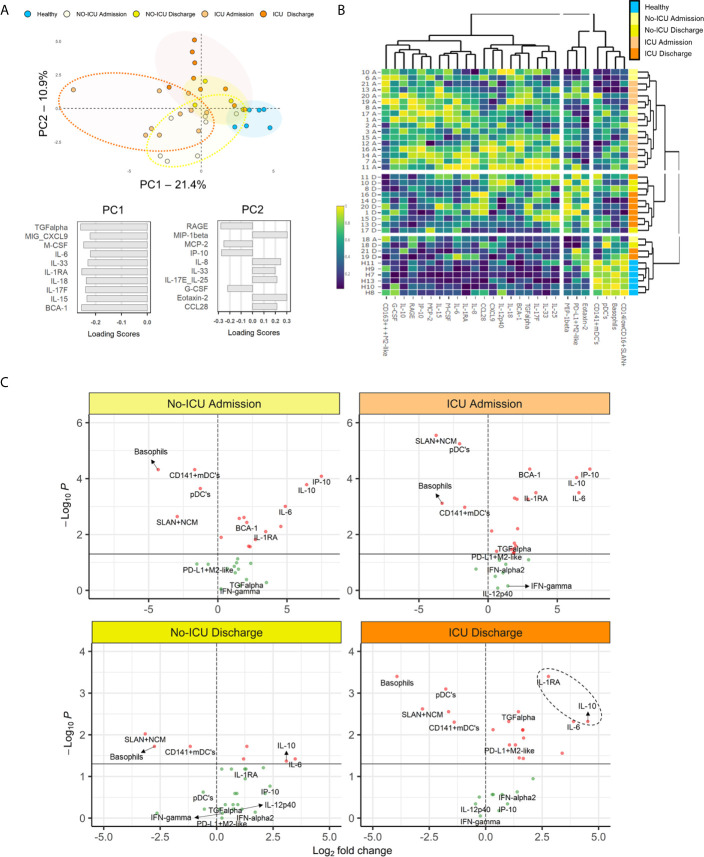
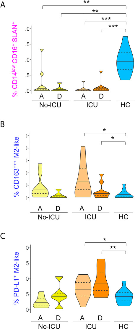
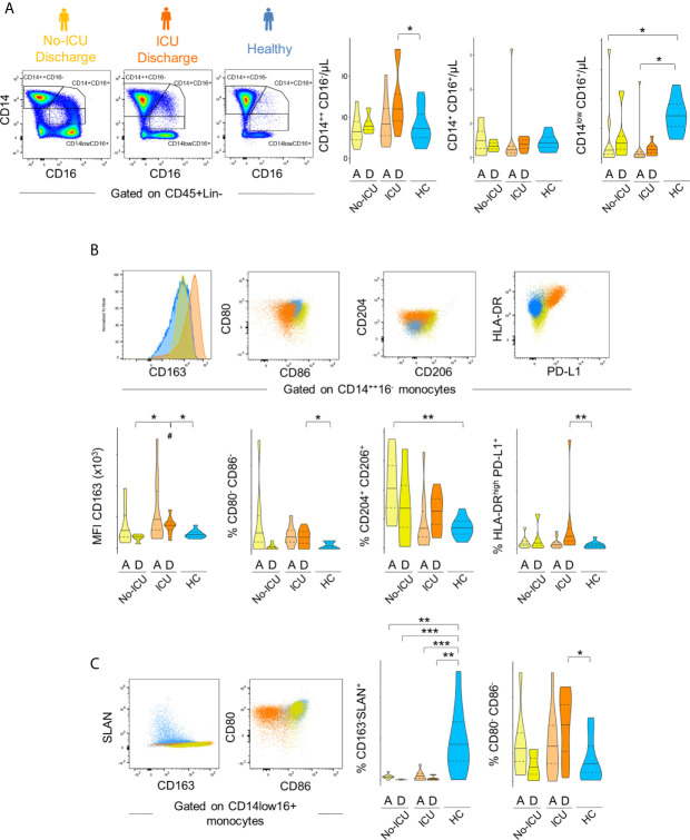
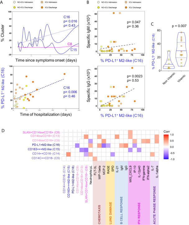
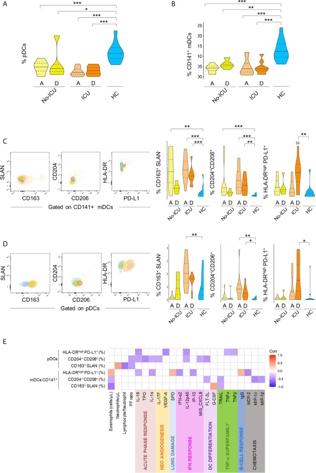
After more than one year since the COVID-19 outbreak, patients with severe disease still constitute the bottleneck of the pandemic management. Aberrant inflammatory responses, ranging from cytokine storm to immune-suppression, were described in COVID-19 and no treatment was demonstrated to change the prognosis significantly. Therefore, there is an urgent need for understanding the underlying pathogenic mechanisms to guide therapeutic interventions. This study was designed to assess myeloid cell activation and phenotype leading to recovery in patients surviving severe COVID-19. We evaluated longitudinally patients with COVID-19 related respiratory insufficiency, stratified according to the need of intensive care unit admission (ICU, n = 11, and No-ICU, n = 9), and age and sex matched healthy controls (HCs, n = 11), by flow cytometry and a wide array of serum inflammatory/immune-regulatory mediators. All patients featured systemic immune-regulatory myeloid cell phenotype as assessed by both unsupervised and supervised analysis of circulating monocyte and dendritic cell subsets. Specifically, we observed a reduction of CD14lowCD16+ monocytes, and reduced expression of CD80, CD86, and Slan. Moreover, mDCs, pDCs, and basophils were significantly reduced, in comparison to healthy subjects. Contemporaneously, both monocytes and DCs showed increased expression of CD163, CD204, CD206, and PD-L1 immune-regulatory markers. The expansion of M2-like monocytes was significantly higher at admission in patients featuring detectable SARS-CoV-2 plasma viral load and it was positively correlated with the levels of specific antibodies. In No-ICU patients, we observed a peak of the alterations at admission and a progressive regression to a phenotype similar to HCs at discharge. Interestingly, in ICU patients, the expression of immuno-suppressive markers progressively increased until discharge. Notably, an increase of M2-like HLA-DRhighPD-L1+ cells in CD14++CD16- monocytes and in dendritic cell subsets was observed at ICU discharge. Furthermore, IFN-γ and IL-12p40 showed a decline over time in ICU patients, while high values of IL1RA and IL-10 were maintained. In conclusion, these results support that timely acquisition of a myeloid cell immune-regulatory phenotype might contribute to recovery in severe systemic SARS-CoV-2 infection and suggest that therapeutic agents favoring an innate immune system regulatory shift may represent the best strategy to be implemented at this stage.

摘要

自 COVID-19 爆发以来已经一年多了,重症患者仍然是疫情管理的瓶颈。COVID-19 中描述了异常的炎症反应,从细胞因子风暴到免疫抑制,目前还没有治疗方法能显著改变预后。因此,迫切需要了解潜在的发病机制,以指导治疗干预。本研究旨在评估导致重症 COVID-19 患者康复的髓系细胞激活和表型。我们通过流式细胞术和一系列血清炎症/免疫调节介质,对因呼吸功能不全而住院的 COVID-19 患者(分为需要入住重症监护病房(ICU)的患者(n=11)和不需要入住 ICU 的患者(n=9),以及年龄和性别匹配的健康对照者(HCs,n=11)进行了纵向评估。所有患者均表现出全身性免疫调节髓系细胞表型,这是通过对循环单核细胞和树突状细胞亚群进行无监督和监督分析得出的。具体来说,我们观察到 CD14lowCD16+单核细胞减少,以及 CD80、CD86 和 Slan 的表达减少。此外,与健康受试者相比,mDC、pDC 和嗜碱性粒细胞显著减少。同时,与单核细胞和 DC 相比,CD163、CD204、CD206 和 PD-L1 等免疫调节标志物的表达增加。在入院时,具有可检测 SARS-CoV-2 血浆病毒载量的患者中,M2 样单核细胞的扩增明显更高,且与特异性抗体水平呈正相关。在不需要 ICU 治疗的患者中,我们观察到入院时的变化达到峰值,并在出院时逐渐恢复到与 HCs 相似的表型。有趣的是,在 ICU 患者中,免疫抑制标志物的表达逐渐增加,直到出院。值得注意的是,在 ICU 出院时,CD14++CD16-单核细胞和树突状细胞亚群中的 HLA-DRhighPD-L1+M2 样细胞增加。此外,IFN-γ和 IL-12p40 的水平随时间下降,而 IL1RA 和 IL-10 的高值保持不变。总之,这些结果支持在严重全身 SARS-CoV-2 感染中及时获得髓系细胞免疫调节表型可能有助于康复,并表明有利于固有免疫系统调节转变的治疗药物可能是现阶段的最佳策略。

https://cdn.ncbi.nlm.nih.gov/pmc/blobs/4698/8265310/51e2a1e18c5b/fimmu-12-691725-g006.jpg
https://cdn.ncbi.nlm.nih.gov/pmc/blobs/4698/8265310/30d27b961ee3/fimmu-12-691725-g001.jpg
https://cdn.ncbi.nlm.nih.gov/pmc/blobs/4698/8265310/abd26f3b70b5/fimmu-12-691725-g002.jpg
https://cdn.ncbi.nlm.nih.gov/pmc/blobs/4698/8265310/b50585561aa4/fimmu-12-691725-g003.jpg
https://cdn.ncbi.nlm.nih.gov/pmc/blobs/4698/8265310/ab6fb83eecc7/fimmu-12-691725-g004.jpg
https://cdn.ncbi.nlm.nih.gov/pmc/blobs/4698/8265310/3b60174cd309/fimmu-12-691725-g005.jpg
https://cdn.ncbi.nlm.nih.gov/pmc/blobs/4698/8265310/51e2a1e18c5b/fimmu-12-691725-g006.jpg

相似文献

1
Severe COVID-19 Recovery Is Associated with Timely Acquisition of a Myeloid Cell Immune-Regulatory Phenotype.严重 COVID-19 康复与髓系细胞免疫调节表型的及时获得有关。
Front Immunol. 2021 Jun 23;12:691725. doi: 10.3389/fimmu.2021.691725. eCollection 2021.
2
Increased sCD163 and sCD14 Plasmatic Levels and Depletion of Peripheral Blood Pro-Inflammatory Monocytes, Myeloid and Plasmacytoid Dendritic Cells in Patients With Severe COVID-19 Pneumonia.严重 COVID-19 肺炎患者的 sCD163 和 sCD14 血浆水平升高及外周血促炎单核细胞、髓系和浆细胞样树突状细胞耗竭。
Front Immunol. 2021 Feb 26;12:627548. doi: 10.3389/fimmu.2021.627548. eCollection 2021.
3
Reduced Expression of Autophagy Markers and Expansion of Myeloid-Derived Suppressor Cells Correlate With Poor T Cell Response in Severe COVID-19 Patients.自噬标志物表达降低和髓系来源抑制细胞扩增与重症 COVID-19 患者 T 细胞反应不良相关。
Front Immunol. 2021 Feb 22;12:614599. doi: 10.3389/fimmu.2021.614599. eCollection 2021.
4
Monocytes and Macrophages, Targets of Severe Acute Respiratory Syndrome Coronavirus 2: The Clue for Coronavirus Disease 2019 Immunoparalysis.单核细胞和巨噬细胞:严重急性呼吸综合征冠状病毒 2 的作用靶点:2019 冠状病毒病免疫麻痹的线索。
J Infect Dis. 2021 Aug 2;224(3):395-406. doi: 10.1093/infdis/jiab044.
5
Critical COVID-19 is associated with distinct leukocyte phenotypes and transcriptome patterns.危重新冠肺炎与独特的白细胞表型和转录组模式相关。
J Intern Med. 2021 Sep;290(3):677-692. doi: 10.1111/joim.13310. Epub 2021 Jun 3.
6
SARS-CoV-2 infection induces mixed M1/M2 phenotype in circulating monocytes and alterations in both dendritic cell and monocyte subsets.SARS-CoV-2 感染诱导循环单核细胞中 M1/M2 表型混合,并改变树突状细胞和单核细胞亚群。
PLoS One. 2020 Dec 31;15(12):e0241097. doi: 10.1371/journal.pone.0241097. eCollection 2020.
7
Respiratory and systemic monocytes, dendritic cells, and myeloid-derived suppressor cells in COVID-19: Implications for disease severity.COVID-19 中的呼吸和全身单核细胞、树突状细胞和髓系来源的抑制性细胞:对疾病严重程度的影响。
J Intern Med. 2023 Feb;293(2):130-143. doi: 10.1111/joim.13559. Epub 2022 Aug 31.
8
Elevated frequencies of CD14HLA-DR MDSCs in COVID-19 patients.新冠病毒肺炎患者中CD14+HLA-DR髓系来源抑制细胞频率升高。
Aging (Albany NY). 2021 Feb 26;13(5):6236-6246. doi: 10.18632/aging.202571.
9
Suppressive Monocytes Impair MAIT Cells Response via IL-10 in Patients with Severe COVID-19.严重 COVID-19 患者中抑制性单核细胞通过 IL-10 抑制 MAIT 细胞反应。
J Immunol. 2021 Oct 1;207(7):1848-1856. doi: 10.4049/jimmunol.2100228. Epub 2021 Aug 27.
10
Disease-duration based comparison of subsets of immune cells in SARS CoV-2 infected patients presenting with mild or severe symptoms identifies prognostic markers for severity.基于疾病持续时间的 SARS-CoV-2 感染轻症和重症患者免疫细胞亚群比较,鉴定出疾病严重程度的预后标志物。
Immun Inflamm Dis. 2021 Jun;9(2):419-434. doi: 10.1002/iid3.402. Epub 2021 Jan 16.

引用本文的文献

1
Temporal multi-omics analysis of COVID-19 in end-stage kidney disease.终末期肾病患者新冠病毒病的时间多组学分析
Cell Genom. 2025 Aug 13;5(8):100918. doi: 10.1016/j.xgen.2025.100918. Epub 2025 Jun 17.
2
The longitudinal characterization of immune responses in COVID-19 patients reveals novel prognostic signatures for disease severity, patients' survival and long COVID.对 COVID-19 患者的免疫反应进行纵向特征分析,揭示了疾病严重程度、患者生存和长新冠的新预后特征。
Front Immunol. 2024 Jul 29;15:1381091. doi: 10.3389/fimmu.2024.1381091. eCollection 2024.
3
COVID-19 PBMCs are doubly harmful, through LDN-mediated lung epithelial damage and monocytic impaired responsiveness to live exposure.

本文引用的文献

1
Increased sCD163 and sCD14 Plasmatic Levels and Depletion of Peripheral Blood Pro-Inflammatory Monocytes, Myeloid and Plasmacytoid Dendritic Cells in Patients With Severe COVID-19 Pneumonia.严重 COVID-19 肺炎患者的 sCD163 和 sCD14 血浆水平升高及外周血促炎单核细胞、髓系和浆细胞样树突状细胞耗竭。
Front Immunol. 2021 Feb 26;12:627548. doi: 10.3389/fimmu.2021.627548. eCollection 2021.
2
SARS-CoV-2 engages inflammasome and pyroptosis in human primary monocytes.严重急性呼吸综合征冠状病毒2(SARS-CoV-2)在人原代单核细胞中激活炎性小体并引发细胞焦亡。
Cell Death Discov. 2021 Mar 1;7(1):43. doi: 10.1038/s41420-021-00428-w.
3
COVID-19 PBMCs 通过 LDN 介导的肺上皮损伤和单核细胞对活病毒暴露反应受损而产生双重危害。
Front Immunol. 2024 May 21;15:1398369. doi: 10.3389/fimmu.2024.1398369. eCollection 2024.
4
Abnormal functional lymphoid tolerance and enhanced myeloid exocytosis are characteristics of resting and stimulated PBMCs in cystic fibrosis patients.异常的功能性淋巴细胞耐受性和增强的髓细胞胞吐作用是囊性纤维化患者静息和受刺激外周血单个核细胞的特征。
Front Immunol. 2024 Feb 26;15:1360716. doi: 10.3389/fimmu.2024.1360716. eCollection 2024.
5
COVID-19. Biology, pathophysiology, and immunology: a pathologist view.新型冠状病毒肺炎(COVID-19)的生物学、病理生理学和免疫学:病理学家的视角。
Pathologica. 2023 Oct;115(5):248-256. doi: 10.32074/1591-951X-954.
6
Basophils absence predicts poor prognosis and indicates immunosuppression of patients in intensive care units.嗜碱性粒细胞缺失预示着预后不良,并表明重症监护病房患者存在免疫抑制。
Sci Rep. 2023 Oct 28;13(1):18533. doi: 10.1038/s41598-023-45865-y.
7
The Role of Toll-like Receptor-4 in Macrophage Imbalance in Lethal COVID-19 Lung Disease, and Its Correlation with Galectin-3.Toll 样受体 4 在致死性 COVID-19 肺部疾病中巨噬细胞失衡中的作用及其与半乳糖凝集素-3 的相关性。
Int J Mol Sci. 2023 Aug 26;24(17):13259. doi: 10.3390/ijms241713259.
8
Multi-omic Profiling Reveals Early Immunological Indicators for Identifying COVID-19 Progressors.多组学分析揭示了用于识别COVID-19进展者的早期免疫指标。
bioRxiv. 2023 May 26:2023.05.25.542297. doi: 10.1101/2023.05.25.542297.
9
Lung Expression of Macrophage Markers CD68 and CD163, Angiotensin Converting Enzyme 2 (ACE2), and Caspase-3 in COVID-19.新冠肺炎肺组织中巨噬细胞标志物 CD68 和 CD163、血管紧张素转化酶 2(ACE2)和半胱氨酸蛋白酶-3 的表达
Medicina (Kaunas). 2023 Apr 6;59(4):714. doi: 10.3390/medicina59040714.
10
Explanatory predictive model for COVID-19 severity risk employing machine learning, shapley addition, and LIME.采用机器学习、Shapley 加法和 LIME 算法对 COVID-19 严重程度风险进行解释性预测模型。
Sci Rep. 2023 Apr 4;13(1):5481. doi: 10.1038/s41598-023-31542-7.
Dual Nature of Type I Interferons in SARS-CoV-2-Induced Inflammation.
I 型干扰素在 SARS-CoV-2 诱导的炎症中的双重性质。
Trends Immunol. 2021 Apr;42(4):312-322. doi: 10.1016/j.it.2021.02.003. Epub 2021 Feb 12.
4
Single-cell proteomic and transcriptomic analysis of macrophage heterogeneity using SCoPE2.单细胞蛋白质组学和转录组学分析使用 SCoPE2 分析巨噬细胞异质性。
Genome Biol. 2021 Jan 27;22(1):50. doi: 10.1186/s13059-021-02267-5.
5
Monocytes and Macrophages, Targets of Severe Acute Respiratory Syndrome Coronavirus 2: The Clue for Coronavirus Disease 2019 Immunoparalysis.单核细胞和巨噬细胞:严重急性呼吸综合征冠状病毒 2 的作用靶点:2019 冠状病毒病免疫麻痹的线索。
J Infect Dis. 2021 Aug 2;224(3):395-406. doi: 10.1093/infdis/jiab044.
6
Does tissue imprinting restrict macrophage plasticity?组织印迹是否限制巨噬细胞的可塑性?
Nat Immunol. 2021 Feb;22(2):118-127. doi: 10.1038/s41590-020-00849-2. Epub 2021 Jan 18.
7
Correlation analysis between disease severity and inflammation-related parameters in patients with COVID-19: a retrospective study.新型冠状病毒肺炎患者疾病严重程度与炎症相关参数的相关性分析:一项回顾性研究。
BMC Infect Dis. 2020 Dec 21;20(1):963. doi: 10.1186/s12879-020-05681-5.
8
Cytokine Storm.细胞因子风暴
N Engl J Med. 2020 Dec 3;383(23):2255-2273. doi: 10.1056/NEJMra2026131.
9
Seroprevalence of anti-SARS-CoV-2 antibodies in COVID-19 patients and healthy volunteers up to 6 months post disease onset.新型冠状病毒肺炎患者和健康志愿者发病后 6 个月内抗 SARS-CoV-2 抗体的血清阳性率。
Eur J Immunol. 2020 Dec;50(12):2025-2040. doi: 10.1002/eji.202048970. Epub 2020 Nov 10.
10
Frontline Science: COVID-19 infection induces readily detectable morphologic and inflammation-related phenotypic changes in peripheral blood monocytes.前沿科学:COVID-19 感染可诱导外周血单核细胞中易于检测到的形态和炎症相关表型变化。
J Leukoc Biol. 2021 Jan;109(1):13-22. doi: 10.1002/JLB.4HI0720-470R. Epub 2020 Oct 11.