Department of Gynecology and Obstetrics, The First People's Hospital of Zunyi and Third Affiliated Hospital of Zunyi Medical University, Zunyi, 563000 Guizhou, China.
Oncology Department, The First People's Hospital of Zunyi and Third Affiliated Hospital of Zunyi Medical University, Zunyi, 563000 Guizhou, China.
Biomed Res Int. 2021 Jun 29;2021:7273846. doi: 10.1155/2021/7273846. eCollection 2021.
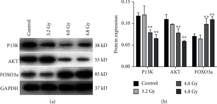
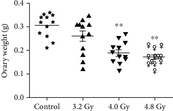
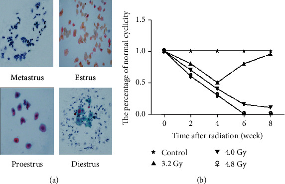
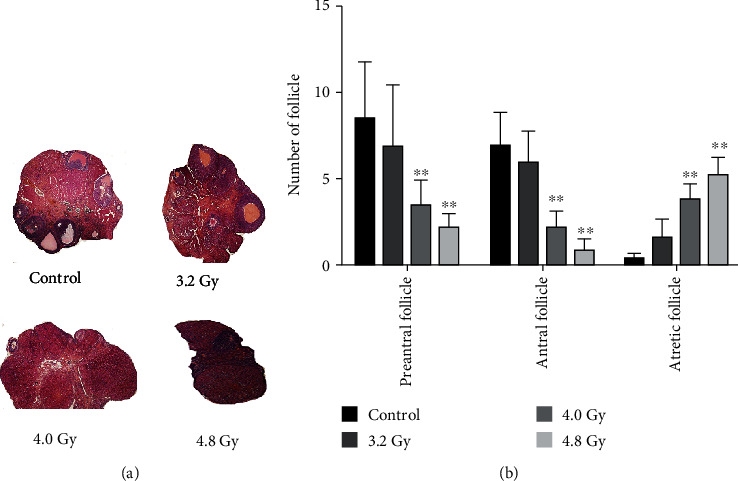
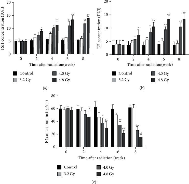
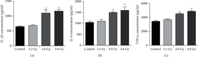
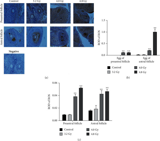
This study is aimed to investigate the mechanisms of radiation-induced mouse models of premature ovarian insufficiency (POI). Wistar female rats were grouped into the control, 3.2 Gy, 4.0 Gy, and 4.8 Gy groups. Overall ovarian functions were assessed with the H&E staining and ELISA. Proinflammatory cytokine secretion was analyzed ELISA, and the reactive oxygen species (ROS) levels were analyzed with immunohistochemistry. Protein expressions were analyzed by Western blot analysis. The 4.0 Gy and 4.8 Gy groups had significantly lower ovarian weight coefficients than the control and 3.2 Gy groups (after only one irradiation therapy). The 3.2 Gy radiation group induced periodic disturbance and hormone change at 4 weeks after radiation. In the 4.0 Gy and 4.8 Gy groups, the preantral follicles and antral follicles were decreased, while Atresia follicles were increased. E2 was decreased, while FSH and LH secretions were increased. The ovaries in the 4.0 Gy group were not completely atrophied, and some preantral follicles remained. Ovarian atrophy and follicular Atresia were found in the 4.8 Gy group. Inflammatory and oxidative markers were upregulated. PI3K and AKT were downregulated in the 4.0 Gy and 4.8 Gy groups, while FOXO3a was upregulated. Ovarian injuries may lead to oxidative damages and inflammatory injuries, downregulate the expression of P13k and Akt, upregulate the expression of FOXO3a, and lead to follicular atresia in the ovary.
本研究旨在探讨辐射诱导的小鼠卵巢早衰(POI)模型的机制。将 Wistar 雌性大鼠分为对照组、3.2Gy 组、4.0Gy 组和 4.8Gy 组。采用 H&E 染色和 ELISA 评估整体卵巢功能。采用 ELISA 分析促炎细胞因子分泌,采用免疫组织化学分析活性氧(ROS)水平。采用 Western blot 分析蛋白表达。与对照组和 3.2Gy 组相比,仅接受一次放射治疗的 4.0Gy 和 4.8Gy 组的卵巢重量系数明显降低。3.2Gy 照射组在照射后 4 周引起周期性紊乱和激素变化。在 4.0Gy 和 4.8Gy 组中,原始卵泡和腔前卵泡减少,而闭锁卵泡增加。E2 降低,而 FSH 和 LH 分泌增加。4.0Gy 组的卵巢未完全萎缩,仍有一些原始卵泡存在。4.8Gy 组出现卵巢萎缩和卵泡闭锁。炎症和氧化标志物上调。在 4.0Gy 和 4.8Gy 组中,PI3K 和 Akt 下调,而 FOXO3a 上调。卵巢损伤可能导致氧化损伤和炎症损伤,下调 P13k 和 Akt 的表达,上调 FOXO3a 的表达,导致卵巢中卵泡闭锁。