Department of Physiology, Medical College of Wisconsin, Milwaukee, Wisconsin.
Department of Biomedical Engineering, Medical College of Wisconsin, Milwaukee, Wisconsin.
Kidney360. 2020 Oct;1(10):1105-1115. doi: 10.34067/kid.0001212020. Epub 2020 Oct 29.
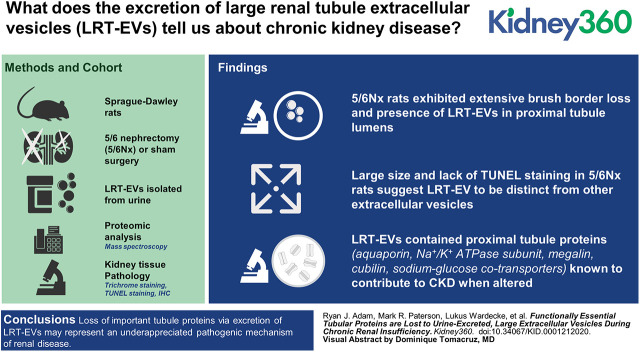
The 5/6 nephrectomy (5/6Nx) rat model recapitulates many elements of human CKD. Within weeks of surgery, 5/6Nx rats spontaneously exhibit proximal tubular damage, including the production of very large extracellular vesicles and brush border shedding. We hypothesized that production and elimination of these structures, termed large renal tubular extracellular vesicles (LRT-EVs), into the urine represents a pathologic mechanism by which essential tubule proteins are lost.
LRT-EVs were isolated from 5/6Nx rat urine 10 weeks after surgery. LRT-EV diameters were measured. LRT-EV proteomic analysis was performed by tandem mass spectrometry. Data are available the ProteomeXchange Consortium with identifier PXD019207. Kidney tissue pathology was evaluated by trichrome staining, TUNEL staining, and immunohistochemistry.
LRT-EV size and a lack of TUNEL staining in 5/6Nx rats suggest LRT-EVs to be distinct from exosomes, microvesicles, and apoptotic bodies. LRT-EVs contained many proximal tubule proteins that, upon disruption, are known to contribute to CKD pathologic hallmarks. Select proteins included aquaporin 1, 16 members of the solute carrier family, basolateral Na/K-ATPase subunit ATP1A1, megalin, cubilin, and sodium-glucose cotransporters (SLC5A1 and SLC5A2). Histologic analysis confirmed the presence of apical membrane proteins in LRT-EVs and brush border loss in 5/6Nx rats.
This study provides comprehensive proteomic analysis of a previously unreported category of extracellular vesicles associated with chronic renal stress. Because LRT-EVs contain proteins responsible for essential renal functions known to be compromised in CKD, their formation and excretion may represent an underappreciated pathogenic mechanism.
5/6 肾切除(5/6Nx)大鼠模型重现了许多人类 CKD 的特征。手术后数周内,5/6Nx 大鼠自发出现近端肾小管损伤,包括产生非常大的细胞外囊泡和刷状缘脱落。我们假设这些结构的产生和消除,称为大肾小管细胞外囊泡(LRT-EVs),进入尿液代表了一种重要的肾小管蛋白丢失的病理机制。
在手术后 10 周,从 5/6Nx 大鼠尿液中分离 LRT-EVs。测量 LRT-EV 的直径。通过串联质谱法进行 LRT-EV 蛋白质组分析。数据可在 ProteomeXchange 联盟中找到,标识符为 PXD019207。通过三色染色、TUNEL 染色和免疫组织化学评估肾脏组织病理学。
LRT-EV 的大小和 5/6Nx 大鼠中缺乏 TUNEL 染色表明 LRT-EVs 与外泌体、微泡和凋亡体不同。LRT-EVs 包含许多已知会导致 CKD 病理特征的近端肾小管蛋白。一些选定的蛋白质包括水通道蛋白 1、溶质载体家族的 16 个成员、基底外侧 Na/K-ATP 酶亚基 ATP1A1、巨球蛋白、内因子和钠-葡萄糖共转运体(SLC5A1 和 SLC5A2)。组织学分析证实 LRT-EVs 中存在顶膜蛋白和 5/6Nx 大鼠的刷状缘丢失。
这项研究提供了与慢性肾应激相关的一种以前未报道的细胞外囊泡类别全面的蛋白质组分析。由于 LRT-EVs 包含已知在 CKD 中受损的负责重要肾脏功能的蛋白质,因此它们的形成和排泄可能代表一种被低估的致病机制。