• 文献检索
  • 文档翻译
  • 深度研究
  • 学术资讯
  • Suppr Zotero 插件Zotero 插件
  • 邀请有礼
  • 套餐&价格
  • 历史记录
应用&插件
Suppr Zotero 插件Zotero 插件浏览器插件Mac 客户端Windows 客户端微信小程序
定价
高级版会员购买积分包购买API积分包
服务
文献检索文档翻译深度研究API 文档MCP 服务
关于我们
关于 Suppr公司介绍联系我们用户协议隐私条款
关注我们

Suppr 超能文献

核心技术专利:CN118964589B侵权必究
粤ICP备2023148730 号-1Suppr @ 2026

文献检索

告别复杂PubMed语法,用中文像聊天一样搜索,搜遍4000万医学文献。AI智能推荐,让科研检索更轻松。

立即免费搜索

文件翻译

保留排版,准确专业,支持PDF/Word/PPT等文件格式,支持 12+语言互译。

免费翻译文档

深度研究

AI帮你快速写综述,25分钟生成高质量综述,智能提取关键信息,辅助科研写作。

立即免费体验

隐股静脉再次高位结扎术中观察到的新生血管形成的组织病理学

Histopathology of Neovascularisation Observed During Saphenofemoral Re-Do High Ligation.

作者信息

Ehrler Michaela, Heim Dominik, Mouton Wolfgang G

机构信息

Department of Surgery, Spitalzentrum Biel, Switzerland.

Department of Surgery, Klinik Hohmad, Switzerland.

出版信息

EJVES Vasc Forum. 2021 Jun 17;52:17-19. doi: 10.1016/j.ejvsvf.2021.06.005. eCollection 2021.

DOI:10.1016/j.ejvsvf.2021.06.005
PMID:34278369
原文链接:https://pmc.ncbi.nlm.nih.gov/articles/PMC8271110/
Abstract

OBJECTIVE

The primary aim of this study was to assess the histopathological criteria of neovascularisation following saphenofemoral high ligation with regard to the delineation of the pathophysiology of the process. The secondary aims were to describe the perivenous morphological changes and to present cost effective agents to histopathologically diagnose neovascularisation.

METHODS

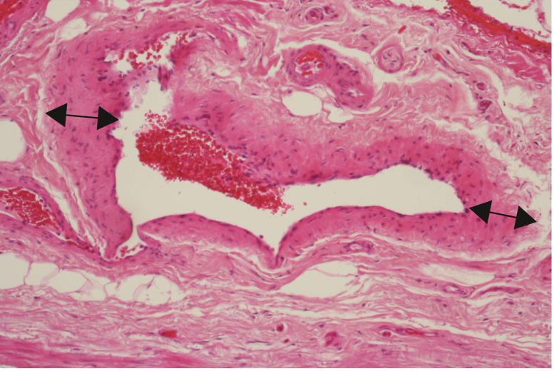
In a prospective study design, vein samples of consecutive patients with recurrent varicose veins in the groin undergoing surgery were collected. The samples were analysed by a vascular histopathologist with a light microscope using standard staining techniques.

RESULTS

The study population comprised 35 patients, 24 of whom were female (69%). Histopathologically, 28 samples (80%) showed typical aspects of neovascularisation. The remaining seven specimens (20%) showed thickened residual veins. An irregular vascular network, increasing perivenous collagen and elastic fibres and perivenous lymph nodes were observed. Present venous valves were the main criterion for residual veins. A surprising finding was the presence of scar tissue in the views of reparative incomplete new valves. Standard staining agents were sufficient to make the diagnosis of neovascularisation in 73% of the samples and reduced the cost by 30% compared with the regular use of specific markers.

CONCLUSION

The histopathological analysis of operative specimens may clarify whether a varicose vein recurrence is the result of neovascularisation or some other cause. Although interesting for research, academic interest, and classification, this may be of very limited clinical relevance for the patient.

摘要

目的

本研究的主要目的是评估大隐静脉高位结扎术后新生血管形成的组织病理学标准,以明确该过程的病理生理学。次要目的是描述静脉周围的形态学变化,并提出具有成本效益的组织病理学诊断新生血管形成的方法。

方法

采用前瞻性研究设计,收集连续腹股沟复发性静脉曲张手术患者的静脉样本。样本由血管组织病理学家使用标准染色技术在光学显微镜下进行分析。

结果

研究人群包括35例患者,其中24例为女性(69%)。组织病理学检查显示,28个样本(80%)呈现典型的新生血管形成特征。其余7个样本(20%)显示残留静脉增厚。观察到不规则的血管网络、静脉周围胶原纤维和弹性纤维增加以及静脉周围淋巴结。现存的静脉瓣膜是残留静脉的主要标准。一个令人惊讶的发现是,在修复不完全的新瓣膜视野中存在瘢痕组织。标准染色剂足以对73%的样本做出新生血管形成的诊断,与常规使用特定标记物相比,成本降低了30%。

结论

手术标本的组织病理学分析可以明确静脉曲张复发是新生血管形成还是其他原因所致。尽管这对于研究、学术兴趣和分类很有意义,但对患者的临床相关性可能非常有限。

https://cdn.ncbi.nlm.nih.gov/pmc/blobs/94d7/8271110/c3dab25bb74f/gr1.jpg

相似文献

1
Histopathology of Neovascularisation Observed During Saphenofemoral Re-Do High Ligation.隐股静脉再次高位结扎术中观察到的新生血管形成的组织病理学
EJVES Vasc Forum. 2021 Jun 17;52:17-19. doi: 10.1016/j.ejvsvf.2021.06.005. eCollection 2021.
2
Clinicopathological evidence that neovascularisation is a cause of recurrent varicose veins.新生血管形成是复发性静脉曲张病因的临床病理证据。
Eur J Vasc Endovasc Surg. 1998 May;15(5):412-5. doi: 10.1016/s1078-5884(98)80202-5.
3
Neovascularisation after surgery for recurrent saphenofemoral incompetence: does surgical dissection technique matter?复发性隐股静脉功能不全手术后的新生血管形成:手术解剖技术重要吗?
Vasa. 2011 Jul;40(4):296-301. doi: 10.1024/0301-1526/a000118.
4
[Neovascularisation as a cause of recurrence after varicose veins operation].[新生血管形成作为静脉曲张手术后复发的一个原因]
Rozhl Chir. 2006 Aug;85(8):399-403.
5
Closure of the cribriform fascia: an efficient anatomical barrier against postoperative neovascularisation at the saphenofemoral junction? A prospective study.筛状筋膜的封闭:隐股交界处术后新生血管形成的有效解剖屏障?一项前瞻性研究。
Eur J Vasc Endovasc Surg. 2007 Sep;34(3):361-6. doi: 10.1016/j.ejvs.2007.03.020. Epub 2007 May 21.
6
Recurrent varicose veins: causes and neovascularisation. A 17-year experience.复发性静脉曲张:病因与新生血管形成。17年的经验总结。
Int Angiol. 2015 Feb;34(1):53-9. Epub 2014 Oct 7.
7
Randomised trial of flush saphenofemoral ligation for primary great saphenous varicose veins.原发性大隐静脉曲张高位结扎剥脱术的随机试验
Eur J Vasc Endovasc Surg. 2008 Oct;36(4):477-84. doi: 10.1016/j.ejvs.2008.06.022. Epub 2008 Aug 20.
8
Neovascularization and recurrent varicose veins: more histologic and ultrasound evidence.新生血管形成与复发性静脉曲张:更多组织学和超声证据
J Vasc Surg. 2004 Aug;40(2):296-302. doi: 10.1016/j.jvs.2004.04.031.
9
Saphenofemoral venous channels associated with recurrent varicose veins are not neovascular.与复发性静脉曲张相关的隐股静脉通道并非新生血管。
Eur J Vasc Endovasc Surg. 2004 Dec;28(6):590-4. doi: 10.1016/j.ejvs.2004.09.011.
10
Endovenous ablation (radiofrequency and laser) and foam sclerotherapy versus open surgery for great saphenous vein varices.大隐静脉曲张的腔内消融(射频和激光)及泡沫硬化疗法与开放手术的比较
Cochrane Database Syst Rev. 2014 Jul 30(7):CD005624. doi: 10.1002/14651858.CD005624.pub3.

引用本文的文献

1
Endoglin and Other Angiogenesis Markers in Recurrent Varicose Veins.复发性静脉曲张中的内皮糖蛋白及其他血管生成标志物
J Pers Med. 2022 Mar 25;12(4):528. doi: 10.3390/jpm12040528.

本文引用的文献

1
Editor's Choice - Management of Chronic Venous Disease: Clinical Practice Guidelines of the European Society for Vascular Surgery (ESVS).编辑推荐——慢性静脉疾病的管理:欧洲血管外科学会(ESVS)临床实践指南
Eur J Vasc Endovasc Surg. 2015 Jun;49(6):678-737. doi: 10.1016/j.ejvs.2015.02.007. Epub 2015 Apr 25.
2
Endovenous ablation (radiofrequency and laser) and foam sclerotherapy versus open surgery for great saphenous vein varices.大隐静脉曲张的腔内消融(射频和激光)及泡沫硬化疗法与开放手术的比较
Cochrane Database Syst Rev. 2014 Jul 30(7):CD005624. doi: 10.1002/14651858.CD005624.pub3.
3
Validity of duplex-ultrasound in identifying the cause of groin recurrence after varicose vein surgery.
双功超声在确定静脉曲张手术后腹股沟复发原因中的有效性。
J Vasc Surg. 2009 Apr;49(4):968-72. doi: 10.1016/j.jvs.2008.10.058. Epub 2009 Feb 26.
4
Neovascularization and recurrent varicose veins: more histologic and ultrasound evidence.新生血管形成与复发性静脉曲张:更多组织学和超声证据
J Vasc Surg. 2004 Aug;40(2):296-302. doi: 10.1016/j.jvs.2004.04.031.
5
The role of postoperative neovascularisation in recurrence of varicose veins: from historical background to today's evidence.术后新生血管形成在静脉曲张复发中的作用:从历史背景到当今证据
Acta Chir Belg. 2004 Jun;104(3):283-9. doi: 10.1080/00015458.2004.11679555.
6
Recurrent varicose veins after surgery: a new appraisal of a common and complex problem in vascular surgery.
Eur J Vasc Endovasc Surg. 2004 Mar;27(3):275-82. doi: 10.1016/j.ejvs.2003.12.006.
7
Clinicopathological evidence that neovascularisation is a cause of recurrent varicose veins.新生血管形成是复发性静脉曲张病因的临床病理证据。
Eur J Vasc Endovasc Surg. 1998 May;15(5):412-5. doi: 10.1016/s1078-5884(98)80202-5.