• 文献检索
  • 文档翻译
  • 深度研究
  • 学术资讯
  • Suppr Zotero 插件Zotero 插件
  • 邀请有礼
  • 套餐&价格
  • 历史记录
应用&插件
Suppr Zotero 插件Zotero 插件浏览器插件Mac 客户端Windows 客户端微信小程序
定价
高级版会员购买积分包购买API积分包
服务
文献检索文档翻译深度研究API 文档MCP 服务
关于我们
关于 Suppr公司介绍联系我们用户协议隐私条款
关注我们

Suppr 超能文献

核心技术专利:CN118964589B侵权必究
粤ICP备2023148730 号-1Suppr @ 2026

文献检索

告别复杂PubMed语法,用中文像聊天一样搜索,搜遍4000万医学文献。AI智能推荐,让科研检索更轻松。

立即免费搜索

文件翻译

保留排版,准确专业,支持PDF/Word/PPT等文件格式,支持 12+语言互译。

免费翻译文档

深度研究

AI帮你快速写综述,25分钟生成高质量综述,智能提取关键信息,辅助科研写作。

立即免费体验

利用动态对比增强磁共振成像的主成分分析对胶质母细胞瘤中的肿瘤微环境酸度进行定量分析。

Quantification of tumor microenvironment acidity in glioblastoma using principal component analysis of dynamic susceptibility contrast enhanced MR imaging.

作者信息

Akbari Hamed, Kazerooni Anahita Fathi, Ware Jeffrey B, Mamourian Elizabeth, Anderson Hannah, Guiry Samantha, Sako Chiharu, Raymond Catalina, Yao Jingwen, Brem Steven, O'Rourke Donald M, Desai Arati S, Bagley Stephen J, Ellingson Benjamin M, Davatzikos Christos, Nabavizadeh Ali

机构信息

Department of Radiology, Perelman School of Medicine, Hospital of University of Pennsylvania, University of Pennsylvania, Philadelphia, PA, USA.

Center for Biomedical Image Computing and Analytics, Perelman School of Medicine, University of Pennsylvania, Philadelphia, PA, USA.

出版信息

Sci Rep. 2021 Jul 22;11(1):15011. doi: 10.1038/s41598-021-94560-3.

DOI:10.1038/s41598-021-94560-3
PMID:34294864
原文链接:https://pmc.ncbi.nlm.nih.gov/articles/PMC8298590/
Abstract

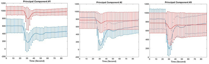
Glioblastoma (GBM) has high metabolic demands, which can lead to acidification of the tumor microenvironment. We hypothesize that a machine learning model built on temporal principal component analysis (PCA) of dynamic susceptibility contrast-enhanced (DSC) perfusion MRI can be used to estimate tumor acidity in GBM, as estimated by pH-sensitive amine chemical exchange saturation transfer echo-planar imaging (CEST-EPI). We analyzed 78 MRI scans in 32 treatment naïve and post-treatment GBM patients. All patients were imaged with DSC-MRI, and pH-weighting that was quantified from CEST-EPI estimation of the magnetization transfer ratio asymmetry (MTR) at 3 ppm. Enhancing tumor (ET), non-enhancing core (NC), and peritumoral T2 hyperintensity (namely, edema, ED) were used to extract principal components (PCs) and to build support vector machines regression (SVR) models to predict MTR values using PCs. Our predicted map correlated with MTR values with Spearman's r equal to 0.66, 0.47, 0.67, 0.71, in NC, ET, ED, and overall, respectively (p < 0.006). The results of this study demonstrates that PCA analysis of DSC imaging data can provide information about tumor pH in GBM patients, with the strongest association within the peritumoral regions.

摘要

胶质母细胞瘤(GBM)具有高代谢需求,这会导致肿瘤微环境酸化。我们假设,基于动态磁敏感对比增强(DSC)灌注磁共振成像(MRI)的时间主成分分析(PCA)构建的机器学习模型,可用于估计GBM中的肿瘤酸度,如通过pH敏感胺化学交换饱和转移回波平面成像(CEST-EPI)所估计的那样。我们分析了32例未经治疗和接受过治疗的GBM患者的78次MRI扫描。所有患者均接受DSC-MRI成像,并通过CEST-EPI对3 ppm处的磁化传递率不对称性(MTR)进行估计来量化pH加权。使用强化肿瘤(ET)、非强化核心(NC)和瘤周T2高信号(即水肿,ED)来提取主成分(PC),并构建支持向量机回归(SVR)模型,以使用PC预测MTR值。我们的预测图与MTR值的Spearman相关系数分别在NC、ET、ED和总体中为0.66、0.47、0.67、0.71(p < 0.006)。本研究结果表明,DSC成像数据的PCA分析可以提供GBM患者肿瘤pH的信息,在瘤周区域的相关性最强。

https://cdn.ncbi.nlm.nih.gov/pmc/blobs/3481/8298590/f19185a09cb9/41598_2021_94560_Fig5_HTML.jpg
https://cdn.ncbi.nlm.nih.gov/pmc/blobs/3481/8298590/29b5bcd87eef/41598_2021_94560_Fig1_HTML.jpg
https://cdn.ncbi.nlm.nih.gov/pmc/blobs/3481/8298590/84615e082a92/41598_2021_94560_Fig2_HTML.jpg
https://cdn.ncbi.nlm.nih.gov/pmc/blobs/3481/8298590/a2cb47d54b41/41598_2021_94560_Fig3_HTML.jpg
https://cdn.ncbi.nlm.nih.gov/pmc/blobs/3481/8298590/fb2cbe637d3e/41598_2021_94560_Fig4_HTML.jpg
https://cdn.ncbi.nlm.nih.gov/pmc/blobs/3481/8298590/f19185a09cb9/41598_2021_94560_Fig5_HTML.jpg

相似文献

1
Quantification of tumor microenvironment acidity in glioblastoma using principal component analysis of dynamic susceptibility contrast enhanced MR imaging.利用动态对比增强磁共振成像的主成分分析对胶质母细胞瘤中的肿瘤微环境酸度进行定量分析。
Sci Rep. 2021 Jul 22;11(1):15011. doi: 10.1038/s41598-021-94560-3.
2
pH-weighted amine chemical exchange saturation transfer echoplanar imaging (CEST-EPI) as a potential early biomarker for bevacizumab failure in recurrent glioblastoma.pH 加权胺化学交换饱和传递 EPI(CEST-EPI)作为复发性胶质母细胞瘤贝伐单抗治疗失败的潜在早期生物标志物。
J Neurooncol. 2019 May;142(3):587-595. doi: 10.1007/s11060-019-03132-z. Epub 2019 Feb 26.
3
Association between Tumor Acidity and Hypervascularity in Human Gliomas Using pH-Weighted Amine Chemical Exchange Saturation Transfer Echo-Planar Imaging and Dynamic Susceptibility Contrast Perfusion MRI at 3T.使用 pH 加权胺化学交换饱和传递回波平面成像和 3T 下动态磁敏感对比灌注 MRI 研究人胶质瘤中的肿瘤酸度与富血管性的相关性。
AJNR Am J Neuroradiol. 2019 Jun;40(6):979-986. doi: 10.3174/ajnr.A6063. Epub 2019 May 16.
4
pH-Weighted amine chemical exchange saturation transfer echo planar imaging visualizes infiltrating glioblastoma cells.pH 加权胺化学交换饱和传递回波平面成像可可视化浸润性胶质母细胞瘤细胞。
Neuro Oncol. 2024 Jan 5;26(1):115-126. doi: 10.1093/neuonc/noad150.
5
Simultaneous pH-sensitive and oxygen-sensitive MRI of human gliomas at 3 T using multi-echo amine proton chemical exchange saturation transfer spin-and-gradient echo echo-planar imaging (CEST-SAGE-EPI).使用多回波胺质子化学交换饱和传递自旋梯度回波平面成像(CEST-SAGE-EPI)在 3T 对人神经胶质瘤进行同时 pH 值敏感和氧敏感 MRI 成像。
Magn Reson Med. 2018 Nov;80(5):1962-1978. doi: 10.1002/mrm.27204. Epub 2018 Apr 6.
6
Nuclear overhauser enhancement mediated chemical exchange saturation transfer imaging at 7 Tesla in glioblastoma patients.7特斯拉下胶质母细胞瘤患者的核奥氏效应介导化学交换饱和转移成像
PLoS One. 2014 Aug 11;9(8):e104181. doi: 10.1371/journal.pone.0104181. eCollection 2014.
7
Metabolic characterization of human IDH mutant and wild type gliomas using simultaneous pH- and oxygen-sensitive molecular MRI.利用同时 pH 值和氧敏感性分子 MRI 对 IDH 突变型和野生型人脑胶质瘤进行代谢特征分析。
Neuro Oncol. 2019 Sep 6;21(9):1184-1196. doi: 10.1093/neuonc/noz078.
8
The pH sensitivity of APT-CEST using phosphorus spectroscopy as a reference method.采用磷谱作为参考方法研究 APT-CEST 的 pH 敏感性。
NMR Biomed. 2019 Nov;32(11):e4125. doi: 10.1002/nbm.4125. Epub 2019 Jul 19.
9
Systematic Evaluation of Amide Proton Chemical Exchange Saturation Transfer at 3 T: Effects of Protein Concentration, pH, and Acquisition Parameters.3T下酰胺质子化学交换饱和转移的系统评估:蛋白质浓度、pH值及采集参数的影响
Invest Radiol. 2016 Oct;51(10):635-46. doi: 10.1097/RLI.0000000000000292.
10
Pattern analysis of dynamic susceptibility contrast-enhanced MR imaging demonstrates peritumoral tissue heterogeneity.动态磁敏感对比增强磁共振成像的模式分析显示肿瘤周围组织的异质性。
Radiology. 2014 Nov;273(2):502-10. doi: 10.1148/radiol.14132458. Epub 2014 Jun 19.

引用本文的文献

1
Beyond the Walls of Troy: A Scoping Review on Pharmacological Strategies to Enhance Drug Delivery Across the Blood-Brain Barrier and Blood-Tumor Barrier.超越特洛伊城墙:关于增强药物透过血脑屏障和血肿瘤屏障递送的药理学策略的范围综述
Int J Mol Sci. 2025 Jul 22;26(15):7050. doi: 10.3390/ijms26157050.
2
TAMing Gliomas: Unraveling the Roles of Iba1 and CD163 in Glioblastoma.靶向胶质瘤:揭示Iba1和CD163在胶质母细胞瘤中的作用
Cancers (Basel). 2025 Apr 26;17(9):1457. doi: 10.3390/cancers17091457.
3
Functional biomaterials for biomimetic 3D in vitro tumor microenvironment modeling.

本文引用的文献

1
Human IDH mutant 1p/19q co-deleted gliomas have low tumor acidity as evidenced by molecular MRI and PET: a retrospective study.分子 MRI 和 PET 显示 IDH 突变 1p/19q 共缺失型人脑胶质瘤酸度较低:一项回顾性研究
Sci Rep. 2020 Jul 17;10(1):11922. doi: 10.1038/s41598-020-68733-5.
2
Consensus recommendations for a dynamic susceptibility contrast MRI protocol for use in high-grade gliomas.用于高级别脑胶质瘤的动态对比磁共振成像协议的共识建议。
Neuro Oncol. 2020 Sep 29;22(9):1262-1275. doi: 10.1093/neuonc/noaa141.
3
Cancer Imaging Phenomics via CaPTk: Multi-Institutional Prediction of Progression-Free Survival and Pattern of Recurrence in Glioblastoma.
用于仿生3D体外肿瘤微环境建模的功能性生物材料。
In Vitro Model. 2023 Jan 27;2(1-2):1-23. doi: 10.1007/s44164-023-00043-2. eCollection 2023 Apr.
4
The peritumoral brain zone in glioblastoma: a review of the pretreatment approach.胶质母细胞瘤的瘤周脑区:预处理方法综述
Pol J Radiol. 2024 Oct 11;89:e480-e487. doi: 10.5114/pjr/192044. eCollection 2024.
5
Differentiation between primary central nervous system lymphomas and gliomas according to pharmacokinetic parameters derived from dynamic contrast-enhanced magnetic resonance imaging.根据动态对比增强磁共振成像得出的药代动力学参数鉴别原发性中枢神经系统淋巴瘤和胶质瘤。
Heliyon. 2024 Jun 6;10(12):e32619. doi: 10.1016/j.heliyon.2024.e32619. eCollection 2024 Jun 30.
6
Peptide-Hitchhiking for the Development of Nanosystems in Glioblastoma.肽搭乘用于神经胶质瘤中纳米系统的发展。
ACS Nano. 2024 Jul 2;18(26):16359-16394. doi: 10.1021/acsnano.4c01790. Epub 2024 Jun 11.
7
Current Status and Future Perspective in Glioma Invasion Research.神经胶质瘤侵袭研究的现状与未来展望
Brain Sci. 2024 Mar 26;14(4):309. doi: 10.3390/brainsci14040309.
8
Peritumoral brain zone in glioblastoma: biological, clinical and mechanical features.胶质母细胞瘤瘤周脑区:生物学、临床和力学特征。
Front Immunol. 2024 Feb 29;15:1347877. doi: 10.3389/fimmu.2024.1347877. eCollection 2024.
9
Statistical plots in oncologic imaging, a primer for neuroradiologists.肿瘤影像学中的统计图表:神经放射科医师入门
Neuroradiol J. 2024 Aug;37(4):418-433. doi: 10.1177/19714009231193158. Epub 2023 Aug 2.
10
The peritumoral brain zone in glioblastoma: where we are and where we are going.胶质母细胞瘤的瘤周脑区:我们的现状与未来展望。
J Neurosci Res. 2023 Feb;101(2):199-216. doi: 10.1002/jnr.25134. Epub 2022 Oct 27.
通过CaPTk实现癌症影像组学:多机构预测胶质母细胞瘤的无进展生存期和复发模式
JCO Clin Cancer Inform. 2020 Mar;4:234-244. doi: 10.1200/CCI.19.00121.
4
Histopathology-validated machine learning radiographic biomarker for noninvasive discrimination between true progression and pseudo-progression in glioblastoma.基于组织病理学验证的机器学习放射影像学生物标志物,用于无创性鉴别胶质母细胞瘤的真性进展和假性进展。
Cancer. 2020 Jun 1;126(11):2625-2636. doi: 10.1002/cncr.32790. Epub 2020 Mar 4.
5
Imaging signatures of glioblastoma molecular characteristics: A radiogenomics review.胶质母细胞瘤分子特征的影像学特征:放射基因组学综述。
J Magn Reson Imaging. 2020 Jul;52(1):54-69. doi: 10.1002/jmri.26907. Epub 2019 Aug 27.
6
Arterial Spin Labeling and Dynamic Susceptibility Contrast-enhanced MR Imaging for evaluation of arteriovenous shunting and tumor hypoxia in glioblastoma.动脉自旋标记和动态对比增强磁共振成像在评估脑胶质母细胞瘤动静脉分流和肿瘤缺氧中的应用。
Sci Rep. 2019 Jun 19;9(1):8747. doi: 10.1038/s41598-019-45312-x.
7
Association between Tumor Acidity and Hypervascularity in Human Gliomas Using pH-Weighted Amine Chemical Exchange Saturation Transfer Echo-Planar Imaging and Dynamic Susceptibility Contrast Perfusion MRI at 3T.使用 pH 加权胺化学交换饱和传递回波平面成像和 3T 下动态磁敏感对比灌注 MRI 研究人胶质瘤中的肿瘤酸度与富血管性的相关性。
AJNR Am J Neuroradiol. 2019 Jun;40(6):979-986. doi: 10.3174/ajnr.A6063. Epub 2019 May 16.
8
Metabolic characterization of human IDH mutant and wild type gliomas using simultaneous pH- and oxygen-sensitive molecular MRI.利用同时 pH 值和氧敏感性分子 MRI 对 IDH 突变型和野生型人脑胶质瘤进行代谢特征分析。
Neuro Oncol. 2019 Sep 6;21(9):1184-1196. doi: 10.1093/neuonc/noz078.
9
pH-weighted amine chemical exchange saturation transfer echoplanar imaging (CEST-EPI) as a potential early biomarker for bevacizumab failure in recurrent glioblastoma.pH 加权胺化学交换饱和传递 EPI(CEST-EPI)作为复发性胶质母细胞瘤贝伐单抗治疗失败的潜在早期生物标志物。
J Neurooncol. 2019 May;142(3):587-595. doi: 10.1007/s11060-019-03132-z. Epub 2019 Feb 26.
10
CBTRUS Statistical Report: Primary Brain and Other Central Nervous System Tumors Diagnosed in the United States in 2011-2015.CBTRUS统计报告:2011 - 2015年美国原发性脑肿瘤及其他中枢神经系统肿瘤诊断情况
Neuro Oncol. 2018 Oct 1;20(suppl_4):iv1-iv86. doi: 10.1093/neuonc/noy131.