• 文献检索
  • 文档翻译
  • 深度研究
  • 学术资讯
  • Suppr Zotero 插件Zotero 插件
  • 邀请有礼
  • 套餐&价格
  • 历史记录
应用&插件
Suppr Zotero 插件Zotero 插件浏览器插件Mac 客户端Windows 客户端微信小程序
定价
高级版会员购买积分包购买API积分包
服务
文献检索文档翻译深度研究API 文档MCP 服务
关于我们
关于 Suppr公司介绍联系我们用户协议隐私条款
关注我们

Suppr 超能文献

核心技术专利:CN118964589B侵权必究
粤ICP备2023148730 号-1Suppr @ 2026

文献检索

告别复杂PubMed语法,用中文像聊天一样搜索,搜遍4000万医学文献。AI智能推荐,让科研检索更轻松。

立即免费搜索

文件翻译

保留排版,准确专业,支持PDF/Word/PPT等文件格式,支持 12+语言互译。

免费翻译文档

深度研究

AI帮你快速写综述,25分钟生成高质量综述,智能提取关键信息,辅助科研写作。

立即免费体验

钙调蛋白激酶 II 介导的视网膜神经节细胞保护后视力的保存。

Preservation of vision after CaMKII-mediated protection of retinal ganglion cells.

机构信息

Department of Ophthalmology, Icahn School of Medicine at Mount Sinai, New York, NY 10029, USA.

Department of Neuroscience, Yale University School of Medicine, New Haven, CT 06511, USA.

出版信息

Cell. 2021 Aug 5;184(16):4299-4314.e12. doi: 10.1016/j.cell.2021.06.031. Epub 2021 Jul 22.

DOI:10.1016/j.cell.2021.06.031
PMID:34297923
原文链接:https://pmc.ncbi.nlm.nih.gov/articles/PMC8530265/
Abstract

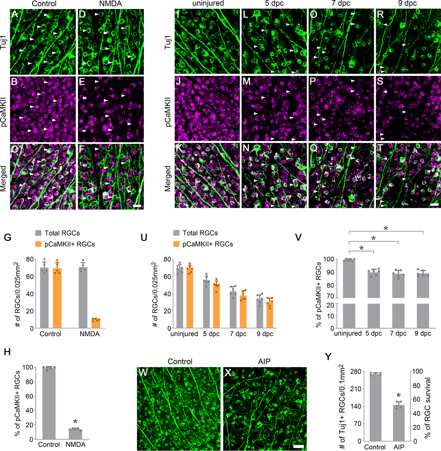
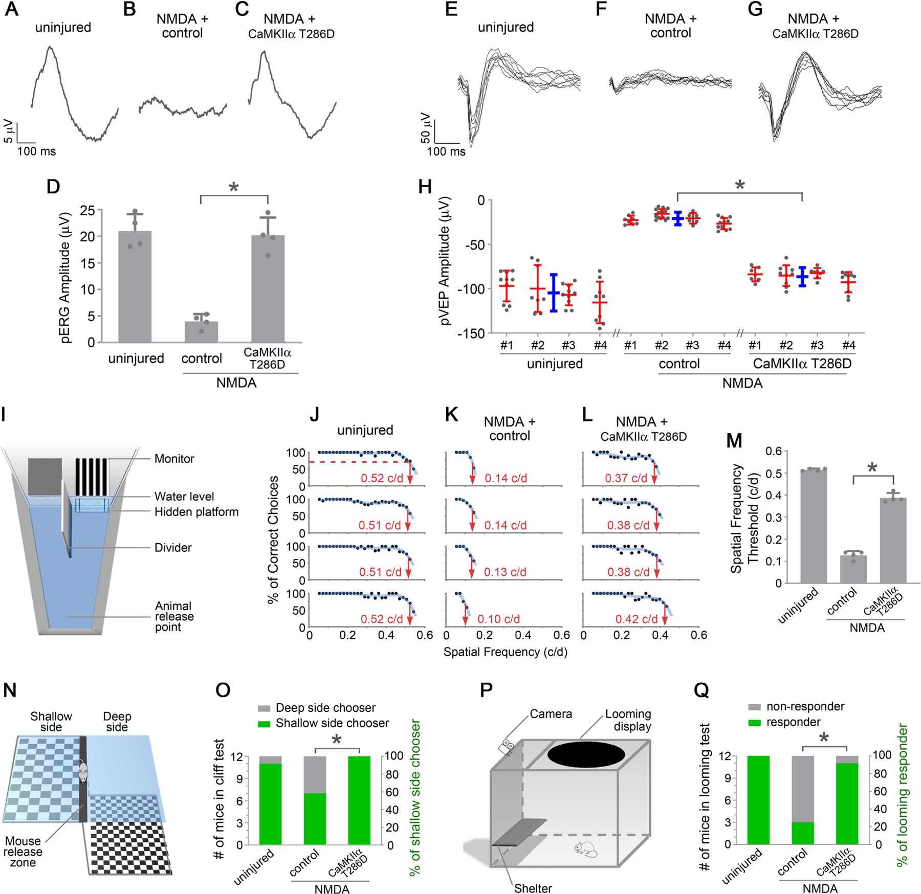
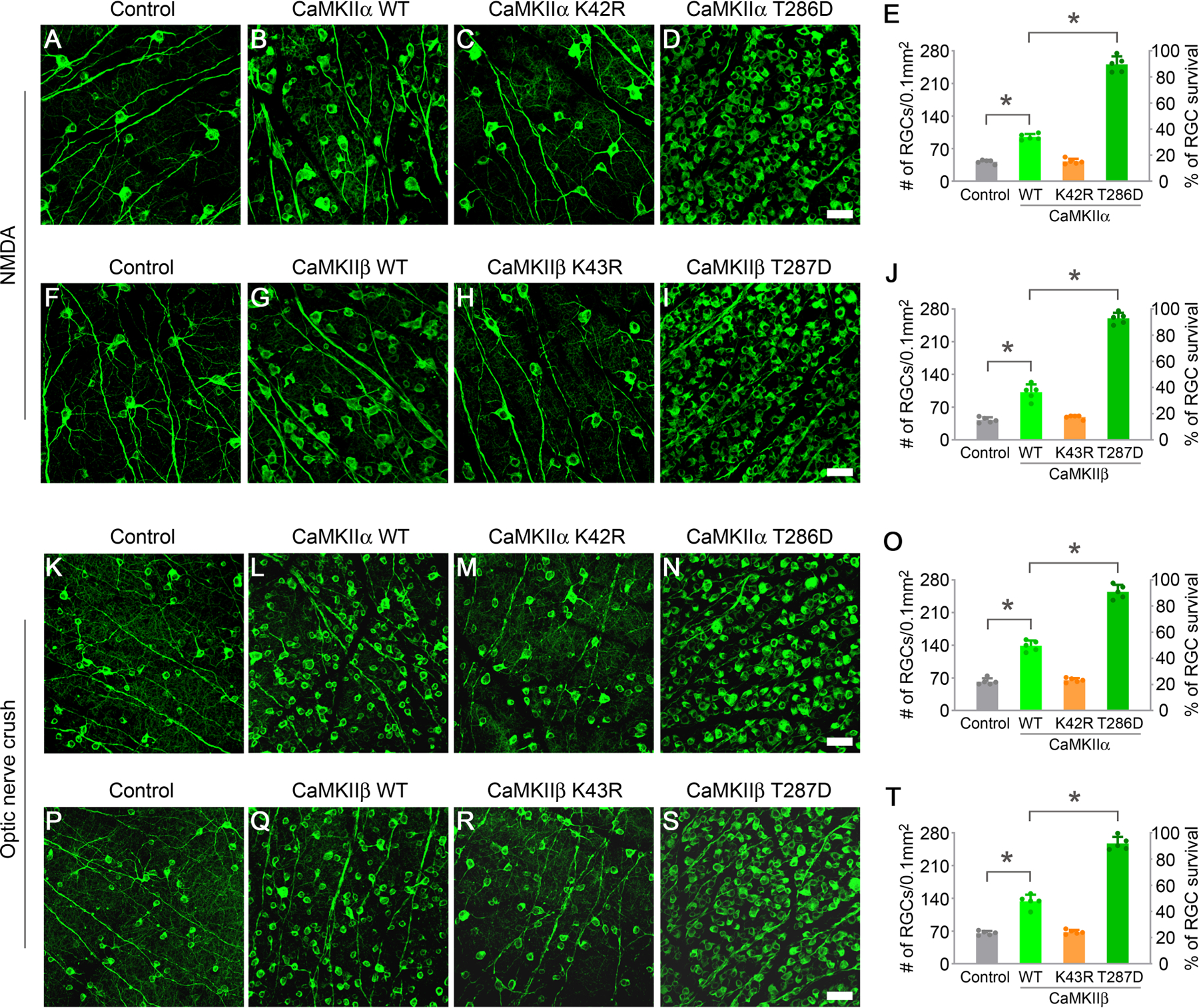
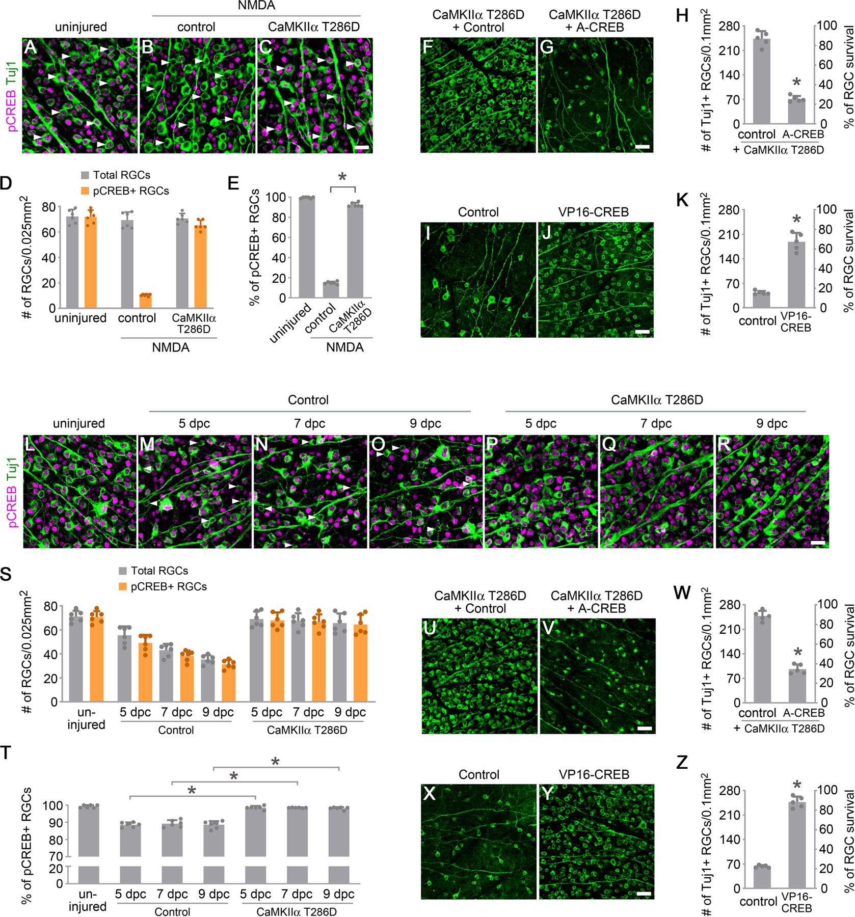
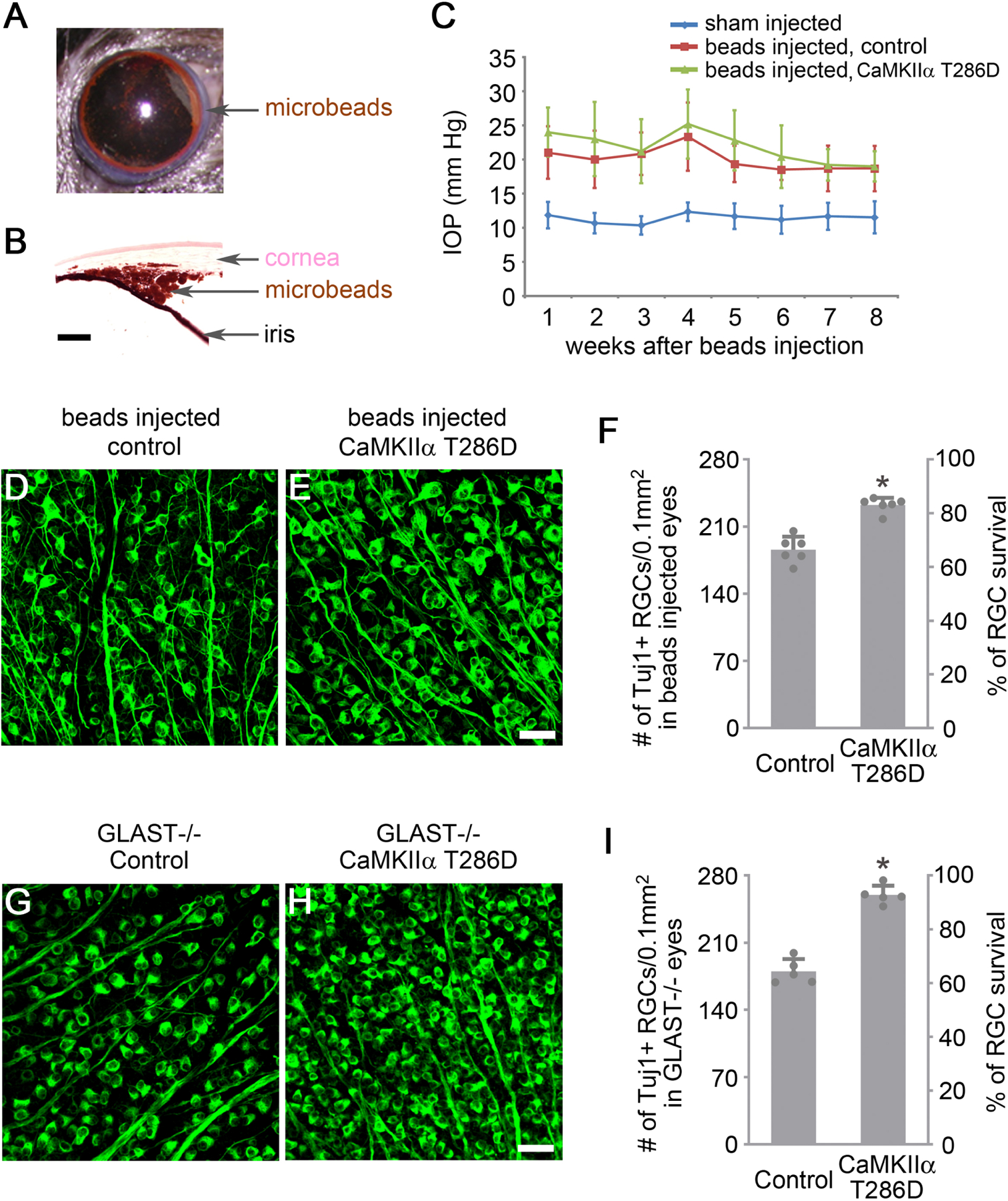
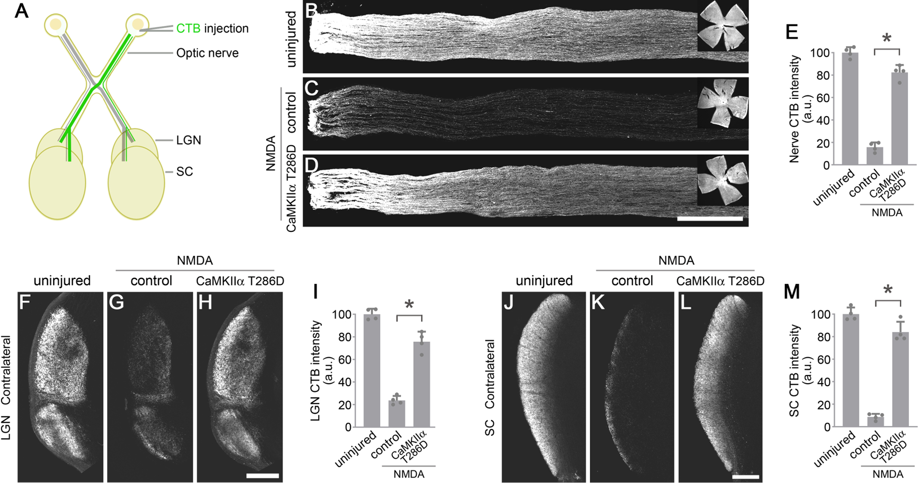
Retinal ganglion cells (RGCs) are the sole output neurons that transmit visual information from the retina to the brain. Diverse insults and pathological states cause degeneration of RGC somas and axons leading to irreversible vision loss. A fundamental question is whether manipulation of a key regulator of RGC survival can protect RGCs from diverse insults and pathological states, and ultimately preserve vision. Here, we report that CaMKII-CREB signaling is compromised after excitotoxic injury to RGC somas or optic nerve injury to RGC axons, and reactivation of this pathway robustly protects RGCs from both injuries. CaMKII activity also promotes RGC survival in the normal retina. Further, reactivation of CaMKII protects RGCs in two glaucoma models where RGCs degenerate from elevated intraocular pressure or genetic deficiency. Last, CaMKII reactivation protects long-distance RGC axon projections in vivo and preserves visual function, from the retina to the visual cortex, and visually guided behavior.

摘要

视网膜神经节细胞(RGCs)是唯一将视觉信息从视网膜传递到大脑的输出神经元。各种损伤和病理状态导致 RGC 体和轴突变性,导致不可逆转的视力丧失。一个基本的问题是,是否可以通过操纵 RGC 存活的关键调节因子来保护 RGC 免受各种损伤和病理状态的影响,并最终保护视力。在这里,我们报告说,在 RGC 体的兴奋毒性损伤或 RGC 轴突的视神经损伤后,CaMKII-CREB 信号通路受到损害,而该通路的重新激活可强烈保护 RGC 免受这两种损伤。CaMKII 活性也促进正常视网膜中的 RGC 存活。此外,CaMKII 的重新激活可保护两种青光眼模型中的 RGC,这些青光眼模型中的 RGC 因眼内压升高或遗传缺陷而变性。最后,CaMKII 的重新激活可保护体内长距离 RGC 轴突投射,并维持从视网膜到视皮层的视觉功能和视觉引导行为。

https://cdn.ncbi.nlm.nih.gov/pmc/blobs/ee67/8530265/04bbaf9c1180/nihms-1721071-f0007.jpg
https://cdn.ncbi.nlm.nih.gov/pmc/blobs/ee67/8530265/994a32399646/nihms-1721071-f0001.jpg
https://cdn.ncbi.nlm.nih.gov/pmc/blobs/ee67/8530265/625ccd292df8/nihms-1721071-f0002.jpg
https://cdn.ncbi.nlm.nih.gov/pmc/blobs/ee67/8530265/301c6a692170/nihms-1721071-f0003.jpg
https://cdn.ncbi.nlm.nih.gov/pmc/blobs/ee67/8530265/497ed398c0b4/nihms-1721071-f0004.jpg
https://cdn.ncbi.nlm.nih.gov/pmc/blobs/ee67/8530265/5c3314ec24d8/nihms-1721071-f0005.jpg
https://cdn.ncbi.nlm.nih.gov/pmc/blobs/ee67/8530265/cf3c60b3b2d7/nihms-1721071-f0006.jpg
https://cdn.ncbi.nlm.nih.gov/pmc/blobs/ee67/8530265/04bbaf9c1180/nihms-1721071-f0007.jpg

相似文献

1
Preservation of vision after CaMKII-mediated protection of retinal ganglion cells.钙调蛋白激酶 II 介导的视网膜神经节细胞保护后视力的保存。
Cell. 2021 Aug 5;184(16):4299-4314.e12. doi: 10.1016/j.cell.2021.06.031. Epub 2021 Jul 22.
2
Role of hypoxia-inducible factor-1α in preconditioning-induced protection of retinal ganglion cells in glaucoma.缺氧诱导因子-1α在青光眼预处理诱导的视网膜神经节细胞保护中的作用
Mol Vis. 2013 Nov 23;19:2360-72. eCollection 2013.
3
Ca/Calmodulin-Dependent Protein Kinase II Enhances Retinal Ganglion Cell Survival But Suppresses Axon Regeneration after Optic Nerve Injury.钙/钙调蛋白依赖性蛋白激酶 II 增强视网膜神经节细胞存活,但抑制视神经损伤后的轴突再生。
eNeuro. 2024 Mar 28;11(3). doi: 10.1523/ENEURO.0478-23.2024. Print 2024 Mar.
4
Role of SARM1 and DR6 in retinal ganglion cell axonal and somal degeneration following axonal injury.SARM1 和 DR6 在轴突损伤后视网膜神经节细胞轴突和体部变性中的作用。
Exp Eye Res. 2018 Jun;171:54-61. doi: 10.1016/j.exer.2018.03.007. Epub 2018 Mar 8.
5
Together JUN and DDIT3 (CHOP) control retinal ganglion cell death after axonal injury.JUN 和 DDIT3(CHOP)共同控制轴突损伤后的视网膜神经节细胞死亡。
Mol Neurodegener. 2017 Oct 2;12(1):71. doi: 10.1186/s13024-017-0214-8.
6
Retinal ganglion cell morphology after optic nerve crush and experimental glaucoma.视神经挤压和实验性青光眼后视网膜神经节细胞形态。
Invest Ophthalmol Vis Sci. 2012 Jun 22;53(7):3847-57. doi: 10.1167/iovs.12-9712.
7
Interleukin-4 protects retinal ganglion cells and promotes axon regeneration.白细胞介素-4可保护视网膜神经节细胞并促进轴突再生。
Cell Commun Signal. 2024 Apr 22;22(1):236. doi: 10.1186/s12964-024-01604-y.
8
Neuroprotection of retinal ganglion cells by a novel gene therapy construct that achieves sustained enhancement of brain-derived neurotrophic factor/tropomyosin-related kinase receptor-B signaling.新型基因治疗构建体实现脑源性神经营养因子/原肌球蛋白相关激酶受体-B 信号的持续增强,对视网膜神经节细胞起到神经保护作用。
Cell Death Dis. 2018 Sep 26;9(10):1007. doi: 10.1038/s41419-018-1041-8.
9
Protection of mouse retinal ganglion cell axons and soma from glaucomatous and ischemic injury by cytoplasmic overexpression of Nmnat1.通过细胞质过表达 Nmnat1 保护小鼠视网膜神经节细胞轴突和体免受青光眼和缺血性损伤。
Invest Ophthalmol Vis Sci. 2013 Jan 2;54(1):25-36. doi: 10.1167/iovs.12-10861.
10
Intravitreal delivery of human NgR-Fc decoy protein regenerates axons after optic nerve crush and protects ganglion cells in glaucoma models.玻璃体内注射人NgR-Fc诱饵蛋白可使视神经挤压伤后轴突再生,并在青光眼模型中保护神经节细胞。
Invest Ophthalmol Vis Sci. 2015 Feb 5;56(2):1357-66. doi: 10.1167/iovs.14-15472.

引用本文的文献

1
Hidden Markov models reveal behavioral state dynamics in depth-related locomotion in mice.隐马尔可夫模型揭示了小鼠深度相关运动中的行为状态动态。
PLoS One. 2025 Aug 26;20(8):e0329367. doi: 10.1371/journal.pone.0329367. eCollection 2025.
2
Spatial Distribution and Morphology of CaMKII-Expressing Amacrine Cells in Marmoset, Macaque, and Human Retina.狨猴、猕猴和人类视网膜中表达钙调蛋白激酶II的无长突细胞的空间分布和形态
J Comp Neurol. 2025 Jul;533(7):e70078. doi: 10.1002/cne.70078.
3
Efficacy and safety of stem cells in the treatment of glaucoma: systematic review and meta-analysis based on animal experiments.

本文引用的文献

1
Mouse γ-Synuclein Promoter-Mediated Gene Expression and Editing in Mammalian Retinal Ganglion Cells.鼠 γ-突触核蛋白启动子介导的哺乳动物视网膜神经节细胞中的基因表达和编辑。
J Neurosci. 2020 May 13;40(20):3896-3914. doi: 10.1523/JNEUROSCI.0102-20.2020. Epub 2020 Apr 16.
2
Single-Cell Profiles of Retinal Ganglion Cells Differing in Resilience to Injury Reveal Neuroprotective Genes.单细胞分析揭示了对损伤具有不同抵抗力的视网膜神经节细胞的特征,并发现了神经保护基因。
Neuron. 2019 Dec 18;104(6):1039-1055.e12. doi: 10.1016/j.neuron.2019.11.006. Epub 2019 Nov 26.
3
Using Looming Visual Stimuli to Evaluate Mouse Vision.
干细胞治疗青光眼的疗效与安全性:基于动物实验的系统评价与Meta分析
Front Pharmacol. 2025 Jul 1;16:1587440. doi: 10.3389/fphar.2025.1587440. eCollection 2025.
4
ATF2 phosphorylation is a core transcriptional driver of neuron apoptosis.激活转录因子2(ATF2)磷酸化是神经元凋亡的核心转录驱动因素。
bioRxiv. 2025 May 8:2023.09.27.559856. doi: 10.1101/2023.09.27.559856.
5
Activation of Sigma 1 Receptor Preserves Visual Function After Optic Nerve Injury.σ1受体激活可在视神经损伤后保留视觉功能。
Invest Ophthalmol Vis Sci. 2025 Jul 1;66(9):13. doi: 10.1167/iovs.66.9.13.
6
Single-cell RNA sequencing reveals anterograde trans-synaptic degeneration and exacerbated synaptic remodeling in myopia.单细胞RNA测序揭示近视中顺行性跨突触变性和加剧的突触重塑。
Exp Mol Med. 2025 Jul 3. doi: 10.1038/s12276-025-01489-y.
7
HOCPCA Exerts Neuroprotection on Retinal Ganglion Cells by Binding to CaMKIIα and Modulating Oxidative Stress and Neuroinflammation in Experimental Glaucoma.HOCPCA通过与CaMKIIα结合并调节实验性青光眼中的氧化应激和神经炎症对视网膜神经节细胞发挥神经保护作用。
Neurosci Bull. 2025 Jun 2. doi: 10.1007/s12264-025-01417-0.
8
Ultrasound-driven wireless piezoelectric hydrogel synergizes with cotransplantation of NSCs-hUCMSCs for structural and functional recovery in spinal cord injury.超声驱动的无线压电水凝胶与神经干细胞-人脐带间充质干细胞共移植协同作用,促进脊髓损伤的结构和功能恢复。
Mater Today Bio. 2025 Apr 26;32:101805. doi: 10.1016/j.mtbio.2025.101805. eCollection 2025 Jun.
9
CaMK2A/CREB pathway activation is associated with enhanced mitophagy and neuronal apoptosis in diabetic retinopathy.CaMK2A/CREB信号通路激活与糖尿病视网膜病变中自噬增强及神经元凋亡有关。
Sci Rep. 2025 Apr 11;15(1):12516. doi: 10.1038/s41598-025-97371-y.
10
Challenging glaucoma with emerging therapies: an overview of advancements against the silent thief of sight.用新兴疗法挑战青光眼:针对视力“隐形窃贼”的进展概述
Front Med (Lausanne). 2025 Mar 26;12:1527319. doi: 10.3389/fmed.2025.1527319. eCollection 2025.
使用逼近视觉刺激评估小鼠视力。
J Vis Exp. 2019 Jun 13(148). doi: 10.3791/59766.
4
Return of function after CNS axon regeneration: Lessons from injury-responsive intrinsically photosensitive and alpha retinal ganglion cells.中枢神经系统轴突再生后的功能恢复:损伤反应性固有光敏性和α视网膜神经节细胞的启示。
Prog Retin Eye Res. 2019 Jul;71:57-67. doi: 10.1016/j.preteyeres.2018.11.006. Epub 2018 Nov 17.
5
Loss of Shp2 Rescues BDNF/TrkB Signaling and Contributes to Improved Retinal Ganglion Cell Neuroprotection.Shp2 缺失可挽救 BDNF/TrkB 信号转导,并有助于改善视网膜神经节细胞的神经保护。
Mol Ther. 2019 Feb 6;27(2):424-441. doi: 10.1016/j.ymthe.2018.09.019. Epub 2018 Oct 4.
6
Downregulation of splicing regulator RBFOX1 compromises visual depth perception.剪接调控因子 RBFOX1 的下调损害了视觉深度知觉。
PLoS One. 2018 Jul 12;13(7):e0200417. doi: 10.1371/journal.pone.0200417. eCollection 2018.
7
A midline thalamic circuit determines reactions to visual threat.中脑丘脑回路决定对视觉威胁的反应。
Nature. 2018 May;557(7704):183-189. doi: 10.1038/s41586-018-0078-2. Epub 2018 May 2.
8
Neuroprotection in Glaucoma: Animal Models and Clinical Trials.青光眼的神经保护:动物模型和临床试验。
Annu Rev Vis Sci. 2017 Sep 15;3:91-120. doi: 10.1146/annurev-vision-102016-061422. Epub 2017 Jul 21.
9
Regenerating optic pathways from the eye to the brain.从眼睛到大脑的视神经通路再生。
Science. 2017 Jun 9;356(6342):1031-1034. doi: 10.1126/science.aal5060.
10
Contributions of Retinal Ganglion Cells to Subcortical Visual Processing and Behaviors.视网膜神经节细胞对皮质下视觉处理和行为的贡献。
Annu Rev Vis Sci. 2015 Nov 24;1:291-328. doi: 10.1146/annurev-vision-082114-035502.