Yang Jianchuan, Wei Hong, Lin Yucheng, Lin Ning, Wu Songsong, Yu Xunbin
Department of Ultrasonography, Fujian Provincial Hospital, Shengli Clinical Medical College, Fujian Medical University, Fuzhou, China.
Department of Cadre Health Care Office, Fujian Provincial Hospital, Shengli Clinical Medical College, Fujian Medical University, Fuzhou, China.
Front Oncol. 2021 Jul 9;11:565196. doi: 10.3389/fonc.2021.565196. eCollection 2021.
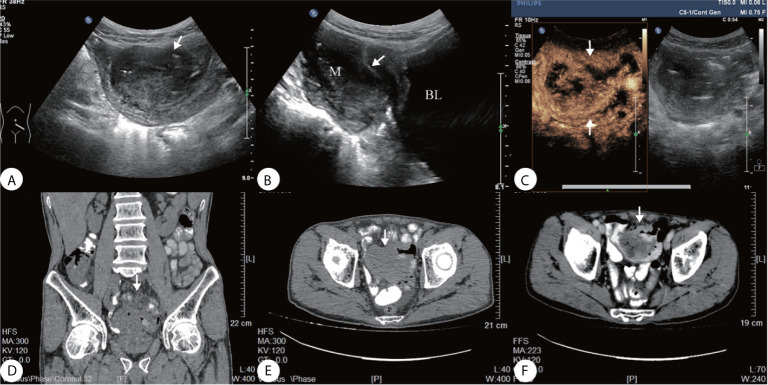
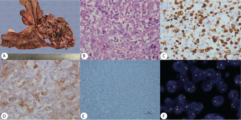
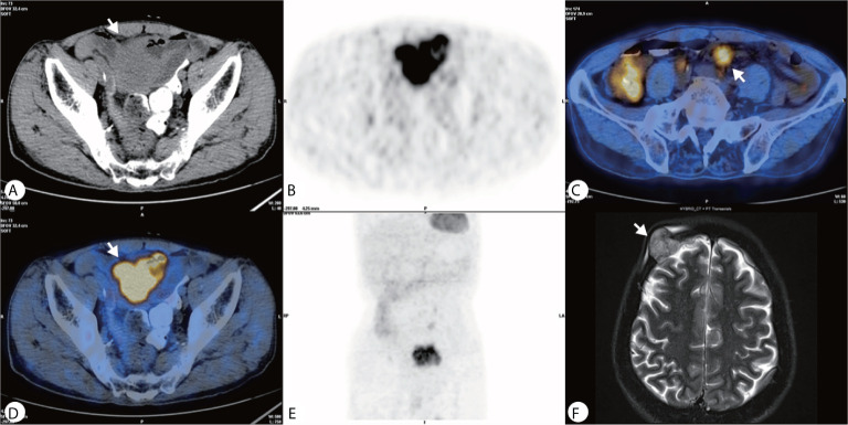
Extraosseous Ewing's sarcoma (EES) is a malignant tumor that is classified as a rare disease. EES is common in children and adolescents, with a rarer incidence being present in the elderly. ES of the primary intestine is rare, with only a few reports described in the literature. Here we report a case of a 69-year-old male patient who was experiencing abdominal pain for over 3 months. Ultrasonography (US) revealed a solid hypoechoic lesion with multiple irregular necrotic areas in the left lower abdomen close to the dome of the bladder. Contrast-enhanced ultrasonography (CEUS) showed that the lesion exhibited heterogeneous enhancement and quick peripheral enhanced tissue wash-out classifying this mass as malignant. PET-CT showed a high metabolic mass in the lower abdomen, multiple metabolic nodules in the mesentery, considered as a small intestinal stromal tumor with lymph nodes metastasis, and that a diagnosis of lymphoma should be excluded. Esophagogastroduodenoscopy performed at another hospital 1 month prior to CT showed an abnormal density in the pelvic cavity that was considered as a colonic diverticulum with an abscess. The endoscopy showed no obvious occupying lesions. The mass was removed and postoperative pathology confirmed ES of the small intestine. The patient avoided receiving chemotherapy. After 2 months, skull metastasis was diagnosed and surgical intervention was done. His survival was only six months after the second surgery. To our knowledge, our case is the first report of ultrasound and CEUS manifestation of EES in the small intestine in elderly.
骨外尤文肉瘤(EES)是一种被归类为罕见病的恶性肿瘤。EES在儿童和青少年中较为常见,在老年人中发病率较低。原发性肠道尤文肉瘤很罕见,文献中仅有少数病例报道。在此,我们报告一例69岁男性患者,其腹痛超过3个月。超声检查(US)显示左下腹靠近膀胱顶部有一个实性低回声病变,伴有多个不规则坏死区域。超声造影(CEUS)显示该病变表现为不均匀强化,且周边强化组织快速消退,将该肿块归类为恶性。PET-CT显示下腹部有一个高代谢肿块,肠系膜有多个代谢结节,考虑为小肠间质瘤伴淋巴结转移,且应排除淋巴瘤诊断。在CT检查前1个月,另一家医院进行的食管胃十二指肠镜检查显示盆腔有异常密度影,考虑为结肠憩室伴脓肿。内镜检查未发现明显占位性病变。切除该肿块后,术后病理证实为小肠尤文肉瘤。患者未接受化疗。2个月后,诊断为颅骨转移并进行了手术干预。第二次手术后他仅存活了6个月。据我们所知,我们的病例是老年患者小肠尤文肉瘤超声及CEUS表现的首例报道。