Matsumoto Ayumi, Matsui Isao, Katsuma Yusuke, Yasuda Seiichi, Shimada Karin, Namba-Hamano Tomoko, Sakaguchi Yusuke, Kaimori Jun-Ya, Takabatake Yoshitsugu, Inoue Kazunori, Isaka Yoshitaka
Department of Nephrology, Osaka University Graduate School of Medicine, Osaka, Japan.
Department of Inter-Organ Communication Research in Kidney Disease, Osaka University Graduate School of Medicine, Osaka, Japan.
Kidney Int Rep. 2021 May 1;6(7):1923-1938. doi: 10.1016/j.ekir.2021.04.021. eCollection 2021 Jul.
Foot process effacement and mitochondrial fission associate with kidney disease pathogenesis. Electron microscopy is the gold-standard method for their visualization, but the observable area of electron microscopy is smaller than light microscopy. It is important to develop alternative ways to quantitatively evaluate these microstructural changes because the lesion site of renal diseases can be focal.
We analyzed elastica-Masson trichrome (EMT) and periodic acid-Schiff (PAS) stained kidney sections using structured illumination microscopy (SIM).
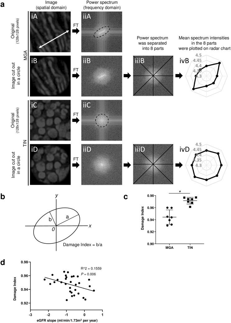
EMT staining revealed three-dimensional (3D) structures of foot process, whereas ponceau xylidine acid fuchsin azophloxine solution induced fluorescence. Conversion of foot process images into their constituent frequencies by Fourier transform showed that the concentric square of (1/4)-(1/16) in the power spectra (PS) included information for normal periodic structures of foot processes. Foot process integrity, assessed by PS, negatively correlated with proteinuria. EMT-stained sections revealed fragmented mitochondria in mice with mitochondrial injuries and patients with tubulointerstitial nephritis; Fourier transform quantified associated mitochondrial injury. Quantified mitochondrial damage in patients with immunoglobulin A (IgA) nephropathy predicted a decline in estimated glomerular filtration rate (eGFR) after kidney biopsy but did not correlate with eGFR at biopsy. PAS-stained sections, excited by a 640 nm laser, combined with the coefficient of variation values, quantified subtle changes in the basement membranes of patients with membranous nephropathy stage I.
Kidney microstructures are quantified from sections prepared in clinical practice using SIM.
足突消失和线粒体裂变与肾脏疾病的发病机制相关。电子显微镜是观察它们的金标准方法,但电子显微镜的可观察区域小于光学显微镜。由于肾脏疾病的病变部位可能是局灶性的,因此开发替代方法来定量评估这些微观结构变化很重要。
我们使用结构照明显微镜(SIM)分析了弹性-Masson三色(EMT)和过碘酸-希夫(PAS)染色的肾脏切片。
EMT染色揭示了足突的三维(3D)结构,而丽春红二甲苯胺酸性品红偶氮间苯二酚溶液可诱导荧光。通过傅里叶变换将足突图像转换为其组成频率,结果表明功率谱(PS)中(1/4)-(1/16)的同心正方形包含足突正常周期性结构的信息。通过PS评估的足突完整性与蛋白尿呈负相关。EMT染色切片显示线粒体损伤小鼠和肾小管间质性肾炎患者的线粒体碎片化;傅里叶变换对相关的线粒体损伤进行了量化。免疫球蛋白A(IgA)肾病患者的线粒体损伤量化结果预测了肾活检后估计肾小球滤过率(eGFR)的下降,但与活检时的eGFR无关。用640nm激光激发的PAS染色切片与变异系数值相结合,量化了I期膜性肾病患者基底膜的细微变化。
使用SIM从临床实践中制备的切片对肾脏微观结构进行量化。