Sahu Biswanath, Anand Rama, Kumar Sandeep, Solanki Ravi Shankar, Mehra Pravesh, Jain Manjula
Department of Radio-Diagnosis, Lady Hardinge Medical College and Associated Hospitals, New Delhi, India.
Department of Dental and Oral Surgery, Lady Hardinge Medical College and Associated Hospitals, New Delhi, India.
Indian J Radiol Imaging. 2021 Jan;31(1):210-223. doi: 10.1055/s-0041-1729767. Epub 2021 May 23.
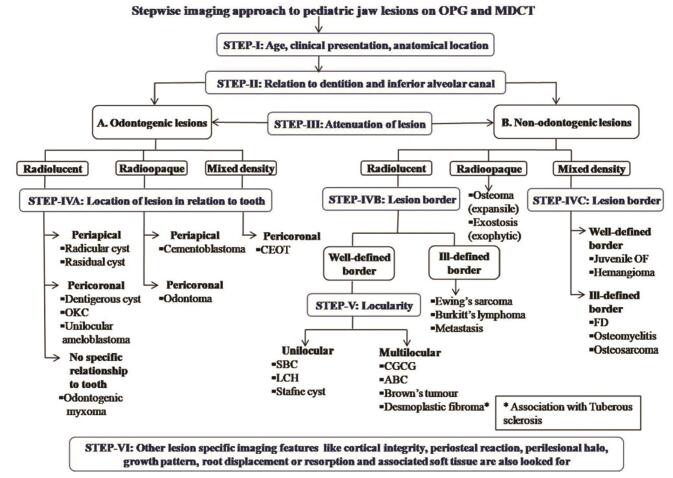
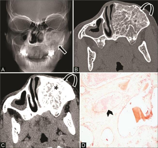
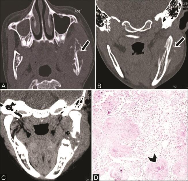
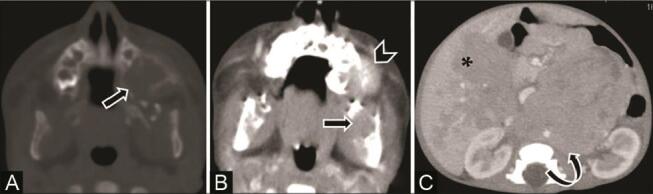
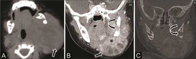
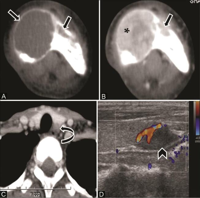
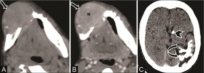
Jaw lesions in the pediatric population, although infrequently encountered in clinical practice, can cause functional impairment and cosmetic disfiguring. It is further complicated by the difficulty in diagnosis due to complex anatomy and facial developmental process during infancy and childhood. Intraosseous pediatric jaw lesions may vary from odontogenic to nonodontogenic types with nonspecific clinical features in most cases. They deserve careful attention by a systematic approach to provide a relevant diagnosis or differential diagnosis for timely management. Imaging plays a major role in diagnosis with orthopantomograph being the foremost investigation, followed by cross-sectional imaging, essentially computed tomography as a problem-solving tool. This article highlights the imaging spectrum of various jaw lesions in the pediatric population with a pattern-based approach for radiological diagnosis.
儿科患者的颌骨病变在临床实践中虽不常见,但可导致功能障碍和外观损毁。由于婴儿期和儿童期复杂的解剖结构及面部发育过程,诊断困难使情况更加复杂。大多数情况下,小儿颌骨骨内病变从牙源性到非牙源性类型各异,临床特征不具特异性。它们值得通过系统方法予以仔细关注,以便为及时治疗提供相关诊断或鉴别诊断。影像学在诊断中起主要作用,全景曲面断层片是首要检查手段,其次是断层成像,主要是计算机断层扫描作为问题解决工具。本文采用基于模式的方法对儿科患者各种颌骨病变的影像学特征进行综述,以用于放射学诊断。