Ma Zhuoran, Wang Feifei, Zhong Yeteng, Salazar Felix, Li Jiachen, Zhang Mingxi, Ren Fuqiang, Wu Anna M, Dai Hongjie
Department of Chemistry and Bio-X, Stanford University Stanford, CA 94305 (USA).
Department of Molecular Imaging and Therapy, Beckman Research, Institute of the City of Hope, Duarte, CA (USA).
Angew Chem Weinheim Bergstr Ger. 2020 Nov 9;132(46):20733-20741. doi: 10.1002/ange.202008083. Epub 2020 Jul 17.
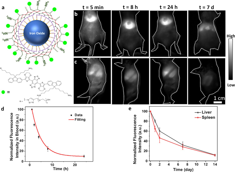
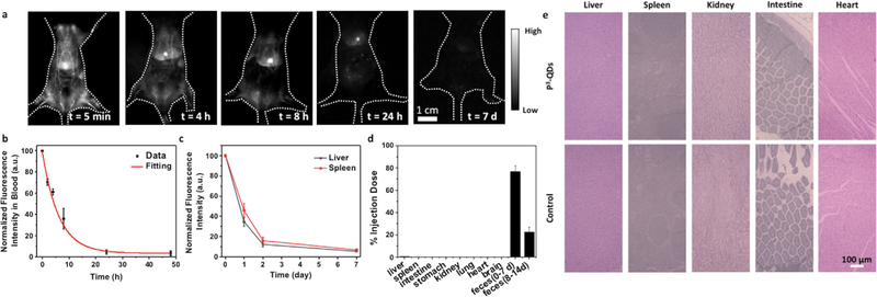
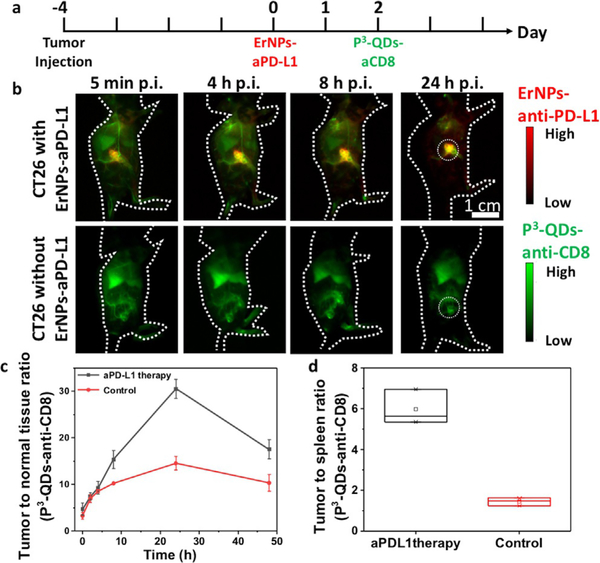
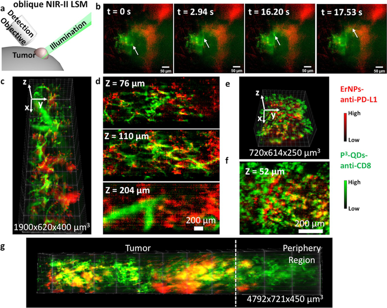
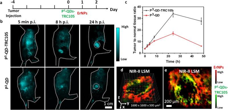
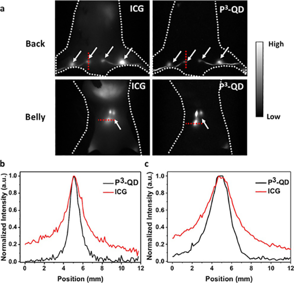
Most NIR-IIb fluorophores are nanoparticle-based probes with long retention ( ≈ 1 month or longer) in the body. Here, we applied a novel cross-linked coating to functionalize core/shell lead sulfide/cadmium sulfide quantum dots (PbS/CdS QDs) emitting at ≈ 1600 nm. The coating was comprised of an amphiphilic polymer followed by three crosslinked amphiphilic polymeric layers (P coating), imparting high biocompatibility and > 90% excretion of QDs within 2 weeks of intravenous administration. The P-QDs were conjugated to an engineered anti-CD8 diabody (Cys-diabody) for in vivo molecular imaging of CD8 + cytotoxic T lymphocytes (CTLs) in response to anti-PD-L1 therapy. Two-plex molecular imaging in combination with down-conversion Er nanoparticles (ErNPs) was performed for real-time in vivo monitoring of PD-L1 positive tumor cells and CTLs with cellular resolution by non-invasive NIR-IIb light sheet microscopy. Imaging of angiogenesis in the tumor microenvironment and of lymph nodes deep in the body with a signal-to-background ratio of up to ≈ 170 was also achieved using P-QDs.
大多数近红外二区(NIR-IIb)荧光团是基于纳米颗粒的探针,在体内具有较长的保留时间(约1个月或更长)。在此,我们应用了一种新型交联涂层对发射波长约为1600 nm的核/壳硫化铅/硫化镉量子点(PbS/CdS QDs)进行功能化。该涂层由一种两亲聚合物和随后的三层交联两亲聚合物层(P涂层)组成,赋予了高生物相容性,并且在静脉注射后2周内量子点的排泄率>90%。将P-QDs与工程化抗CD8双体(Cys-双体)偶联,用于对抗PD-L1治疗作出反应的CD8 + 细胞毒性T淋巴细胞(CTL)进行体内分子成像。通过非侵入性近红外二区光片显微镜,结合下转换铒纳米颗粒(ErNPs)进行双分子成像,以细胞分辨率对PD-L1阳性肿瘤细胞和CTL进行实时体内监测。使用P-QDs还实现了肿瘤微环境中血管生成以及体内深部淋巴结的成像,信号与背景比高达约170。