Biosciences and Physiopathology Program, Universidade Estadual de Maringá, Maringá, Brazil.
Department of Clinical Analysis and Biomedicine, Universidade Estadual de Maringá, Maringá, Brazil.
Front Cell Infect Microbiol. 2021 Jul 14;11:687499. doi: 10.3389/fcimb.2021.687499. eCollection 2021.
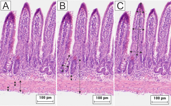
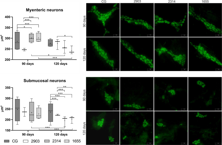
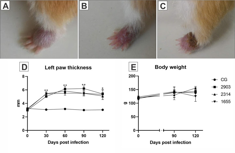
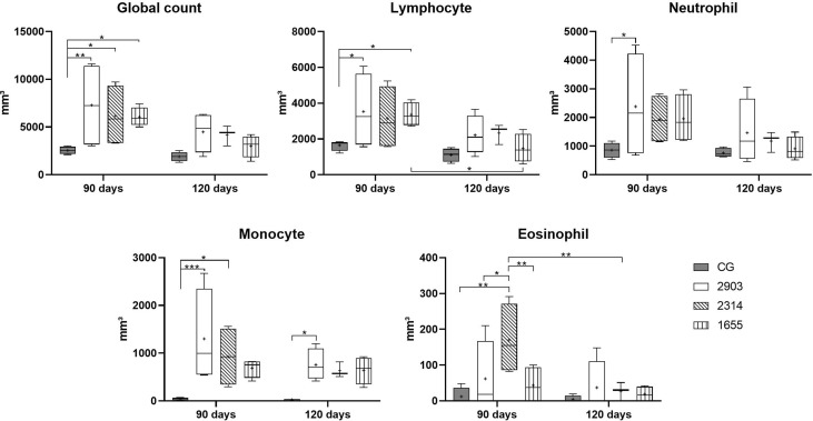
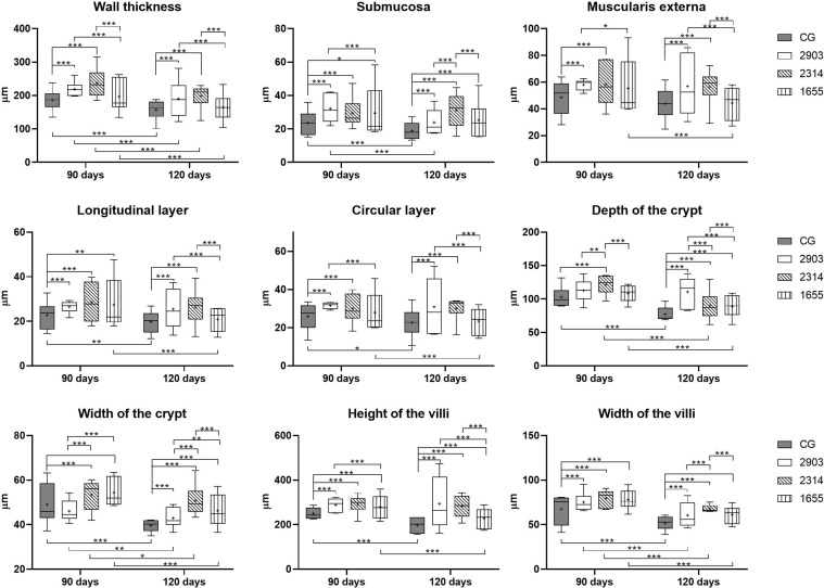
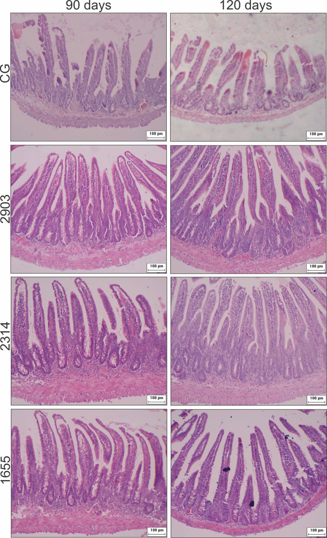
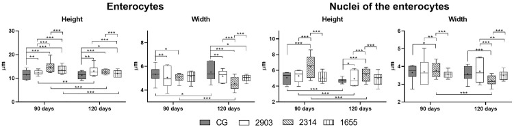
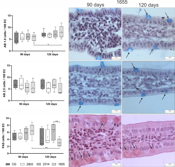
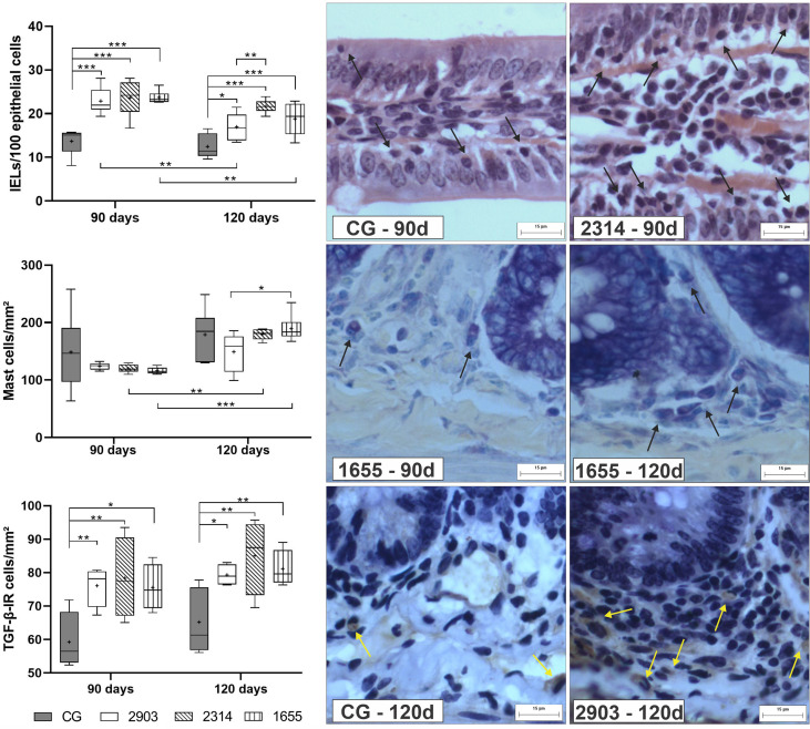
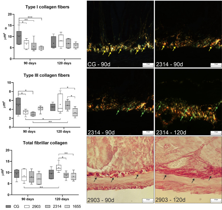
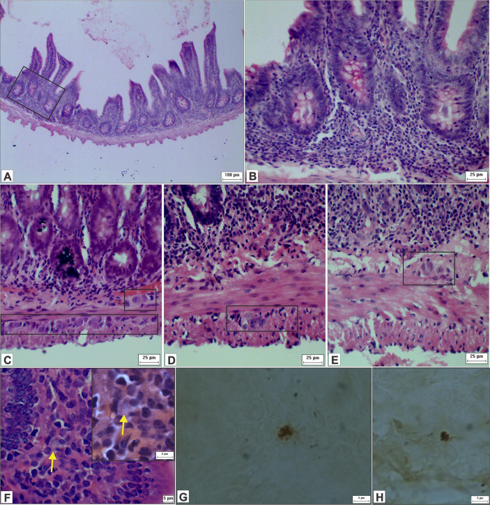
is one of the main causes of cutaneous leishmaniasis in the Americas. This species presents genetic polymorphism that can cause destructive lesions in oral, nasal, and oropharyngeal tracts. In a previous study, the parasite caused several histopathological changes to hamster ileums. Our study evaluates immune response components, morphological changes, and effects on neurons in the ileums of hamsters infected by three different strains of in two infection periods. For the experiment, we separated hamsters into four groups: a control group and three infected groups. Infected hamsters were euthanized 90- or 120-days post infection. We used three strains of : the reference MHOM/BR/1975/M2903 and two strains isolated from patients who had different responses to Glucantime treatment (MHOM/BR/2003/2314 and MHOM/BR/2000/1655). After laparotomy, ileums were collected for histological processing, biochemical analysis, and evaluation of neurons in the myenteric and submucosal plexuses of the enteric nervous system (ENS). The results demonstrated the increase of blood leukocytes after the infection. Optical microscopy analysis showed histopathological changes with inflammatory infiltrates, edemas, ganglionitis, and amastigotes in the ileums of infected hamsters. We observed changes in the organ histoarchitecture of infected hamsters when compared to control groups, such as thicker muscular and submucosa layers, deeper and wider crypts, and taller and broader villi. The number of intraepithelial lymphocytes and TGF-β-immunoreactive cells increased in all infected groups when compared to the control groups. Mast cells increased with longer infection periods. The infection also caused remodeling of intestinal collagen and morphometry of myenteric and submucosal plexus neurons; but this effect was dependent on infection duration. Our results show that infection caused time-dependent alterations in hamster ileums. This was demonstrated by the reduction of inflammatory cells and the increase of tissue regeneration factors at 120 days of infection. The infected groups demonstrated different profiles in organ histoarchitecture, migration of immune cells, and morphometry of ENS neurons. These findings suggest that the small intestine (or at least the ileum) is a target organ for infection, as the infection caused changes that were dependent on duration and strain.
是美洲地区皮肤利什曼病的主要原因之一。该物种具有遗传多态性,可导致口腔、鼻腔和口咽道的破坏性病变。在之前的一项研究中,寄生虫导致仓鼠回肠出现多种组织病理学变化。我们的研究评估了三种不同株 在两个感染期感染仓鼠后,对回肠的免疫反应成分、形态变化和对神经元的影响。为此实验,我们将仓鼠分为四组:对照组和三组感染组。感染后的仓鼠在感染后 90 或 120 天被安乐死。我们使用了三种:参考 MHOM/BR/1975/M2903 株和从对 Glucantime 治疗反应不同的患者中分离出的两种株(MHOM/BR/2003/2314 和 MHOM/BR/2000/1655)。剖腹手术后,收集回肠进行组织学处理、生化分析和评估肠神经系统(ENS)的肌间和黏膜下神经丛中的神经元。结果显示感染后白细胞增多。光镜分析显示,感染仓鼠的回肠有炎症浸润、水肿、神经节炎和 内阿米巴的组织病理学变化。与对照组相比,我们观察到感染仓鼠的器官组织学结构发生了变化,如肌层和黏膜下层较厚、隐窝较深较宽、绒毛较高较宽。与对照组相比,所有感染组的上皮内淋巴细胞和 TGF-β 免疫反应细胞数量均增加。肥大细胞随着感染时间的延长而增加。感染还导致肠胶原重塑和肌间和黏膜下神经丛神经元的形态计量学改变;但这种影响取决于感染持续时间。我们的结果表明, 感染导致仓鼠回肠的时间依赖性改变。感染 120 天时,炎症细胞减少,组织再生因子增加,证明了这一点。感染组在器官组织学、免疫细胞迁移和 ENS 神经元形态计量学方面表现出不同的特征。这些发现表明,小肠(或至少回肠)是 感染的靶器官,因为感染引起的变化取决于持续时间和株。