Xiong Fei, Wu Guan-Hua, Wang Bing, Chen Yong-Jun
Department of Biliary-Pancreatic Surgery, Tongji Hospital, Tongji Medical College, Huazhong University of Science and Technology, Wuhan, 430074, Hubei, China.
Cancer Cell Int. 2021 Aug 4;21(1):411. doi: 10.1186/s12935-021-02117-1.
Altered Plastin-3 (PLS3; an actin-binding protein) expression was associated with human carcinogenesis, including pancreatic ductal adenocarcinoma (PDA). This study first assessed differentially expressed genes (DEGs) and then bioinformatically and experimentally confirmed PLS3 to be able to predict PDA prognosis and distinguish PDA from diffuse large B-cell lymphoma.
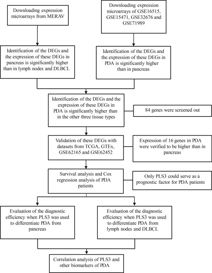
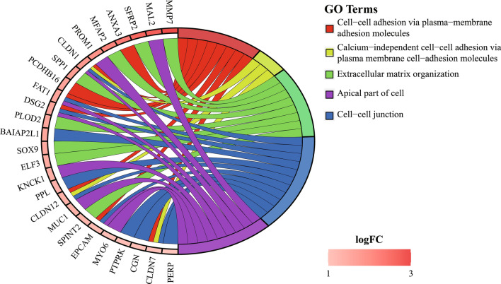
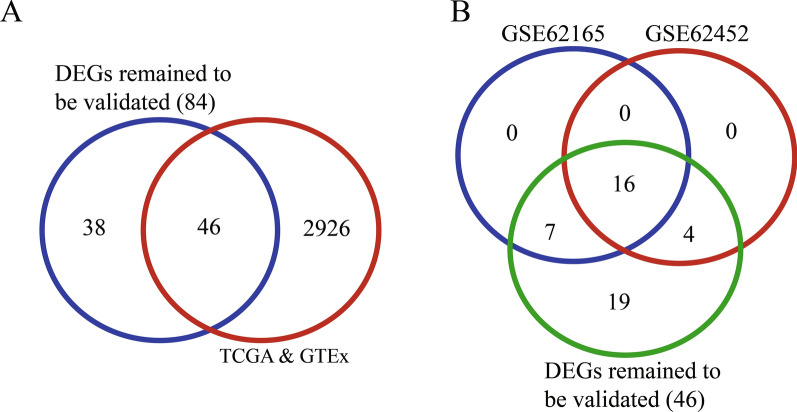
This study screened multiple online databases and revealed DEGs among PDA, normal pancreas, diffuse large B-cell lymphoma (DLBCL), and normal lymph node tissues and then focused on PLS3. These DEGs were analyzed for Gene Ontology (GO) terms, Kaplan-Meier curves, and the log-rank test to characterize their association with PDA prognosis. The receiver operating characteristic curve (ROC) was plotted, and Spearman's tests were performed. Differential PLS3 expression in different tissue specimens (n = 30) was evaluated by reverse transcription quantitative polymerase chain reaction (RT-qPCR).
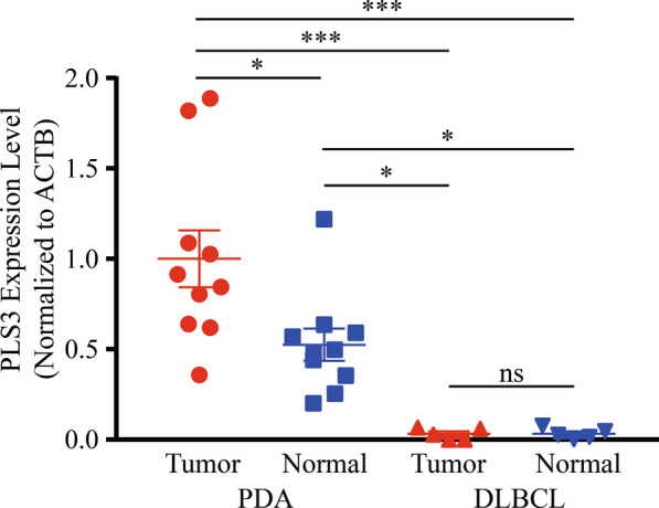
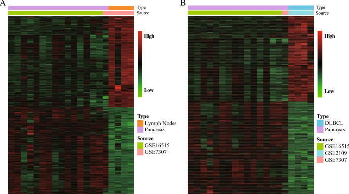
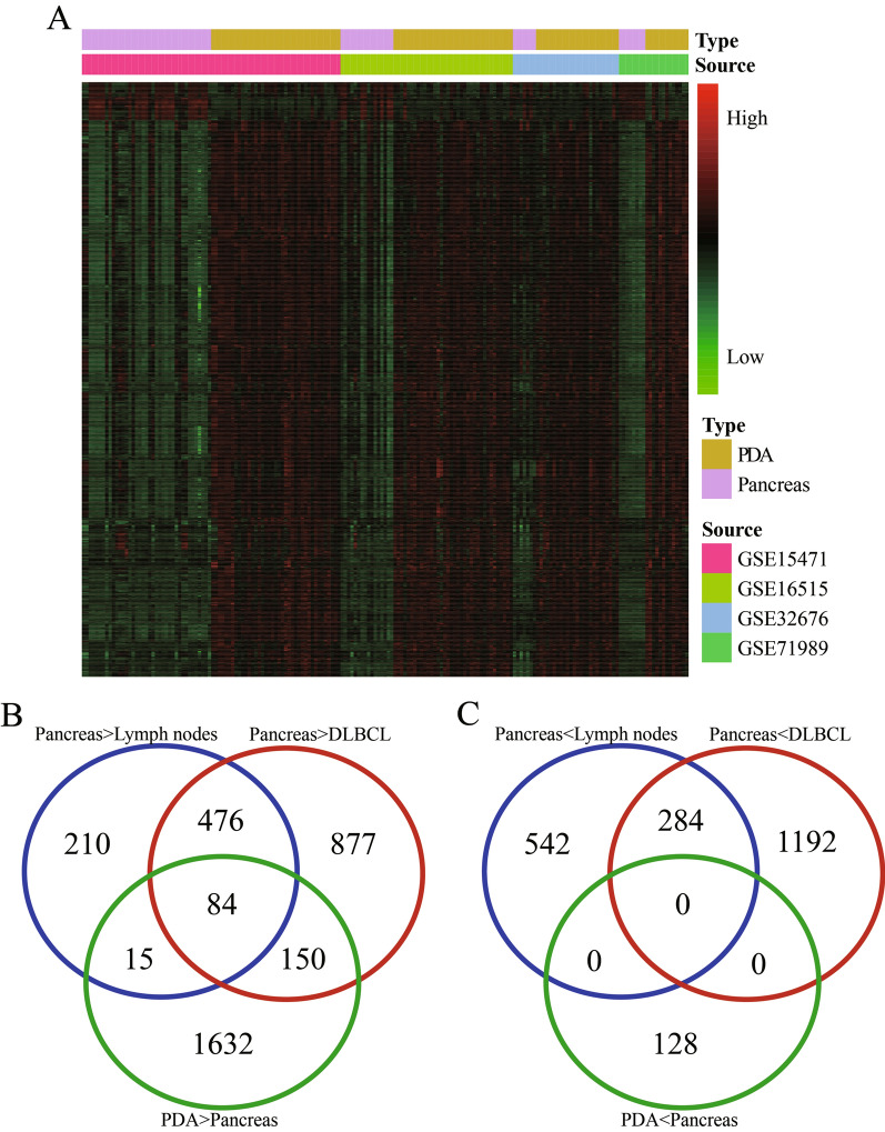
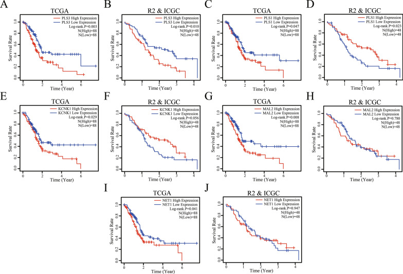
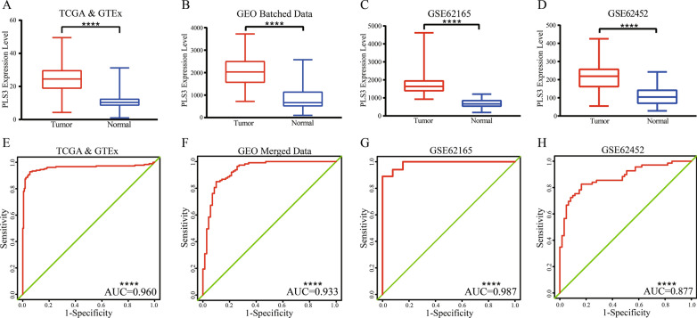
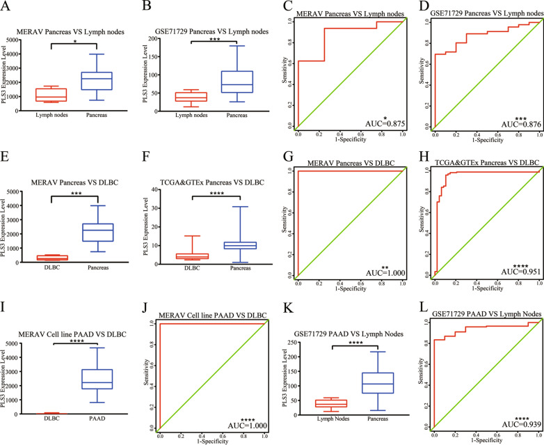
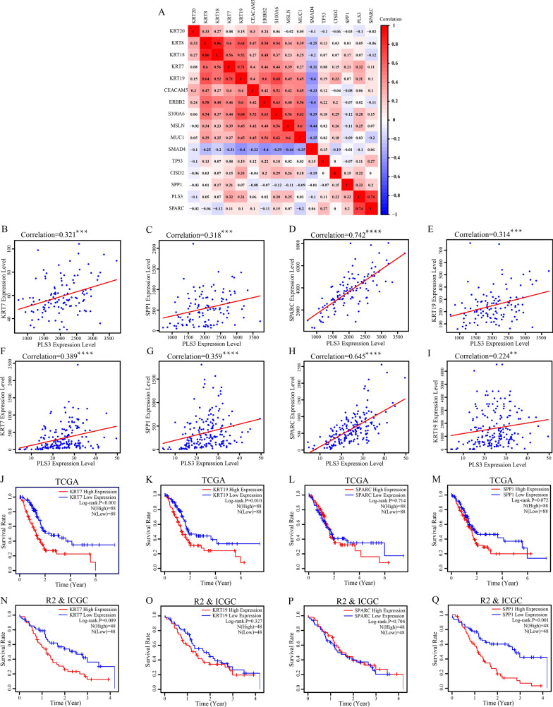
There were a great number of DEGs between PDA and lymph node, between PDA and DLBCL, and between PDA and normal pancreatic tissues. Five DEGs (NET1, KCNK1, MAL2, PLS1, and PLS3) were associated with poor overall survival of PDA patients, but only PLS3 was further verified by the R2 and ICGC datasets. The ROC analysis showed a high PLS3 AUC (area under the curve) value for PDA diagnosis, while PLS3 was able to distinguish PDA from DLBCL. The results of Spearman's analysis showed that PLS3 expression was associated with levels of KRT7, SPP1, and SPARC. Differential PLS3 expression in different tissue specimens was further validated by RT-qPCR.
Altered PLS3 expression was useful in diagnosis and prognosis of PDA as well as to distinguish PDA from DLBCL.
丝束蛋白-3(PLS3;一种肌动蛋白结合蛋白)表达改变与人类致癌作用相关,包括胰腺导管腺癌(PDA)。本研究首先评估差异表达基因(DEG),然后通过生物信息学和实验证实PLS3能够预测PDA预后,并将PDA与弥漫性大B细胞淋巴瘤区分开来。
本研究筛选了多个在线数据库,揭示了PDA、正常胰腺、弥漫性大B细胞淋巴瘤(DLBCL)和正常淋巴结组织中的DEG,然后聚焦于PLS3。对这些DEG进行基因本体(GO)术语、Kaplan-Meier曲线和对数秩检验分析,以表征它们与PDA预后的关联。绘制受试者工作特征曲线(ROC),并进行Spearman检验。通过逆转录定量聚合酶链反应(RT-qPCR)评估不同组织标本(n = 30)中PLS3的差异表达。
PDA与淋巴结、PDA与DLBCL以及PDA与正常胰腺组织之间存在大量DEG。五个DEG(NET1、KCNK1、MAL2、PLS1和PLS3)与PDA患者的总体生存率差相关,但只有PLS3通过R2和ICGC数据集得到进一步验证。ROC分析显示,PLS3的曲线下面积(AUC)值对PDA诊断具有较高的诊断价值,而PLS3能够将PDA与DLBCL区分开来。Spearman分析结果表明,PLS3表达与KRT7、SPP1和SPARC水平相关。RT-qPCR进一步验证了不同组织标本中PLS3的差异表达。
PLS3表达改变在PDA的诊断和预后以及将PDA与DLBCL区分开来方面具有重要作用。