Patil Shankargouda, Baeshen Hosam Ali
Department of Maxillofacial Surgery and Diagnostic Sciences, Division of Oral Pathology, College of Dentistry, Jazan University, Jazan, Saudi Arabia.
Department of Orthodontics, College of dentistry, King Abdulaziz University, Saudi Arabia.
Saudi J Biol Sci. 2021 Aug;28(8):4613-4618. doi: 10.1016/j.sjbs.2021.04.068. Epub 2021 May 1.
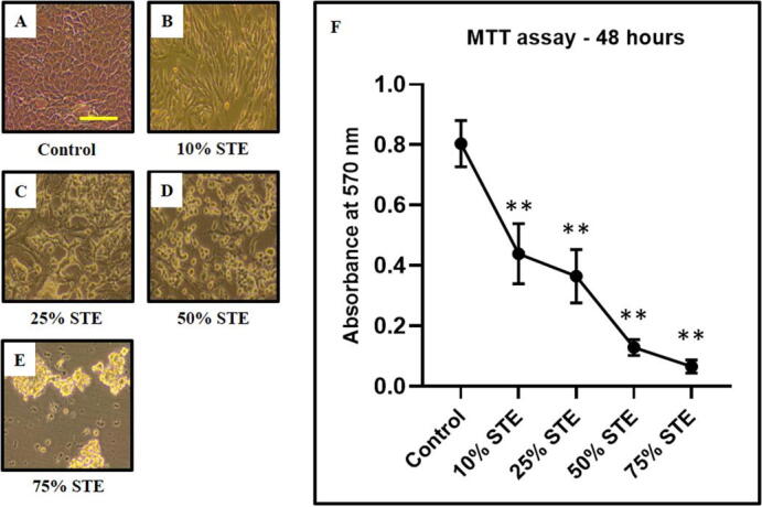
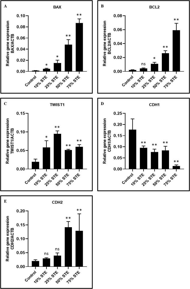
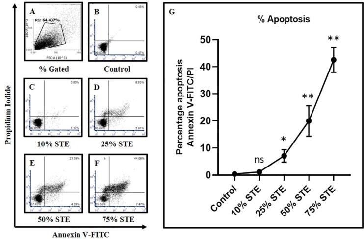
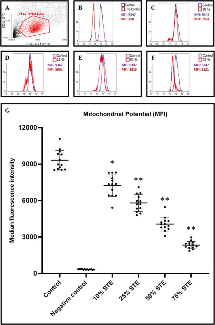
Smokeless tobacco habits are detrimental to oral health. A correlation between tobacco use and local epithelial tissue damage exists. Yet, the underlying cellular mechanism is not precisely characterized. This study assessed the dose-dependent action of Smokeless tobacco extract on gingival epithelial cells. Gingival tissue was taken from 5 healthy donors. Gingival epithelial cells were isolated by an enzymatic method and cultured up to passage 2. The cultured cells were treated with smokeless tobacco extract at 10%, 25%, 50%, and 75% volume concentration. After 48 h of incubation, MTT assay, Annexin V/PI assay, and DiIC1(5) assay were used to evaluate viability, apoptosis, and mitochondrial potential of the cells. RT-qPCR was used to determine the expression of BAX, BCL2, ECAD, NCAD, and TWIST. The Smokeless tobacco extract reduced cell viability by disrupting the mitochondrial potential and inducing apoptosis. Further, the Smokeless tobacco extract induced a dose-dependent epithelial-mesenchymal-transition in gingival epithelial cells. Apoptotic cellular death caused by tobacco extract on the gingival epithelial system was dependant on the mitochondrial potential of the cell. The results demonstrate that smokeless tobacco causes detrimental metabolic alterations of the periodontium.
This study elucidates the mechanism by which Smokeless tobacco products cause cellular damage to the gingival epithelium. The use of Smokeless tobacco products can lead to major cellular and surface changes in the gingiva and its appearance. The consequences of these changes are not limited to oral cancer but also increases a person's risk for dental and periodontal disease.
无烟烟草习惯对口腔健康有害。烟草使用与局部上皮组织损伤之间存在关联。然而,潜在的细胞机制尚未得到精确表征。本研究评估了无烟烟草提取物对牙龈上皮细胞的剂量依赖性作用。从5名健康供体获取牙龈组织。通过酶法分离牙龈上皮细胞并培养至第2代。将培养的细胞用体积浓度为10%、25%、50%和75%的无烟烟草提取物处理。孵育48小时后,使用MTT法、Annexin V/PI法和DiIC1(5)法评估细胞的活力、凋亡和线粒体电位。RT-qPCR用于测定BAX、BCL2、E-cadherin(E钙黏蛋白)、N-cadherin(N钙黏蛋白)和TWIST的表达。无烟烟草提取物通过破坏线粒体电位和诱导凋亡降低细胞活力。此外,无烟烟草提取物在牙龈上皮细胞中诱导剂量依赖性的上皮-间质转化。烟草提取物对牙龈上皮系统引起的凋亡性细胞死亡取决于细胞的线粒体电位。结果表明,无烟烟草会导致牙周组织有害的代谢改变。
本研究阐明了无烟烟草产品对牙龈上皮造成细胞损伤的机制。使用无烟烟草产品会导致牙龈及其外观出现重大的细胞和表面变化。这些变化的后果不仅限于口腔癌,还会增加一个人患牙齿和牙周疾病的风险。