Department of Biotechnology and Dr. B.C. Guha Centre for Genetic Engineering and Biotechnology, University of Calcutta, WB, India.
PLoS One. 2013 Jul 11;8(7):e68224. doi: 10.1371/journal.pone.0068224. Print 2013.
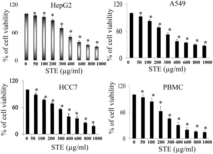
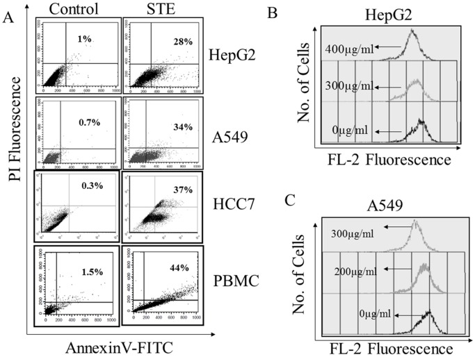
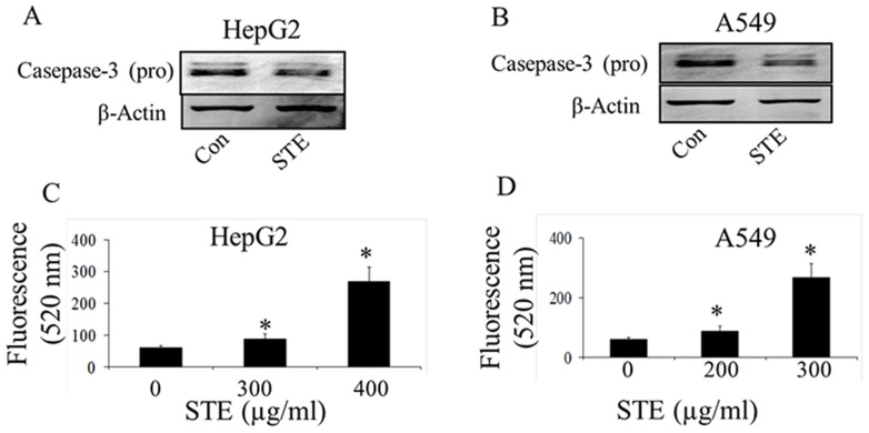
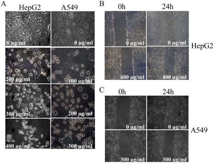
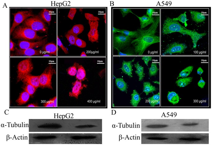
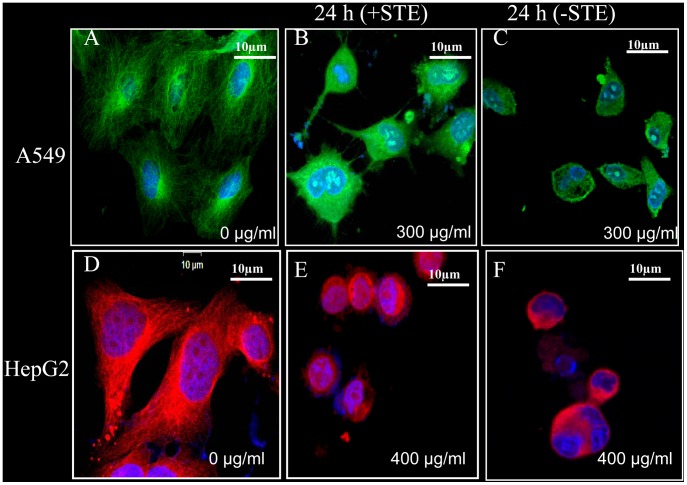
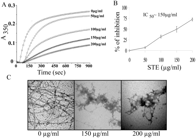
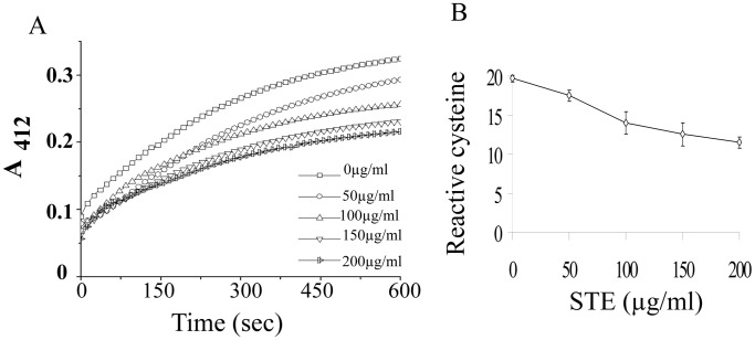
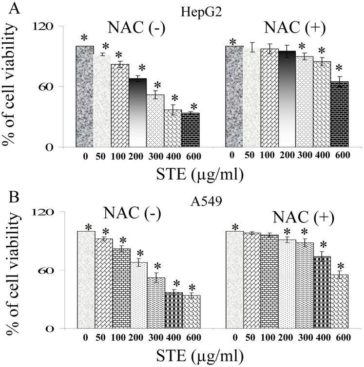
Smokeless tobacco usage is a growing public health problem worldwide. The molecular mechanism(s) underlying smokeless tobacco associated tissue damage remain largely unidentified. In the present study we have tried to explore the effects of aqueous extract of smokeless tobacco (STE) on tubulin-microtubule, the major cytoskeleton protein that maintains cells morphology and participates in cell division. Exposure to STE resulted in dose-dependent cytotoxicity in a variety of mammalian transformed cell lines such as human lung epithelial cells A549, human liver epithelial cells HepG2, and mouse squamous epithelial cells SCC7, [corrected] as well as non-tumorogenic human peripheral blood mononuclear cells PBMC. Cellular morphology of STE-treated cells was altered and the associated disruption of microtubule network indicates that STE targets tubulin-microtubule system in both cell lines. Furthermore it was also observed that STE-treatment resulted in the selective degradation of cellular tubulin, whereas actin remains unaltered. In vitro, polymerization of purified tubulin was inhibited by STE with the IC50 value∼150 µg/ml and this is associated with the loss of reactive cysteine residues of tubulin. Application of thiol-based antioxidant N-acetyl cysteine (NAC) significantly abrogates STE-mediated microtubule damage and associated cytotoxicity in both A549 and HepG2 cells. These results suggest that microtubule damage is one of the key mechanisms of STE-induced cytotoxity in mammalian cells.
无烟烟草制品的使用是一个全球性的公共卫生问题。与无烟烟草相关的组织损伤的分子机制在很大程度上仍未被确定。在本研究中,我们试图探讨无烟烟草水提物(STE)对微管蛋白的影响,微管蛋白是维持细胞形态和参与细胞分裂的主要细胞骨架蛋白。STE 暴露于各种哺乳动物转化细胞系,如人肺上皮细胞 A549、人肝上皮细胞 HepG2 和鼠鳞状上皮细胞 SCC7,以及非肿瘤源性的人外周血单核细胞 PBMC,导致细胞毒性呈剂量依赖性。STE 处理细胞的细胞形态发生改变,微管网络的相关破坏表明 STE 靶向两种细胞系中的微管蛋白-微管系统。此外,还观察到 STE 处理导致细胞微管蛋白的选择性降解,而肌动蛋白保持不变。在体外,STE 抑制纯化微管蛋白的聚合,IC50 值约为 150μg/ml,这与微管蛋白反应性半胱氨酸残基的丧失有关。应用巯基抗氧化剂 N-乙酰半胱氨酸(NAC)显著减轻了 A549 和 HepG2 细胞中 STE 介导的微管损伤和相关细胞毒性。这些结果表明,微管蛋白损伤是 STE 诱导哺乳动物细胞细胞毒性的关键机制之一。