• 文献检索
  • 文档翻译
  • 深度研究
  • 学术资讯
  • Suppr Zotero 插件Zotero 插件
  • 邀请有礼
  • 套餐&价格
  • 历史记录
应用&插件
Suppr Zotero 插件Zotero 插件浏览器插件Mac 客户端Windows 客户端微信小程序
定价
高级版会员购买积分包购买API积分包
服务
文献检索文档翻译深度研究API 文档MCP 服务
关于我们
关于 Suppr公司介绍联系我们用户协议隐私条款
关注我们

Suppr 超能文献

核心技术专利:CN118964589B侵权必究
粤ICP备2023148730 号-1Suppr @ 2026

文献检索

告别复杂PubMed语法,用中文像聊天一样搜索,搜遍4000万医学文献。AI智能推荐,让科研检索更轻松。

立即免费搜索

文件翻译

保留排版,准确专业,支持PDF/Word/PPT等文件格式,支持 12+语言互译。

免费翻译文档

深度研究

AI帮你快速写综述,25分钟生成高质量综述,智能提取关键信息,辅助科研写作。

立即免费体验

扩散张量成像揭示了肌痛性脑脊髓炎/慢性疲劳综合征中的神经元微观结构变化。

Diffusion tensor imaging reveals neuronal microstructural changes in myalgic encephalomyelitis/chronic fatigue syndrome.

作者信息

Thapaliya Kiran, Marshall-Gradisnik Sonya, Staines Donald, Barnden Leighton

机构信息

National Centre for Neuroimmunology and Emerging Diseases, Menzies Health Institute Queensland, Griffith University, Brisbane, Queensland, Australia.

Centre for Advanced Imaging, The University of Queensland, Brisbane, Queensland, Australia.

出版信息

Eur J Neurosci. 2021 Sep;54(6):6214-6228. doi: 10.1111/ejn.15413. Epub 2021 Aug 15.

DOI:10.1111/ejn.15413
PMID:34355438
原文链接:https://pmc.ncbi.nlm.nih.gov/articles/PMC9291819/
Abstract

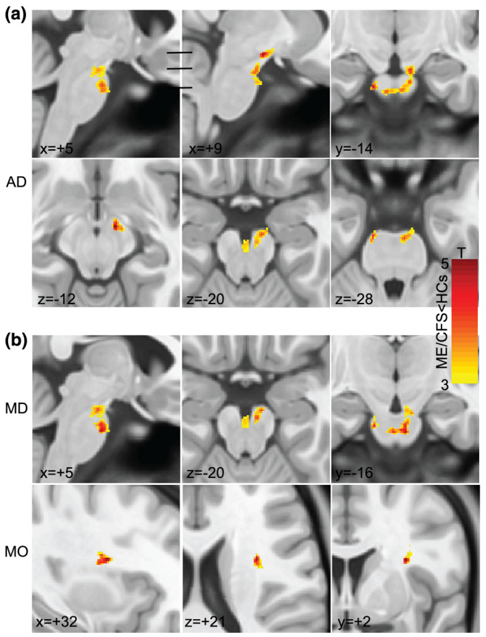
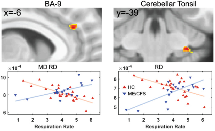
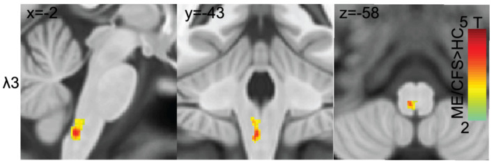
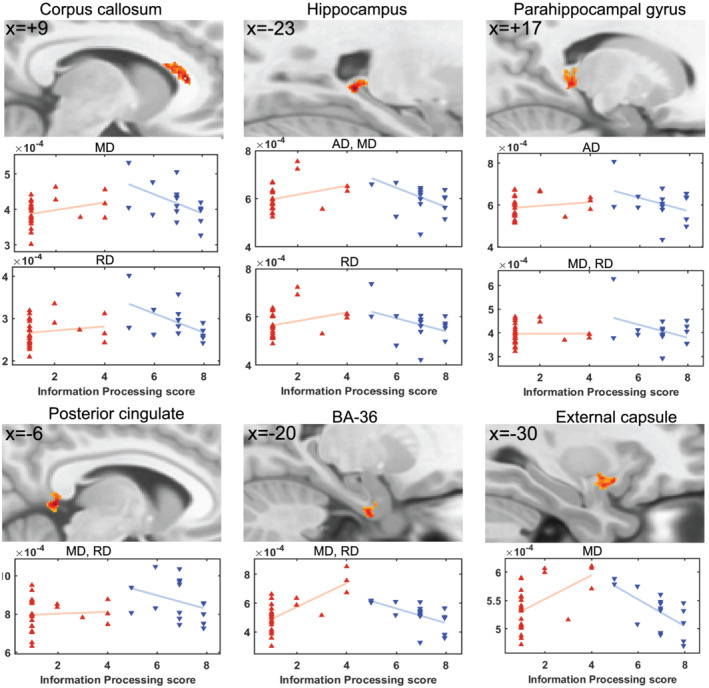
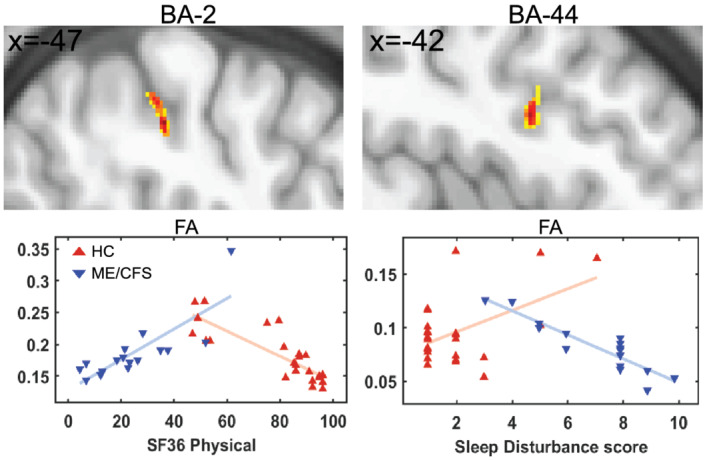
Myalgic encephalomyelitis/chronic fatigue syndrome (ME/CFS) patients suffer from a variety of physical and neurological complaints indicating the central nervous system plays a role in ME/CFS pathophysiology. Diffusion tensor imaging (DTI) has been used to study microstructural changes in neurodegenerative diseases. In this study, we evaluated DTI parameters to investigate microstructural abnormalities in ME/CFS patients. We estimated DTI parameters in 25 ME/CFS patients who met Fukuda criteria (ME/CFS ), 18 ME/CFS patients who met International Consensus Criteria (ICC) (ME/CFS ) only and 26 healthy control (HC) subjects. In addition to voxel-based DTI-parameter group comparisons, we performed voxel-based DTI-parameter interaction-with-group regressions with clinical and autonomic measures to test for abnormal regressions. Group comparisons between ME/CFS and HC detected significant clusters (a) with decreased axial diffusivity (p = .001) and mean diffusivity (p = .01) in the descending cortico-cerebellar tract in the midbrain and pons and (b) with increased transverse diffusivity in the medulla. The mode of anisotropy was significantly decreased (p = .001) in a cluster in the superior longitudinal fasciculus region. Voxel-based group comparisons between ME/CFS and HC did not detect significant clusters. For ME/CFS and HC, DTI parameter interaction-with-group regressions were abnormal for the clinical measures of information processing score, SF36 physical, sleep disturbance score and respiration rate in both grey and white matter regions. Our study demonstrated that DTI parameters are sensitive to microstructural changes in ME/CFS and could potentially act as an imaging biomarker of abnormal pathophysiology in ME/CFS. The study also shows that strict case definitions are essential in investigation of the pathophysiology of ME/CFS.

摘要

肌痛性脑脊髓炎/慢性疲劳综合征(ME/CFS)患者存在多种身体和神经方面的不适,这表明中枢神经系统在ME/CFS的病理生理学中发挥作用。扩散张量成像(DTI)已被用于研究神经退行性疾病中的微观结构变化。在本研究中,我们评估了DTI参数,以调查ME/CFS患者的微观结构异常。我们对25名符合福田标准的ME/CFS患者(ME/CFS )、18名仅符合国际共识标准(ICC)的ME/CFS患者(ME/CFS )和26名健康对照(HC)受试者进行了DTI参数估计。除了基于体素的DTI参数组间比较外,我们还进行了基于体素的DTI参数与组的交互回归分析,并结合临床和自主神经测量指标,以测试异常回归情况。ME/CFS组与HC组之间的组间比较发现了显著的聚类区域:(a)中脑和脑桥的皮质 - 小脑下行束中轴向扩散率降低(p = 0.001)和平均扩散率降低(p = 0.01);(b)延髓中的横向扩散率增加。在上级纵向束区域的一个聚类中,各向异性模式显著降低(p = 0.001)。ME/CFS组与HC组之间基于体素的组间比较未发现显著聚类。对于ME/CFS组和HC组,在灰质和白质区域,DTI参数与组的交互回归在信息处理评分、SF36身体评分、睡眠障碍评分和呼吸频率等临床测量指标方面均表现异常。我们的研究表明,DTI参数对ME/CFS中的微观结构变化敏感,有可能作为ME/CFS异常病理生理学的成像生物标志物。该研究还表明,严格的病例定义对于ME/CFS病理生理学研究至关重要。

https://cdn.ncbi.nlm.nih.gov/pmc/blobs/5ee7/9291819/ff730165053d/EJN-54-6214-g004.jpg
https://cdn.ncbi.nlm.nih.gov/pmc/blobs/5ee7/9291819/d80ba323518a/EJN-54-6214-g003.jpg
https://cdn.ncbi.nlm.nih.gov/pmc/blobs/5ee7/9291819/4070e917ff71/EJN-54-6214-g002.jpg
https://cdn.ncbi.nlm.nih.gov/pmc/blobs/5ee7/9291819/f5624d02021d/EJN-54-6214-g005.jpg
https://cdn.ncbi.nlm.nih.gov/pmc/blobs/5ee7/9291819/c4815d2edfdf/EJN-54-6214-g006.jpg
https://cdn.ncbi.nlm.nih.gov/pmc/blobs/5ee7/9291819/ff730165053d/EJN-54-6214-g004.jpg

相似文献

1
Diffusion tensor imaging reveals neuronal microstructural changes in myalgic encephalomyelitis/chronic fatigue syndrome.扩散张量成像揭示了肌痛性脑脊髓炎/慢性疲劳综合征中的神经元微观结构变化。
Eur J Neurosci. 2021 Sep;54(6):6214-6228. doi: 10.1111/ejn.15413. Epub 2021 Aug 15.
2
Volumetric differences in hippocampal subfields and associations with clinical measures in myalgic encephalomyelitis/chronic fatigue syndrome.脑内海马亚区容积差异及其与肌痛性脑脊髓炎/慢性疲劳综合征临床指标的相关性。
J Neurosci Res. 2022 Jul;100(7):1476-1486. doi: 10.1002/jnr.25048. Epub 2022 Mar 31.
3
What lies beneath: White matter microstructure in pediatric myalgic encephalomyelitis/chronic fatigue syndrome using diffusion MRI.潜在机制:弥散磁共振成像在儿科肌痛性脑脊髓炎/慢性疲劳综合征中的白质微观结构研究。
J Neurosci Res. 2023 Oct;101(10):1572-1585. doi: 10.1002/jnr.25223. Epub 2023 Jun 18.
4
Mapping of pathological change in chronic fatigue syndrome using the ratio of T1- and T2-weighted MRI scans.利用T1加权和T2加权磁共振成像扫描比值对慢性疲劳综合征的病理变化进行映射。
Neuroimage Clin. 2020;28:102366. doi: 10.1016/j.nicl.2020.102366. Epub 2020 Jul 31.
5
Brain abnormalities in myalgic encephalomyelitis/chronic fatigue syndrome: Evaluation by diffusional kurtosis imaging and neurite orientation dispersion and density imaging.肌痛性脑脊髓炎/慢性疲劳综合征的脑异常:扩散峰度成像和神经丝取向弥散和密度成像评估。
J Magn Reson Imaging. 2019 Mar;49(3):818-824. doi: 10.1002/jmri.26247. Epub 2018 Nov 14.
6
Free-water-corrected diffusion and adrenergic/muscarinic antibodies in myalgic encephalomyelitis/chronic fatigue syndrome.肌痛性脑脊髓炎/慢性疲劳综合征中的自由水校正扩散及肾上腺素能/毒蕈碱样抗体
J Neuroimaging. 2023 Sep-Oct;33(5):845-851. doi: 10.1111/jon.13128. Epub 2023 May 27.
7
Connectivity Between Salience and Default Mode Networks and Subcortical Nodes Distinguishes Between Two Classes of Myalgic Encephalomyelitis/Chronic Fatigue Syndrome.突显网络与默认模式网络及皮质下节点之间的连接性可区分两类肌痛性脑脊髓炎/慢性疲劳综合征。
Brain Connect. 2023 Apr;13(3):164-173. doi: 10.1089/brain.2022.0049. Epub 2022 Dec 13.
8
Alteration of Cortical Volume and Thickness in Myalgic Encephalomyelitis/Chronic Fatigue Syndrome.肌痛性脑脊髓炎/慢性疲劳综合征患者皮质体积和厚度的改变
Front Neurosci. 2022 Apr 22;16:848730. doi: 10.3389/fnins.2022.848730. eCollection 2022.
9
Intra brainstem connectivity is impaired in chronic fatigue syndrome.慢性疲劳综合征患者的脑桥内连接受损。
Neuroimage Clin. 2019;24:102045. doi: 10.1016/j.nicl.2019.102045. Epub 2019 Oct 19.
10
Autonomic correlations with MRI are abnormal in the brainstem vasomotor centre in Chronic Fatigue Syndrome.慢性疲劳综合征患者脑干血管运动中枢中,自主神经与磁共振成像的相关性异常。
Neuroimage Clin. 2016 Mar 31;11:530-537. doi: 10.1016/j.nicl.2016.03.017. eCollection 2016.

引用本文的文献

1
Distinct white matter alteration patterns in post-infectious and gradual onset chronic fatigue syndrome revealed by diffusion MRI.扩散磁共振成像揭示感染后和渐发性慢性疲劳综合征中不同的白质改变模式。
Sci Rep. 2025 Jul 7;15(1):24256. doi: 10.1038/s41598-025-09379-z.
2
Review of Neuroimaging Methods in ME/CFS.肌痛性脑脊髓炎/慢性疲劳综合征的神经影像学方法综述
Methods Mol Biol. 2025;2920:257-277. doi: 10.1007/978-1-0716-4498-0_15.
3
Recent research in myalgic encephalomyelitis/chronic fatigue syndrome: an evidence map.肌痛性脑脊髓炎/慢性疲劳综合征的最新研究:证据图谱

本文引用的文献

1
White matter integrity correlates with cognition and disease severity in Fabry disease.脑白质完整性与法布里病的认知功能和疾病严重程度相关。
Brain. 2020 Dec 5;143(11):3331-3342. doi: 10.1093/brain/awaa282.
2
Mapping of pathological change in chronic fatigue syndrome using the ratio of T1- and T2-weighted MRI scans.利用T1加权和T2加权磁共振成像扫描比值对慢性疲劳综合征的病理变化进行映射。
Neuroimage Clin. 2020;28:102366. doi: 10.1016/j.nicl.2020.102366. Epub 2020 Jul 31.
3
Usefulness of diffusion tensor imaging findings as biomarkers for amyotrophic lateral sclerosis.
Health Technol Assess. 2025 Mar 26:1-78. doi: 10.3310/BTBD8846.
4
Exertional Exhaustion (Post-Exertional Malaise, PEM) Evaluated by the Effects of Exercise on Cerebrospinal Fluid Metabolomics-Lipidomics and Serine Pathway in Myalgic Encephalomyelitis/Chronic Fatigue Syndrome.通过运动对肌痛性脑脊髓炎/慢性疲劳综合征患者脑脊液代谢组学-脂质组学及丝氨酸途径的影响评估运动性疲劳(运动后不适,PEM)
Int J Mol Sci. 2025 Feb 1;26(3):1282. doi: 10.3390/ijms26031282.
5
Hippocampal subfield volume alterations and associations with severity measures in long COVID and ME/CFS: A 7T MRI study.长新冠和肌痛性脑脊髓炎/慢性疲劳综合征中海马亚区体积改变及其与严重程度指标的关联:一项7T磁共振成像研究
PLoS One. 2025 Jan 13;20(1):e0316625. doi: 10.1371/journal.pone.0316625. eCollection 2025.
6
Stroop task and practice effects demonstrate cognitive dysfunction in long COVID and myalgic encephalomyelitis / chronic fatigue syndrome.斯特鲁普任务和练习效应表明长新冠和肌痛性脑脊髓炎/慢性疲劳综合征存在认知功能障碍。
Sci Rep. 2024 Nov 5;14(1):26796. doi: 10.1038/s41598-024-75651-3.
7
Subcortical and default mode network connectivity is impaired in myalgic encephalomyelitis/chronic fatigue syndrome.肌痛性脑脊髓炎/慢性疲劳综合征患者的皮质下和默认模式网络连接受损。
Front Neurosci. 2024 Jan 29;17:1318094. doi: 10.3389/fnins.2023.1318094. eCollection 2023.
8
Conceptual foundations of acetylcarnitine supplementation in neuropsychiatric long COVID syndrome: a narrative review.乙酰左旋肉碱在神经精神长新冠综合征中的补充作用的概念基础:叙事性综述。
Eur Arch Psychiatry Clin Neurosci. 2024 Dec;274(8):1829-1845. doi: 10.1007/s00406-023-01734-3. Epub 2024 Jan 3.
9
Biomarkers for myalgic encephalomyelitis/chronic fatigue syndrome (ME/CFS): a systematic review.肌痛性脑脊髓炎/慢性疲劳综合征 (ME/CFS) 的生物标志物:系统评价。
BMC Med. 2023 May 24;21(1):189. doi: 10.1186/s12916-023-02893-9.
10
Brainstem volume changes in myalgic encephalomyelitis/chronic fatigue syndrome and long COVID patients.肌痛性脑脊髓炎/慢性疲劳综合征和长期新冠患者的脑干体积变化
Front Neurosci. 2023 Mar 2;17:1125208. doi: 10.3389/fnins.2023.1125208. eCollection 2023.
弥散张量成像结果作为肌萎缩侧索硬化症生物标志物的效用。
Sci Rep. 2020 Mar 23;10(1):5199. doi: 10.1038/s41598-020-62049-0.
4
Diffusion Tensor Imaging of Microstructural Changes in the Gray and White Matter in Patients With Crigler-Najjar Syndrome Type I.I型克里格勒-纳贾尔综合征患者灰质和白质微观结构变化的扩散张量成像
J Comput Assist Tomogr. 2020 May/Jun;44(3):393-398. doi: 10.1097/RCT.0000000000001008.
5
Intra brainstem connectivity is impaired in chronic fatigue syndrome.慢性疲劳综合征患者的脑桥内连接受损。
Neuroimage Clin. 2019;24:102045. doi: 10.1016/j.nicl.2019.102045. Epub 2019 Oct 19.
6
Complex diffusion-weighted image estimation via matrix recovery under general noise models.基于一般噪声模型的矩阵恢复的复杂扩散加权图像估计。
Neuroimage. 2019 Oct 15;200:391-404. doi: 10.1016/j.neuroimage.2019.06.039. Epub 2019 Jun 18.
7
Hyperintense sensorimotor T1 spin echo MRI is associated with brainstem abnormality in chronic fatigue syndrome.高信号感觉运动 T1 自旋回波 MRI 与慢性疲劳综合征的脑干异常有关。
Neuroimage Clin. 2018;20:102-109. doi: 10.1016/j.nicl.2018.07.011. Epub 2018 Jul 11.
8
Brain abnormalities in myalgic encephalomyelitis/chronic fatigue syndrome: Evaluation by diffusional kurtosis imaging and neurite orientation dispersion and density imaging.肌痛性脑脊髓炎/慢性疲劳综合征的脑异常:扩散峰度成像和神经丝取向弥散和密度成像评估。
J Magn Reson Imaging. 2019 Mar;49(3):818-824. doi: 10.1002/jmri.26247. Epub 2018 Nov 14.
9
Brain function characteristics of chronic fatigue syndrome: A task fMRI study.慢性疲劳综合征的大脑功能特征:一项任务 fMRI 研究。
Neuroimage Clin. 2018 Apr 25;19:279-286. doi: 10.1016/j.nicl.2018.04.025. eCollection 2018.
10
Imaging brain microstructure with diffusion MRI: practicality and applications.用弥散 MRI 成像大脑微观结构:实用性和应用。
NMR Biomed. 2019 Apr;32(4):e3841. doi: 10.1002/nbm.3841. Epub 2017 Nov 29.