LIM 16, Nephrology Department, Hospital das Clínicas HCFMUSP, Universidade de São Paulo, São Paulo 05403-000, Brazil.
Post-Graduation, Universidade Nove de Julho (UNINOVE), São Paulo 01525-000, Brazil.
Toxins (Basel). 2021 Jul 19;13(7):503. doi: 10.3390/toxins13070503.
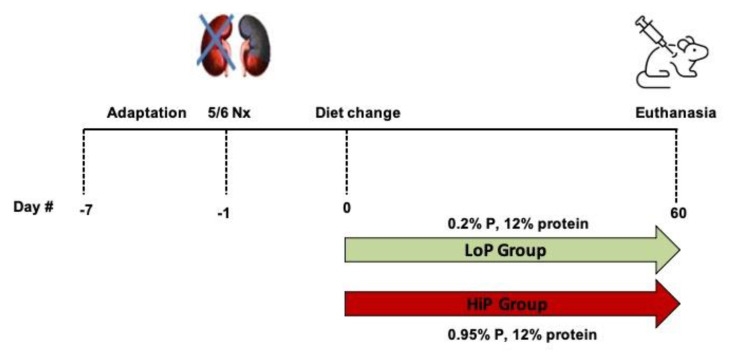
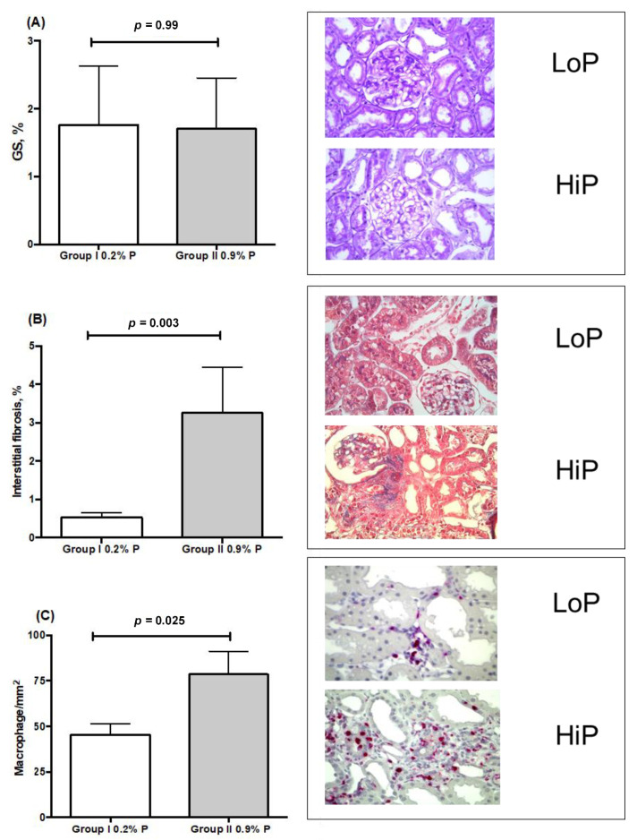
Several factors contribute to renal-function decline in CKD patients, and the role of phosphate content in the diet is still a matter of debate. This study aims to analyze the mechanism by which phosphate, independent of protein, is associated with the progression of CKD. Adult Munich-Wistar rats were submitted to 5/6 nephrectomy (Nx), fed with a low-protein diet, and divided into two groups. Only phosphate content (low phosphate, LoP, 0.2%; high phosphate, HiP, 0.95%) differentiated diets. After sixty days, biochemical parameters and kidney histology were analyzed. The HiP group presented worse renal function, with higher levels of PTH, FGF-23, and fractional excretion of phosphate. In the histological analysis of the kidney tissue, they also showed a higher percentage of interstitial fibrosis, expression of α-actin, PCNA, and renal infiltration by macrophages. The LoP group presented higher expression of beclin-1 in renal tubule cells, a marker of autophagic flux, when compared to the HiP group. Our findings highlight the action of phosphate in the induction of kidney interstitial inflammation and fibrosis, contributing to the progression of renal disease. A possible effect of phosphate on the dysregulation of the renal cell autophagy mechanism needs further investigation with clinical studies.
多种因素可导致慢性肾脏病(CKD)患者肾功能下降,而饮食中磷含量的作用仍存在争议。本研究旨在分析独立于蛋白质以外的磷与 CKD 进展之间的关联机制。成年慕尼黑-维斯塔大鼠接受 5/6 肾切除术(Nx),并给予低蛋白饮食,然后分为两组。两组饮食的唯一区别是磷含量(低磷,LoP,0.2%;高磷,HiP,0.95%)。六十天后,分析生化参数和肾脏组织学。HiP 组肾功能更差,甲状旁腺激素(PTH)、成纤维细胞生长因子 23(FGF-23)和磷的分数排泄更高。在肾脏组织的组织学分析中,HiP 组还显示出更高的间质纤维化百分比、α-肌动蛋白、增殖细胞核抗原(PCNA)的表达和巨噬细胞对肾脏的浸润。与 HiP 组相比,LoP 组肾小管细胞中自噬流标志物 beclin-1 的表达更高。我们的研究结果强调了磷在诱导肾脏间质炎症和纤维化中的作用,这有助于肾脏疾病的进展。需要进一步进行临床研究以探讨磷对肾脏细胞自噬机制失调的可能影响。