• 文献检索
  • 文档翻译
  • 深度研究
  • 学术资讯
  • Suppr Zotero 插件Zotero 插件
  • 邀请有礼
  • 套餐&价格
  • 历史记录
应用&插件
Suppr Zotero 插件Zotero 插件浏览器插件Mac 客户端Windows 客户端微信小程序
定价
高级版会员购买积分包购买API积分包
服务
文献检索文档翻译深度研究API 文档MCP 服务
关于我们
关于 Suppr公司介绍联系我们用户协议隐私条款
关注我们

Suppr 超能文献

核心技术专利:CN118964589B侵权必究
粤ICP备2023148730 号-1Suppr @ 2026

文献检索

告别复杂PubMed语法,用中文像聊天一样搜索,搜遍4000万医学文献。AI智能推荐,让科研检索更轻松。

立即免费搜索

文件翻译

保留排版,准确专业,支持PDF/Word/PPT等文件格式,支持 12+语言互译。

免费翻译文档

深度研究

AI帮你快速写综述,25分钟生成高质量综述,智能提取关键信息,辅助科研写作。

立即免费体验

B 型超声和超声造影在鉴别恶性与良性胸腔积液中的诊断准确性

Diagnostic Accuracy of B-Mode- and Contrast-Enhanced Ultrasound in Differentiating Malignant from Benign Pleural Effusions.

作者信息

Safai Zadeh Ehsan, Weide Johanna, Dietrich Christoph Frank, Trenker Corinna, Koczulla Andreas Rembert, Görg Christian

机构信息

Interdisciplinary Center of Ultrasound Diagnostics, University Hospital Giessen and Marburg, Philipps University Marburg, Baldingerstraße, 35033 Marburg, Germany.

Department Allgemeine Innere Medizin (DAIM), Kliniken Hirslanden Bern, Beau Site, Salem und Permanence, 3018 Bern, Switzerland.

出版信息

Diagnostics (Basel). 2021 Jul 19;11(7):1293. doi: 10.3390/diagnostics11071293.

DOI:10.3390/diagnostics11071293
PMID:34359376
原文链接:https://pmc.ncbi.nlm.nih.gov/articles/PMC8305637/
Abstract

PURPOSE

To evaluate the value of CEUS in differentiating malignant from benign pleural effusions (PEs).

METHODS

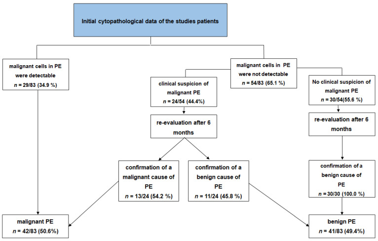
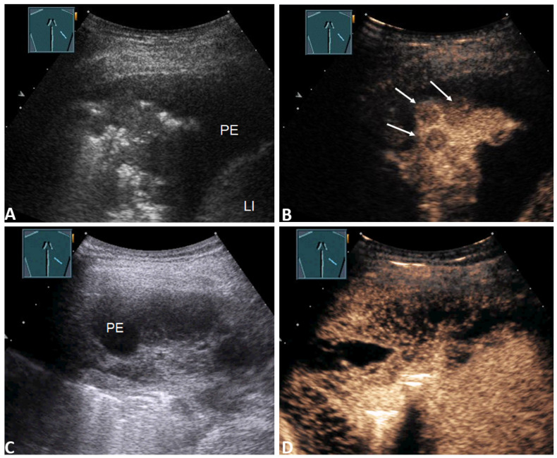
From 2008 to 2017, 83 patients with PEs of unknown cause were examined using B-mode thoracic ultrasound (B-TUS), CEUS, and cytological examination. The extent of enhancement of the pleural thickening, the presence of enhancement of septa or a solid mass within the PE, and the homogeneity of the enhancement in the associated lung consolidation, were examined. Subsequently, the diagnostic value of cytology, B-TUS, and CEUS in differentiating malignant from benign PEs was determined.

RESULTS

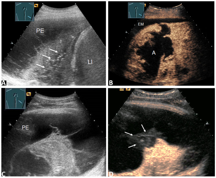
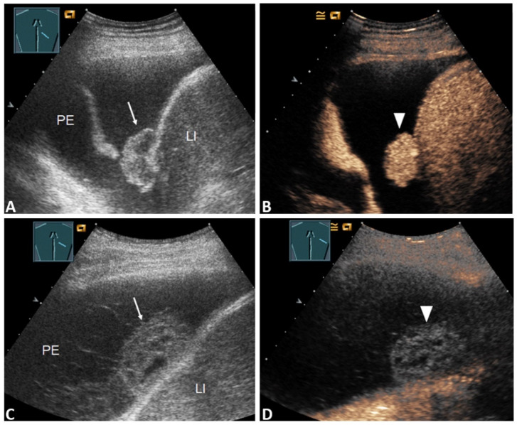
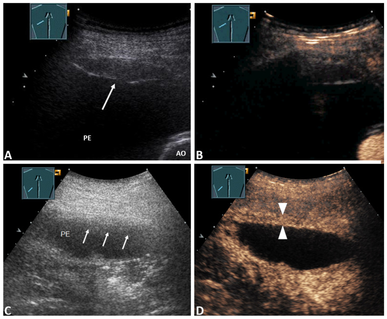
With CEUS, markedly enhanced pleural thickening and inhomogeneous enhanced lung consolidation were significantly more frequently associated with malignancy ( < 0.05). In the subgroup analysis, the use of CEUS increased the sensitivity from 69.2 to 92.3 in patients with initial negative cytology but clinical suspicion of malignant PE; it also increased the specificity from 63.0 to 90.0, the positive predictive value from 69.2 to 92.3, the negative predictive value from 63.0 to 90.0, and the diagnostic accuracy from 66.7 to 87.5, in the evaluation of PE malignancy.

CONCLUSION

The use of clinically based B-TUS and CEUS as a complementary method to cytological evaluation may be beneficial for evaluating a PE of unknown cause. CEUS patterns of enhanced pleural thickening and inhomogeneous enhanced lung consolidation may suggest a malignant PE.

摘要

目的

评估超声造影(CEUS)在鉴别恶性与良性胸腔积液(PEs)中的价值。

方法

2008年至2017年,对83例病因不明的胸腔积液患者进行了B型胸部超声(B-TUS)、CEUS及细胞学检查。观察胸膜增厚的强化程度、胸腔积液内分隔或实性肿块的强化情况以及相关肺实变强化的均匀性。随后,确定细胞学、B-TUS及CEUS在鉴别恶性与良性胸腔积液中的诊断价值。

结果

CEUS检查显示,明显强化的胸膜增厚及不均匀强化的肺实变与恶性病变显著相关(<0.05)。在亚组分析中,对于初始细胞学检查阴性但临床怀疑为恶性胸腔积液的患者,使用CEUS可使敏感性从69.2%提高至92.3%;在评估胸腔积液恶性程度时,还可使特异性从63.0%提高至90.0%,阳性预测值从69.2%提高至92.3%,阴性预测值从63.0%提高至90.0%,诊断准确性从66.7%提高至87.5%。

结论

将基于临床的B-TUS和CEUS作为细胞学评估的补充方法,可能有助于评估病因不明的胸腔积液。胸膜增厚强化及肺实变不均匀强化的CEUS表现可能提示恶性胸腔积液。

https://cdn.ncbi.nlm.nih.gov/pmc/blobs/be96/8305637/d9ce5b4da45c/diagnostics-11-01293-g005.jpg
https://cdn.ncbi.nlm.nih.gov/pmc/blobs/be96/8305637/36baeccdc00b/diagnostics-11-01293-g001.jpg
https://cdn.ncbi.nlm.nih.gov/pmc/blobs/be96/8305637/4a252a70286c/diagnostics-11-01293-g002.jpg
https://cdn.ncbi.nlm.nih.gov/pmc/blobs/be96/8305637/0e61ee06195c/diagnostics-11-01293-g003.jpg
https://cdn.ncbi.nlm.nih.gov/pmc/blobs/be96/8305637/13b5f77da3ee/diagnostics-11-01293-g004.jpg
https://cdn.ncbi.nlm.nih.gov/pmc/blobs/be96/8305637/d9ce5b4da45c/diagnostics-11-01293-g005.jpg

相似文献

1
Diagnostic Accuracy of B-Mode- and Contrast-Enhanced Ultrasound in Differentiating Malignant from Benign Pleural Effusions.B 型超声和超声造影在鉴别恶性与良性胸腔积液中的诊断准确性
Diagnostics (Basel). 2021 Jul 19;11(7):1293. doi: 10.3390/diagnostics11071293.
2
Contrast-enhanced ultrasound is helpful for differentiating benign from malignant parietal pleural lesions.增强超声有助于区分良性和恶性壁层胸膜病变。
J Clin Ultrasound. 2022 Jan;50(1):90-98. doi: 10.1002/jcu.23088. Epub 2021 Oct 19.
3
Thoracic ultrasound in the diagnosis of malignant pleural effusion.胸部超声在恶性胸腔积液诊断中的应用
Thorax. 2009 Feb;64(2):139-43. doi: 10.1136/thx.2008.100545. Epub 2008 Oct 13.
4
Evaluation of parietal pleural adhesion and invasion in subpleural lung cancer: value of B-mode ultrasound and contrast-enhanced ultrasound.胸膜下肺癌脏层胸膜粘连及侵犯的评估:B 型超声与超声造影的价值
Quant Imaging Med Surg. 2024 May 1;14(5):3302-3311. doi: 10.21037/qims-23-1542. Epub 2024 Apr 26.
5
Utility of high-frequency B-mode and contrast-enhanced ultrasound for the differential diagnosis of benign and malignant pleural diseases: a prospective study.高频B型超声与超声造影在鉴别诊断良恶性胸膜疾病中的应用:一项前瞻性研究。
J Thorac Dis. 2022 Oct;14(10):3695-3705. doi: 10.21037/jtd-22-636.
6
Detection of peripheral embolic consolidations using contrast-enhanced ultrasonography in patients with no evidence of pulmonary embolism on computed tomography: A pilot study.在计算机断层扫描未显示肺栓塞证据的患者中,使用对比增强超声检测外周栓子性实变:一项初步研究。
J Clin Ultrasound. 2017 Nov 12;45(9):575-579. doi: 10.1002/jcu.22511. Epub 2017 Jul 5.
7
Quantitative and qualitative analysis of contrast-enhanced ultrasound for differentiating benign and malignant superficial enlarged lymph nodes.超声造影定量及定性分析在鉴别浅表肿大淋巴结良恶性中的应用
Quant Imaging Med Surg. 2024 Sep 1;14(9):6362-6373. doi: 10.21037/qims-24-658. Epub 2024 Aug 16.
8
[Value of contrast-enhanced ultrasound (CEUS) in the differential diagnosis between benign and malignant renal neoplasms].[超声造影(CEUS)在肾良恶性肿瘤鉴别诊断中的价值]
Zhonghua Zhong Liu Za Zhi. 2013 May;35(5):382-5. doi: 10.3760/cma.j.issn.0253-3766.2013.05.013.
9
Contrast-enhanced ultrasound (CEUS) has excellent diagnostic accuracy in differentiating focal liver lesions: results from a Swiss tertiary gastroenterological centre.超声造影(CEUS)在鉴别局灶性肝脏病变方面具有出色的诊断准确性:来自瑞士一家三级胃肠病学中心的结果。
Swiss Med Wkly. 2019 Jun 30;149:w20087. doi: 10.4414/smw.2019.20087. eCollection 2019 Jun 17.
10
Contrast-Enhanced Ultrasound of the Pleural Cavity: A Method to Locate Pleural Catheters and Identify Fibrous Septa.胸腔增强超声:定位胸腔导管和识别纤维性间隔的一种方法。
Ultrasound Med Biol. 2021 May;47(5):1261-1268. doi: 10.1016/j.ultrasmedbio.2021.01.011. Epub 2021 Feb 1.

引用本文的文献

1
Comments and Illustrations of the European Federation of Societies for Ultrasound in Medicine (EFSUMB) Guidelines: Rare Malignant Pulmonal and Pleural Tumors: Primary Pulmonary Sarcoma and Mesothelioma, Imaging Features on Transthoracic Ultrasound.欧洲医学超声学会联合会(EFSUMB)指南评注与插图:罕见的肺和胸膜恶性肿瘤:原发性肺肉瘤和间皮瘤,经胸超声成像特征
Diagnostics (Basel). 2024 Oct 21;14(20):2339. doi: 10.3390/diagnostics14202339.
2
Ultrasound-Assisted and Ultrasound-Guided Thoracentesis: An Educational Review.超声辅助与超声引导下胸腔穿刺术:一项教育综述
Diagnostics (Basel). 2024 May 29;14(11):1124. doi: 10.3390/diagnostics14111124.
3

本文引用的文献

1
Perfusion Patterns of Peripheral Pulmonary Granulomatous Lesions Using Contrast-Enhanced Ultrasound (CEUS) and Their Correlation with Immunohistochemically Detected Vascularization Patterns.使用对比增强超声(CEUS)观察外周肺部肉芽肿性病变的灌注模式及其与免疫组化检测到的血管生成模式的相关性。
J Ultrasound Med. 2022 Mar;41(3):565-574. doi: 10.1002/jum.15730. Epub 2021 May 6.
2
Contrast-Enhanced Ultrasound for Evaluation of Pleural Effusion: A Pictorial Essay.对比增强超声在胸腔积液评估中的应用:影像学研究。
J Ultrasound Med. 2022 Feb;41(2):485-503. doi: 10.1002/jum.15705. Epub 2021 Mar 29.
3
Perfusion Patterns of Peripheral Pulmonary Lesions in COVID-19 Patients Using Contrast-Enhanced Ultrasound (CEUS): A Case Series.
The Value of Contrast-Enhanced Ultrasound (CEUS) in the Evaluation of Central Lung Cancer with Obstructive Atelectasis.
对比增强超声(CEUS)在评估伴有阻塞性肺不张的中央型肺癌中的价值
Diagnostics (Basel). 2024 May 18;14(10):1051. doi: 10.3390/diagnostics14101051.
4
Comparative Evaluation of Chest Ultrasonography and Computed Tomography as Predictors of Malignant Pleural Effusion: A Prospective Study.胸部超声检查与计算机断层扫描作为恶性胸腔积液预测指标的比较评估:一项前瞻性研究
Diagnostics (Basel). 2024 May 17;14(10):1041. doi: 10.3390/diagnostics14101041.
5
Evaluation of parietal pleural adhesion and invasion in subpleural lung cancer: value of B-mode ultrasound and contrast-enhanced ultrasound.胸膜下肺癌脏层胸膜粘连及侵犯的评估:B 型超声与超声造影的价值
Quant Imaging Med Surg. 2024 May 1;14(5):3302-3311. doi: 10.21037/qims-23-1542. Epub 2024 Apr 26.
6
B-mode ultrasound and contrast-enhanced ultrasound for evaluation of pneumonia: A pictorial essay.B超和超声造影在肺炎评估中的应用:图文综述
Australas J Ultrasound Med. 2023 Feb 15;26(2):100-114. doi: 10.1002/ajum.12332. eCollection 2023 May.
7
Utility of high-frequency B-mode and contrast-enhanced ultrasound for the differential diagnosis of benign and malignant pleural diseases: a prospective study.高频B型超声与超声造影在鉴别诊断良恶性胸膜疾病中的应用:一项前瞻性研究。
J Thorac Dis. 2022 Oct;14(10):3695-3705. doi: 10.21037/jtd-22-636.
8
Diagnostic Performance of Point Shear Wave Elastography (pSWE) Using Acoustic Radiation Force Impulse (ARFI) Technology in Mesenteric Masses: A Feasibility Study.使用声辐射力脉冲(ARFI)技术的点剪切波弹性成像(pSWE)在肠系膜肿物中的诊断性能:一项可行性研究
Diagnostics (Basel). 2022 Feb 18;12(2):523. doi: 10.3390/diagnostics12020523.
9
Perfusion Patterns of Peripheral Organizing Pneumonia (POP) Using Contrast-Enhanced Ultrasound (CEUS) and Their Correlation with Immunohistochemically Detected Vascularization Patterns.使用超声造影(CEUS)评估外周型机化性肺炎(POP)的灌注模式及其与免疫组化检测的血管生成模式的相关性
Diagnostics (Basel). 2021 Sep 2;11(9):1601. doi: 10.3390/diagnostics11091601.
10
The Value of Lung Ultrasound to Detect the Early Pleural and Pulmonary Pathologies in Nonhospitalized COVID-19-Suspected Cases in a Population With a Low Prevalence of COVID-19 Infection: A Prospective Study in 297 Subjects.对 COVID-19 感染率低人群中疑似 COVID-19 的非住院患者进行肺部超声检查以早期发现胸膜和肺部病变的价值:297 例患者的前瞻性研究。
J Ultrasound Med. 2022 Jun;41(6):1397-1403. doi: 10.1002/jum.15822. Epub 2021 Sep 4.
COVID-19 患者肺部周边病变的超声造影灌注模式:病例系列研究。
J Ultrasound Med. 2021 Nov;40(11):2403-2411. doi: 10.1002/jum.15624. Epub 2021 Jan 18.
4
Thoracic ultrasound for malignant pleural effusion: a systematic review and meta-analysis.用于恶性胸腔积液的胸部超声检查:一项系统评价和荟萃分析。
ERJ Open Res. 2020 Nov 10;6(4). doi: 10.1183/23120541.00464-2020. eCollection 2020 Oct.
5
Diagnostic Accuracy of Thoracic Ultrasonography to Differentiate Transudative From Exudative Pleural Effusion.胸腔超声对胸水中渗出液与漏出液的鉴别诊断准确性。
Chest. 2020 Aug;158(2):692-697. doi: 10.1016/j.chest.2020.02.051. Epub 2020 Mar 17.
6
Ultrasound-Guided Biopsy of Pleural-Based Pulmonary Lesions by Injection of Contrast-Enhancing Drugs.通过注射造影增强药物对胸膜下肺病变进行超声引导活检。
Front Pharmacol. 2019 Sep 3;10:960. doi: 10.3389/fphar.2019.00960. eCollection 2019.
7
Histological validation of pulmonary infarction detected with contrast-enhanced ultrasound in patients with negative computed tomography pulmonary angiogram: A case series.计算机断层扫描肺动脉造影阴性患者中经对比增强超声检测到的肺梗死的组织学验证:病例系列
J Clin Ultrasound. 2019 Oct;47(8):461-465. doi: 10.1002/jcu.22755. Epub 2019 Jul 22.
8
Pleural Effusion in Adults-Etiology, Diagnosis, and Treatment.成人胸腔积液:病因、诊断与治疗。
Dtsch Arztebl Int. 2019 May 24;116(21):377-386. doi: 10.3238/arztebl.2019.0377.
9
Pleural thickening on screening chest X-rays: a single institutional study.筛查性胸部 X 光片的胸膜增厚:单机构研究。
Respir Res. 2019 Jul 5;20(1):138. doi: 10.1186/s12931-019-1116-9.
10
Diagnostic yield of pleural fluid cytology in malignant effusions: an Australian tertiary centre experience.恶性胸腔积液中胸腔积液细胞学检查的诊断率:澳大利亚一家三级中心的经验
Intern Med J. 2018 Nov;48(11):1318-1324. doi: 10.1111/imj.13991.