• 文献检索
  • 文档翻译
  • 深度研究
  • 学术资讯
  • Suppr Zotero 插件Zotero 插件
  • 邀请有礼
  • 套餐&价格
  • 历史记录
应用&插件
Suppr Zotero 插件Zotero 插件浏览器插件Mac 客户端Windows 客户端微信小程序
定价
高级版会员购买积分包购买API积分包
服务
文献检索文档翻译深度研究API 文档MCP 服务
关于我们
关于 Suppr公司介绍联系我们用户协议隐私条款
关注我们

Suppr 超能文献

核心技术专利:CN118964589B侵权必究
粤ICP备2023148730 号-1Suppr @ 2026

文献检索

告别复杂PubMed语法,用中文像聊天一样搜索,搜遍4000万医学文献。AI智能推荐,让科研检索更轻松。

立即免费搜索

文件翻译

保留排版,准确专业,支持PDF/Word/PPT等文件格式,支持 12+语言互译。

免费翻译文档

深度研究

AI帮你快速写综述,25分钟生成高质量综述,智能提取关键信息,辅助科研写作。

立即免费体验

硒纳米粒子对不同人癌细胞系细胞毒性作用的机制。

Mechanisms of the Cytotoxic Effect of Selenium Nanoparticles in Different Human Cancer Cell Lines.

机构信息

Federal Research Center "Pushchino Scientific Center for Biological Research of the Russian Academy of Sciences", Institute of Cell Biophysics of the Russian Academy of Sciences, 3 Institutskaya St., 142290 Pushchino, Russia.

Prokhorov General Physics Institute of the Russian Academy of Sciences, 38 Vavilove St., 119991 Moscow, Russia.

出版信息

Int J Mol Sci. 2021 Jul 21;22(15):7798. doi: 10.3390/ijms22157798.

DOI:10.3390/ijms22157798
PMID:34360564
原文链接:https://pmc.ncbi.nlm.nih.gov/articles/PMC8346078/
Abstract

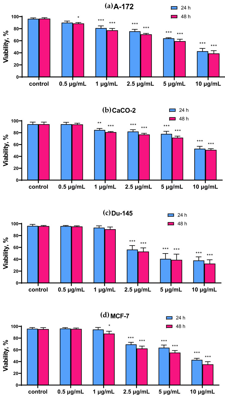
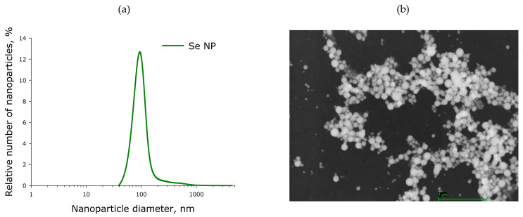
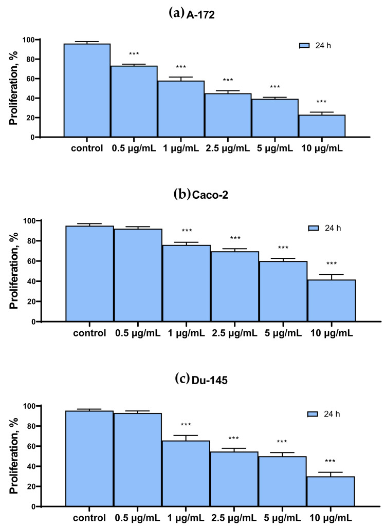
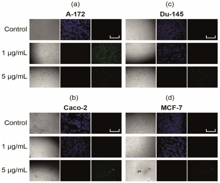
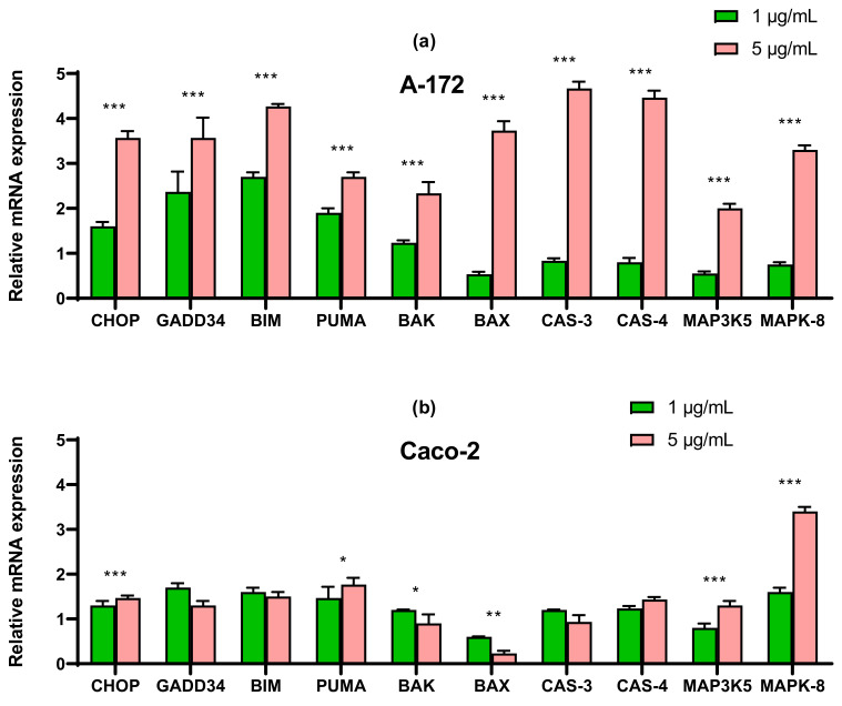
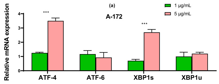
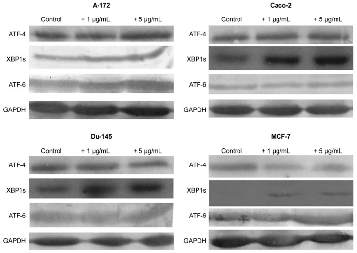
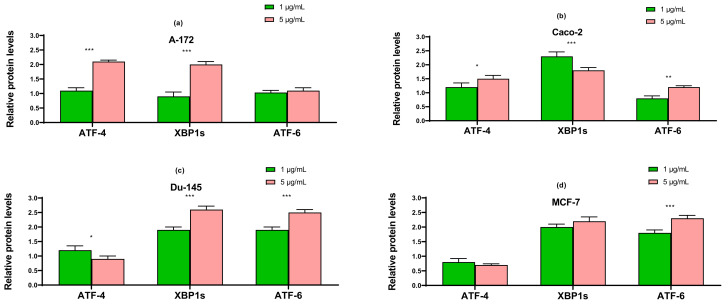
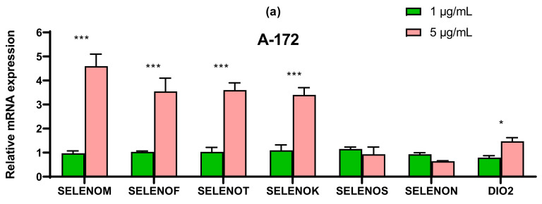
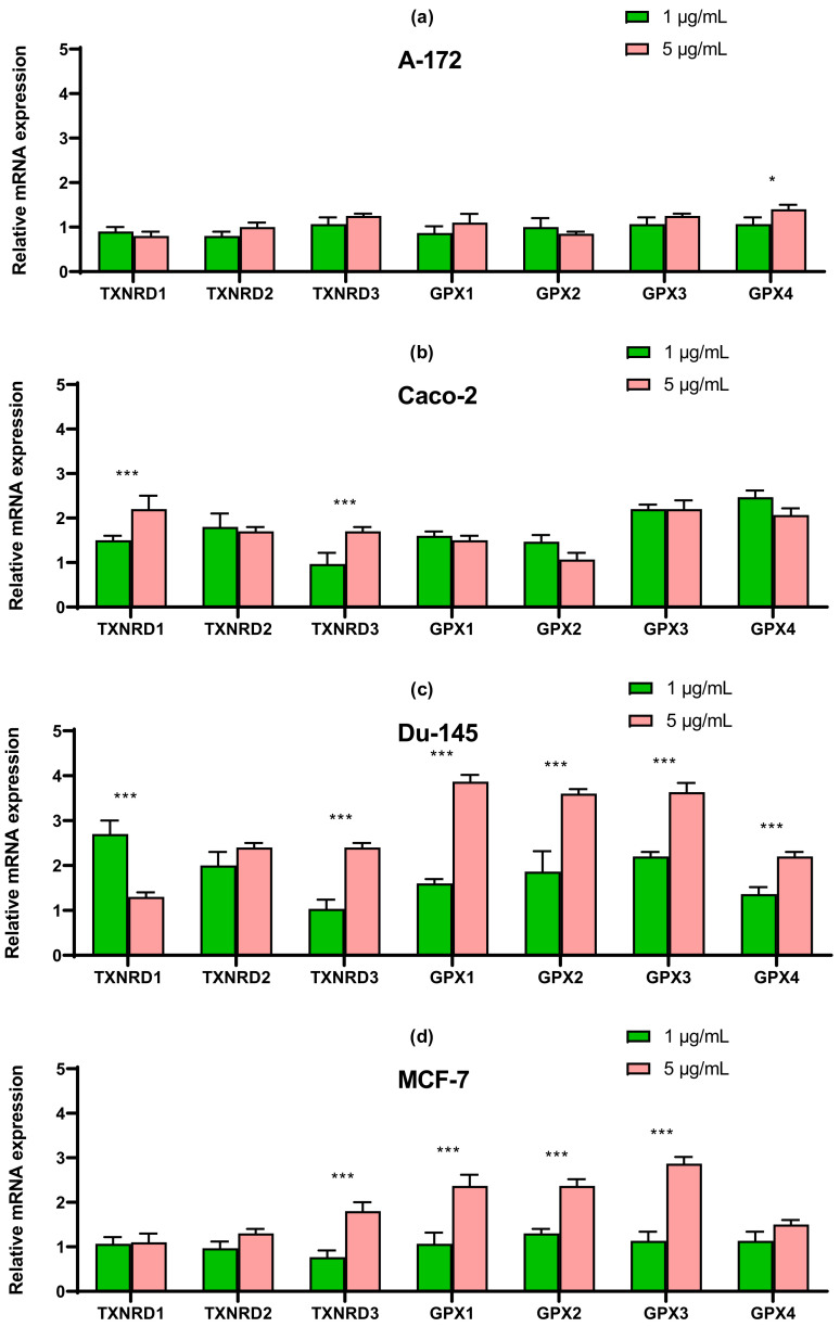
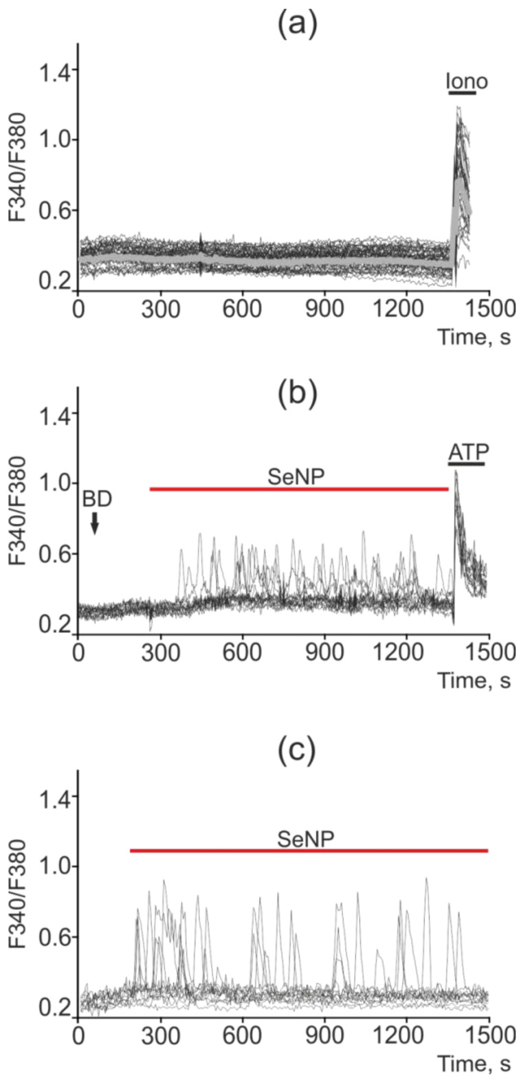
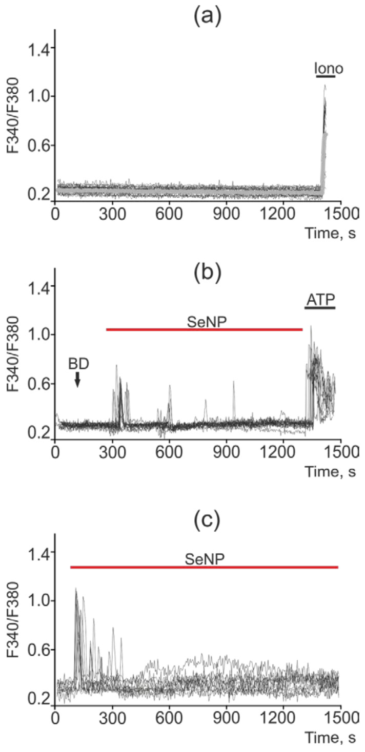
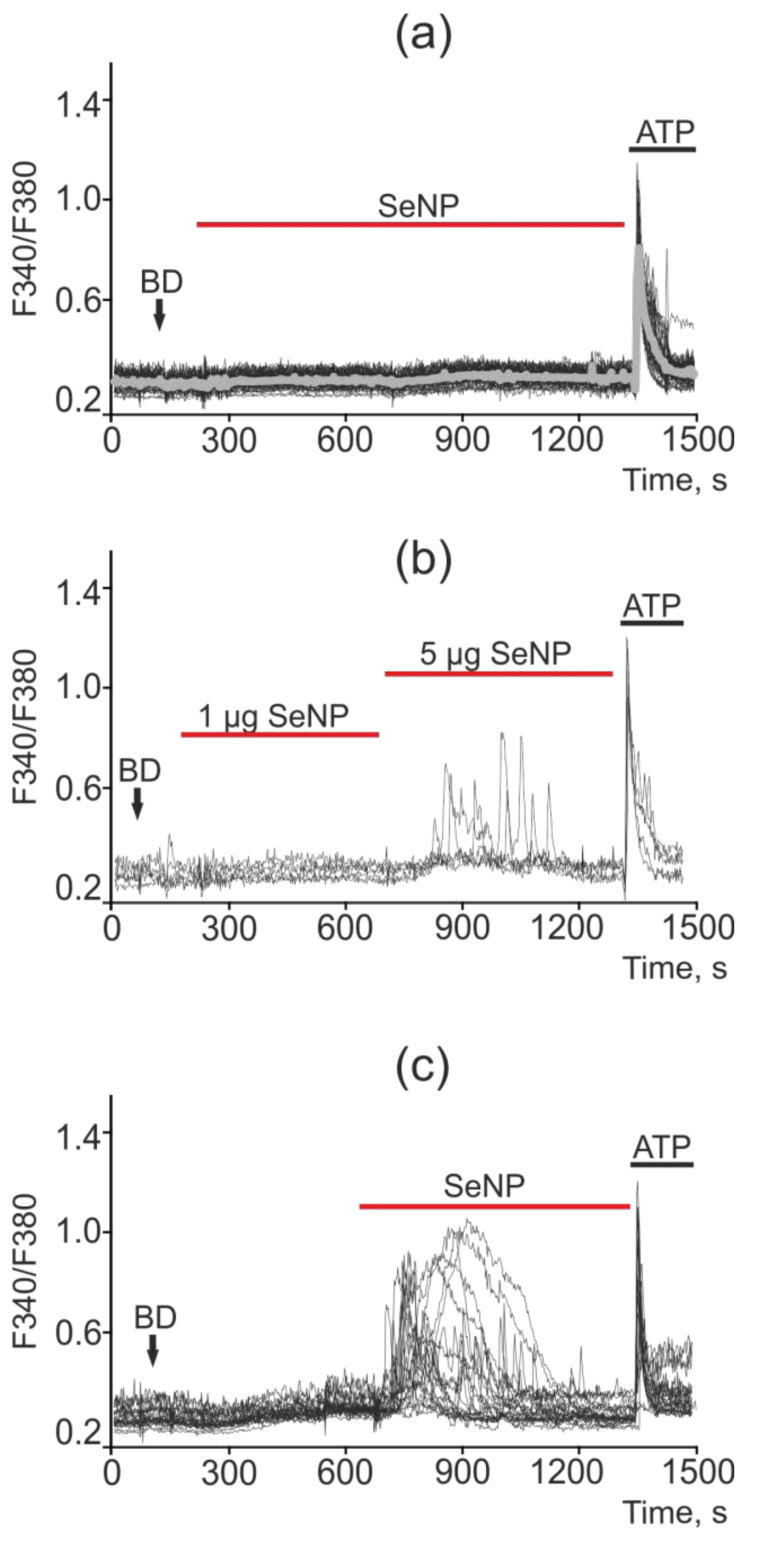
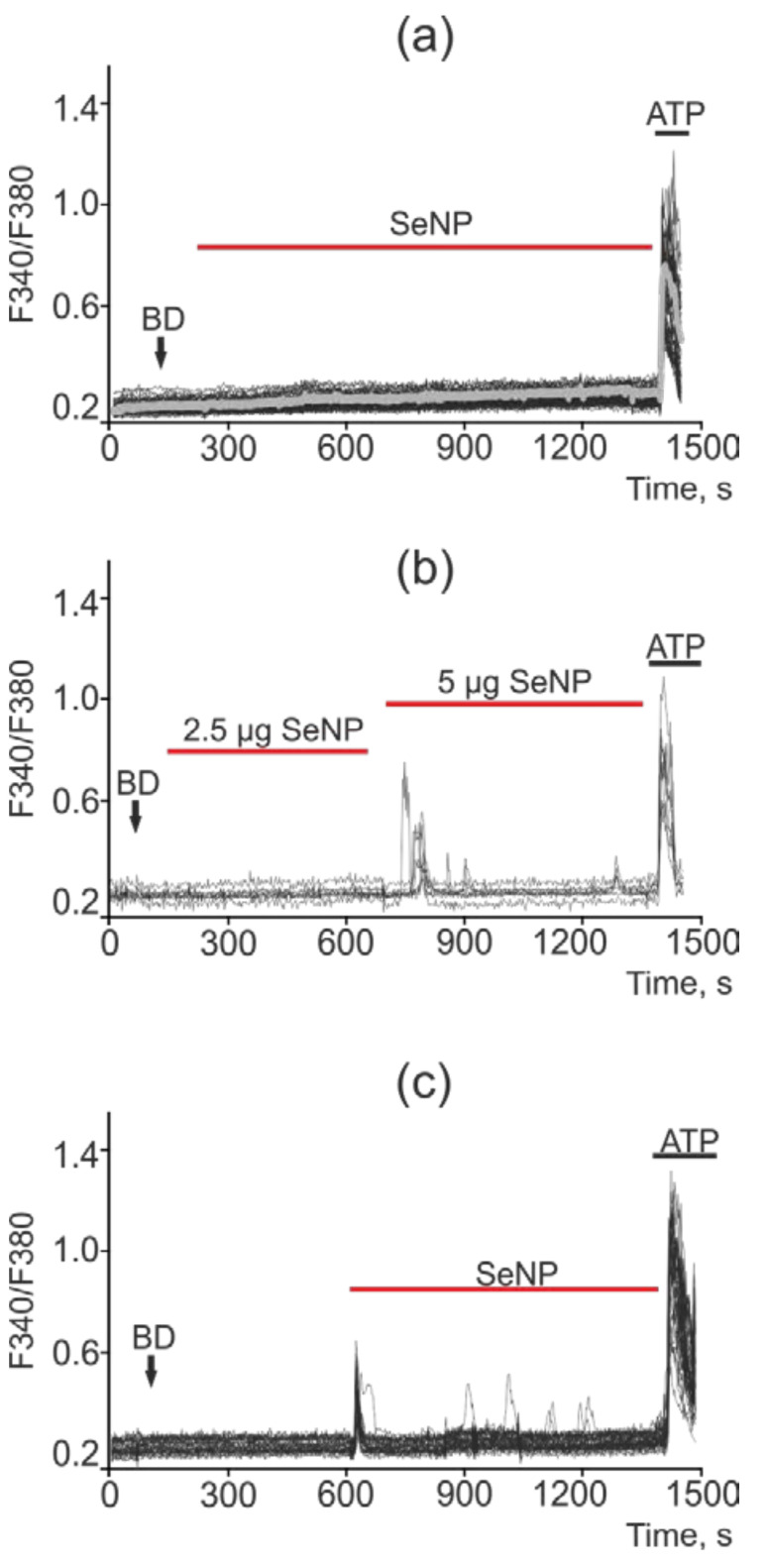
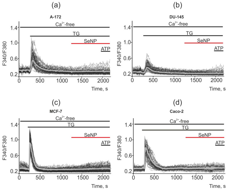
In recent decades, studies on the functional features of Se nanoparticles (SeNP) have gained great popularity due to their high biocompatibility, stability, and pronounced selectivity. A large number of works prove the anticarcinogenic effect of SeNP. In this work, the molecular mechanisms regulating the cytotoxic effects of SeNP, obtained by laser ablation, were studied by the example of four human cancer cell lines: A-172 (glioblastoma), Caco-2, (colorectal adenocarcinoma), DU-145 (prostate carcinoma), MCF-7 (breast adenocarcinoma). It was found that SeNP had different concentration-dependent effects on cancer cells of the four studied human lines. SeNP at concentrations of less than 1 μg/mL had no cytotoxic effect on the studied cancer cells, with the exception of the A-172 cell line, for which 0.5 μg/mL SeNP was the minimum concentration affecting its metabolic activity. It was shown that SeNP concentration-dependently caused cancer cell apoptosis, but not necrosis. In addition, it was found that SeNP enhanced the expression of pro-apoptotic genes in almost all cancer cell lines, with the exception of Caco-2 and activated various pathways of adaptive and pro-apoptotic signaling pathways of UPR. Different effects of SeNP on the expression of ER-resident selenoproteins and selenium-containing glutathione peroxidases and thioredoxin reductases, depending on the cell line, were established. In addition, SeNP triggered Ca signals in all investigated cancer cell lines. Different sensitivity of cancer cell lines to SeNP can determine the induction of the process of apoptosis in them through regulation of the Ca signaling system, mechanisms of ER stress, and activation of various expression patterns of genes encoding pro-apoptotic proteins.

摘要

近几十年来,由于硒纳米颗粒(SeNP)具有高生物相容性、稳定性和显著的选择性,因此对其功能特征的研究备受关注。大量研究证明了 SeNP 的抗癌作用。在这项工作中,通过激光烧蚀获得的 SeNP 的细胞毒性作用的调控分子机制,以四种人类癌细胞系:A-172(胶质母细胞瘤)、Caco-2(结肠直肠腺癌)、DU-145(前列腺癌)和 MCF-7(乳腺腺癌)为例进行了研究。结果发现,SeNP 对四种研究人类细胞系的癌细胞具有不同浓度依赖性的影响。SeNP 的浓度低于 1μg/mL 时,对研究的癌细胞没有细胞毒性作用,除了 A-172 细胞系,其 0.5μg/mL 的 SeNP 是影响其代谢活性的最小浓度。结果表明,SeNP 浓度依赖性地诱导癌细胞凋亡,但不诱导坏死。此外,还发现 SeNP 增强了几乎所有癌细胞系中促凋亡基因的表达,除了 Caco-2 细胞系,并激活了 UPR 的适应性和促凋亡信号通路的各种途径。还建立了 SeNP 对 ER 驻留硒蛋白和含硒谷胱甘肽过氧化物酶和硫氧还蛋白还原酶的表达的依赖性浓度效应,这取决于细胞系。此外,SeNP 在所有研究的癌细胞系中触发了 Ca 信号。癌细胞系对 SeNP 的不同敏感性可以通过调节 Ca 信号系统、内质网应激机制和激活编码促凋亡蛋白的各种基因表达模式来确定它们中凋亡过程的诱导。

https://cdn.ncbi.nlm.nih.gov/pmc/blobs/1afa/8346078/5adc832e6966/ijms-22-07798-g015.jpg
https://cdn.ncbi.nlm.nih.gov/pmc/blobs/1afa/8346078/8131cd6f427a/ijms-22-07798-g001a.jpg
https://cdn.ncbi.nlm.nih.gov/pmc/blobs/1afa/8346078/49082609a942/ijms-22-07798-g002a.jpg
https://cdn.ncbi.nlm.nih.gov/pmc/blobs/1afa/8346078/d41b7c6f2686/ijms-22-07798-g003.jpg
https://cdn.ncbi.nlm.nih.gov/pmc/blobs/1afa/8346078/d51c427a4ecd/ijms-22-07798-g004a.jpg
https://cdn.ncbi.nlm.nih.gov/pmc/blobs/1afa/8346078/8d146dd50c2d/ijms-22-07798-g005a.jpg
https://cdn.ncbi.nlm.nih.gov/pmc/blobs/1afa/8346078/0b977d02dd83/ijms-22-07798-g006.jpg
https://cdn.ncbi.nlm.nih.gov/pmc/blobs/1afa/8346078/6418d7bc9710/ijms-22-07798-g007.jpg
https://cdn.ncbi.nlm.nih.gov/pmc/blobs/1afa/8346078/3464b8c060e9/ijms-22-07798-g008a.jpg
https://cdn.ncbi.nlm.nih.gov/pmc/blobs/1afa/8346078/7a709d3dfce5/ijms-22-07798-g009.jpg
https://cdn.ncbi.nlm.nih.gov/pmc/blobs/1afa/8346078/05c658f53897/ijms-22-07798-g010.jpg
https://cdn.ncbi.nlm.nih.gov/pmc/blobs/1afa/8346078/1d5eecdf5a79/ijms-22-07798-g011.jpg
https://cdn.ncbi.nlm.nih.gov/pmc/blobs/1afa/8346078/efeb3aff026a/ijms-22-07798-g012.jpg
https://cdn.ncbi.nlm.nih.gov/pmc/blobs/1afa/8346078/d9ddacbecd3b/ijms-22-07798-g013.jpg
https://cdn.ncbi.nlm.nih.gov/pmc/blobs/1afa/8346078/5c837b512725/ijms-22-07798-g014.jpg
https://cdn.ncbi.nlm.nih.gov/pmc/blobs/1afa/8346078/5adc832e6966/ijms-22-07798-g015.jpg

相似文献

1
Mechanisms of the Cytotoxic Effect of Selenium Nanoparticles in Different Human Cancer Cell Lines.硒纳米粒子对不同人癌细胞系细胞毒性作用的机制。
Int J Mol Sci. 2021 Jul 21;22(15):7798. doi: 10.3390/ijms22157798.
2
Expression of ER-resident selenoproteins and activation of cancer cells apoptosis mechanisms under ER-stress conditions caused by methylseleninic acid.在甲基硒酸引起的内质网应激条件下,内质网驻留硒蛋白的表达和癌细胞凋亡机制的激活。
Gene. 2020 Sep 10;755:144884. doi: 10.1016/j.gene.2020.144884. Epub 2020 Jun 18.
3
Selenium nanoparticles as a carrier of 5-fluorouracil to achieve anticancer synergism.硒纳米颗粒作为 5-氟尿嘧啶的载体实现抗癌协同作用。
ACS Nano. 2012 Aug 28;6(8):6578-91. doi: 10.1021/nn202452c. Epub 2012 Jul 26.
4
Mechanism of Ca-Dependent Pro-Apoptotic Action of Selenium Nanoparticles, Mediated by Activation of Cx43 Hemichannels.由Cx43半通道激活介导的硒纳米颗粒钙依赖性促凋亡作用机制
Biology (Basel). 2021 Aug 3;10(8):743. doi: 10.3390/biology10080743.
5
A dandelion polysaccharide and its selenium nanoparticles: Structure features and evaluation of anti-tumor activity in zebrafish models.蒲公英多糖及其硒纳米粒子:结构特征及在斑马鱼模型中抗肿瘤活性的评价。
Carbohydr Polym. 2021 Oct 15;270:118365. doi: 10.1016/j.carbpol.2021.118365. Epub 2021 Jun 22.
6
Impact of nano-selenium on nuclear maturation and genes expression profile of buffalo oocytes matured in vitro.纳米硒对水牛卵母细胞体外成熟和基因表达谱的影响。
Mol Biol Rep. 2020 Nov;47(11):8593-8603. doi: 10.1007/s11033-020-05902-9. Epub 2020 Oct 17.
7
Novel walnut peptide-selenium hybrids with enhanced anticancer synergism: facile synthesis and mechanistic investigation of anticancer activity.具有增强抗癌协同作用的新型核桃肽-硒杂化物:简便合成及抗癌活性机制研究
Int J Nanomedicine. 2016 Apr 11;11:1305-21. doi: 10.2147/IJN.S92257. eCollection 2016.
8
Selenium Nanoparticles Synthesized Using (MH191156) Show Antiproliferative and Anti-angiogenic Activity Against Cervical Cancer Cells.使用(MH191156)合成的硒纳米粒子对宫颈癌细胞显示出抗增殖和抗血管生成活性。
Int J Nanomedicine. 2020 Jun 23;15:4523-4540. doi: 10.2147/IJN.S247426. eCollection 2020.
9
Involvement of TRPM2 Channel on Hypoxia-Induced Oxidative Injury, Inflammation, and Cell Death in Retinal Pigment Epithelial Cells: Modulator Action of Selenium Nanoparticles.TRPM2 通道在缺氧诱导的氧化损伤、炎症和视网膜色素上皮细胞死亡中的作用:硒纳米粒子的调节剂作用。
Biol Trace Elem Res. 2021 Apr;199(4):1356-1369. doi: 10.1007/s12011-020-02556-3. Epub 2021 Jan 2.
10
Comparative Analysis of the Cytotoxic Effect of a Complex of Selenium Nanoparticles Doped with Sorafenib, "Naked" Selenium Nanoparticles, and Sorafenib on Human Hepatocyte Carcinoma HepG2 Cells.硒纳米粒子掺杂索拉非尼复合物、“裸”硒纳米粒子和索拉非尼对人肝癌 HepG2 细胞的细胞毒性作用比较分析。
Int J Mol Sci. 2022 Jun 14;23(12):6641. doi: 10.3390/ijms23126641.

引用本文的文献

1
Is There Any Correlation Between Green Synthesis Parameters and the Properties of Obtained Selenium Nanoparticles?绿色合成参数与所得硒纳米颗粒的性质之间是否存在相关性?
Molecules. 2025 Jul 5;30(13):2865. doi: 10.3390/molecules30132865.
2
Targeted nanodelivery systems for personalized cancer therapy.用于个性化癌症治疗的靶向纳米递送系统。
Rep Pract Oncol Radiother. 2025 Feb 19;29(6):776-788. doi: 10.5603/rpor.103524. eCollection 2024.
3
Bee Pollen as a Source of Biopharmaceuticals for Neurodegeneration and Cancer Research: A Scoping Review and Translational Prospects.

本文引用的文献

1
Production and Use of Selenium Nanoparticles as Fertilizers.作为肥料的硒纳米颗粒的生产与应用
ACS Omega. 2020 Jul 10;5(28):17767-17774. doi: 10.1021/acsomega.0c02448. eCollection 2020 Jul 21.
2
Expression of ER-resident selenoproteins and activation of cancer cells apoptosis mechanisms under ER-stress conditions caused by methylseleninic acid.在甲基硒酸引起的内质网应激条件下,内质网驻留硒蛋白的表达和癌细胞凋亡机制的激活。
Gene. 2020 Sep 10;755:144884. doi: 10.1016/j.gene.2020.144884. Epub 2020 Jun 18.
3
Design and Characterization of a Cancer-Targeted Drug Co-Delivery System Composed of Liposomes and Selenium Nanoparticles.
蜂花粉作为神经退行性疾病和癌症研究的生物制药来源:一项范围综述及转化前景
Molecules. 2024 Dec 13;29(24):5893. doi: 10.3390/molecules29245893.
4
Antimicrobial Feature of Nanoparticles in the Antibiotic Resistance Era: From Mechanism to Application.抗生素耐药时代纳米颗粒的抗菌特性:从作用机制到应用
Adv Biomed Res. 2024 Nov 30;13:113. doi: 10.4103/abr.abr_92_24. eCollection 2024.
5
In vivo evaluation of selenium-tellurium based nanoparticles as a novel treatment for bovine mastitis.基于硒碲的纳米颗粒作为牛乳腺炎新疗法的体内评估
J Anim Sci Biotechnol. 2024 Dec 20;15(1):173. doi: 10.1186/s40104-024-01128-y.
6
The Immunomodulatory Effects of Selenium: A Journey from the Environment to the Human Immune System.硒的免疫调节作用:从环境到人体免疫系统的探索。
Nutrients. 2024 Sep 30;16(19):3324. doi: 10.3390/nu16193324.
7
Current Non-Metal Nanoparticle-Based Therapeutic Approaches for Glioblastoma Treatment.当前基于非金属纳米颗粒的胶质母细胞瘤治疗方法
Biomedicines. 2024 Aug 11;12(8):1822. doi: 10.3390/biomedicines12081822.
8
Laser fragmentation of amorphous and crystalline selenium of various morphologies and assessment of their antioxidant and protection properties.不同形态的非晶态和晶态硒的激光破碎及其抗氧化和保护性能评估。
Front Chem. 2024 Aug 9;12:1459477. doi: 10.3389/fchem.2024.1459477. eCollection 2024.
9
Exploring the Link between Oxidative Stress, Selenium Levels, and Obesity in Youth.探讨氧化应激、硒水平与青少年肥胖的关系。
Int J Mol Sci. 2024 Jul 2;25(13):7276. doi: 10.3390/ijms25137276.
10
Molecular Mechanisms of the Therapeutic Effect of Selenium Nanoparticles in Hepatocellular Carcinoma.硒纳米粒子治疗肝细胞癌的分子机制。
Cells. 2024 Jun 26;13(13):1102. doi: 10.3390/cells13131102.
设计并表征一种由脂质体和硒纳米粒子组成的癌症靶向药物共递系统。
J Nanosci Nanotechnol. 2020 Sep 1;20(9):5295-5304. doi: 10.1166/jnn.2020.17882.
4
Nucleolin-targeted selenium nanocomposites with enhanced theranostic efficacy to antagonize glioblastoma.具有增强治疗诊断功效以对抗胶质母细胞瘤的核仁素靶向硒纳米复合材料。
J Mater Chem B. 2017 Apr 28;5(16):3024-3034. doi: 10.1039/c6tb03365b. Epub 2017 Apr 6.
5
Functionalized selenium nanoparticles for targeted siRNA delivery silence Derlin1 and promote antitumor efficacy against cervical cancer.功能化硒纳米颗粒用于靶向 siRNA 递送沉默 Derlin1 并增强宫颈癌的抗肿瘤疗效。
Drug Deliv. 2020 Dec;27(1):15-25. doi: 10.1080/10717544.2019.1667452.
6
Antitumor action of diphenyl diselenide nanocapsules: In vitro assessments and preclinical evidence in an animal model of glioblastoma multiforme.二苯二硒纳米胶囊的抗肿瘤作用:在多形性胶质母细胞瘤动物模型中的体外评估和临床前证据。
J Trace Elem Med Biol. 2019 Sep;55:180-189. doi: 10.1016/j.jtemb.2019.06.010. Epub 2019 Jul 5.
7
Methylseleninic Acid Induces Lipid Peroxidation and Radiation Sensitivity in Head and Neck Cancer Cells.甲基硒酸诱导头颈部癌细胞的脂质过氧化和辐射敏感性。
Int J Mol Sci. 2019 Jan 8;20(1):225. doi: 10.3390/ijms20010225.
8
Therapeutic applications of selenium nanoparticles.硒纳米粒子的治疗应用。
Biomed Pharmacother. 2019 Mar;111:802-812. doi: 10.1016/j.biopha.2018.12.146. Epub 2019 Jan 4.
9
Protective and adaptogenic role of peroxiredoxin 2 (Prx2) in neutralization of oxidative stress induced by ionizing radiation.过氧化物还原酶 2(Prx2)在中和电离辐射诱导的氧化应激中的保护和适应作用。
Free Radic Biol Med. 2019 Apr;134:76-86. doi: 10.1016/j.freeradbiomed.2018.12.032. Epub 2018 Dec 31.
10
Methylseleninic Acid Suppresses Breast Cancer Growth via the JAK2/STAT3 Pathway.甲基亚硒酸通过JAK2/STAT3信号通路抑制乳腺癌生长。
Reprod Sci. 2019 Jun;26(6):829-838. doi: 10.1177/1933719118815582. Epub 2018 Dec 9.