• 文献检索
  • 文档翻译
  • 深度研究
  • 学术资讯
  • Suppr Zotero 插件Zotero 插件
  • 邀请有礼
  • 套餐&价格
  • 历史记录
应用&插件
Suppr Zotero 插件Zotero 插件浏览器插件Mac 客户端Windows 客户端微信小程序
定价
高级版会员购买积分包购买API积分包
服务
文献检索文档翻译深度研究API 文档MCP 服务
关于我们
关于 Suppr公司介绍联系我们用户协议隐私条款
关注我们

Suppr 超能文献

核心技术专利:CN118964589B侵权必究
粤ICP备2023148730 号-1Suppr @ 2026

文献检索

告别复杂PubMed语法,用中文像聊天一样搜索,搜遍4000万医学文献。AI智能推荐,让科研检索更轻松。

立即免费搜索

文件翻译

保留排版,准确专业,支持PDF/Word/PPT等文件格式,支持 12+语言互译。

免费翻译文档

深度研究

AI帮你快速写综述,25分钟生成高质量综述,智能提取关键信息,辅助科研写作。

立即免费体验

线粒体功能障碍和线粒体通透性转换孔 (mPTP) 的改变导致特发性肺纤维化成纤维细胞的抗凋亡。

Mitochondrial Dysfunction and Alterations in Mitochondrial Permeability Transition Pore (mPTP) Contribute to Apoptosis Resistance in Idiopathic Pulmonary Fibrosis Fibroblasts.

机构信息

Laboratorio de Biología Celular, Departamento de Fibrosis Pulmonar, Instituto Nacional de Enfermedades Respiratorias "Ismael Cosío Villegas", Ciudad de México 14080, Mexico.

Laboratorio de Investigación en Enfermedades Reumáticas, Instituto Nacional de Enfermedades Respiratorias "Ismael Cosío Villegas", Ciudad de México 14080, Mexico.

出版信息

Int J Mol Sci. 2021 Jul 23;22(15):7870. doi: 10.3390/ijms22157870.

DOI:10.3390/ijms22157870
PMID:34360637
原文链接:https://pmc.ncbi.nlm.nih.gov/articles/PMC8346102/
Abstract

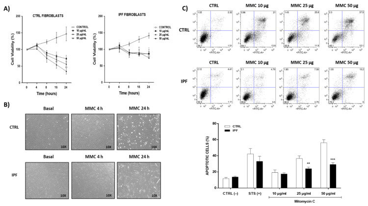
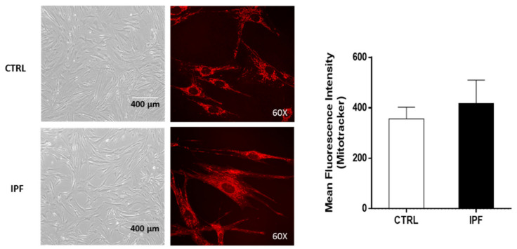
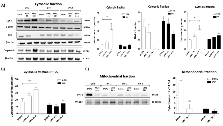
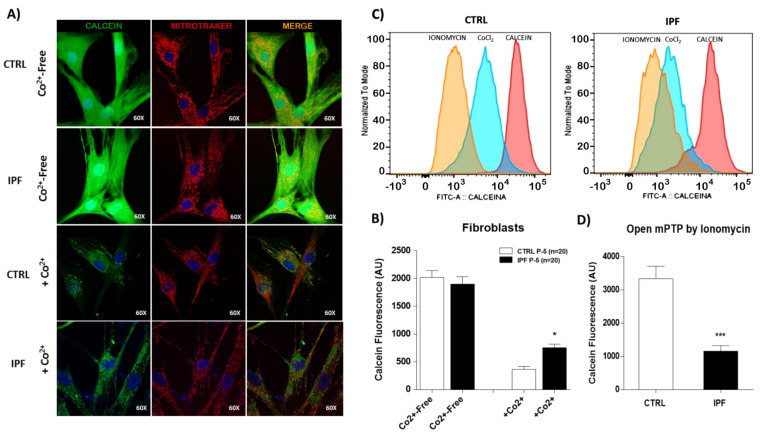
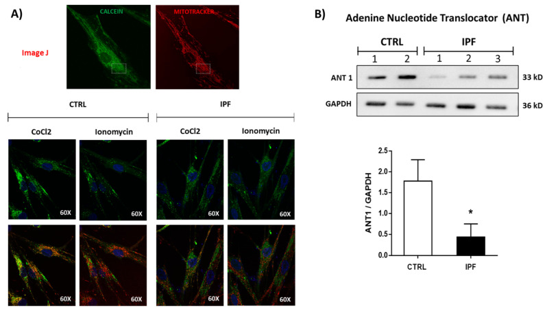
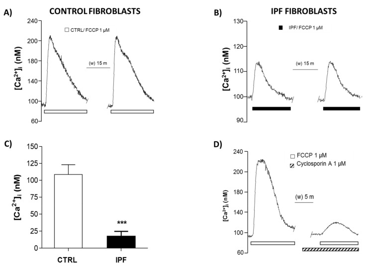
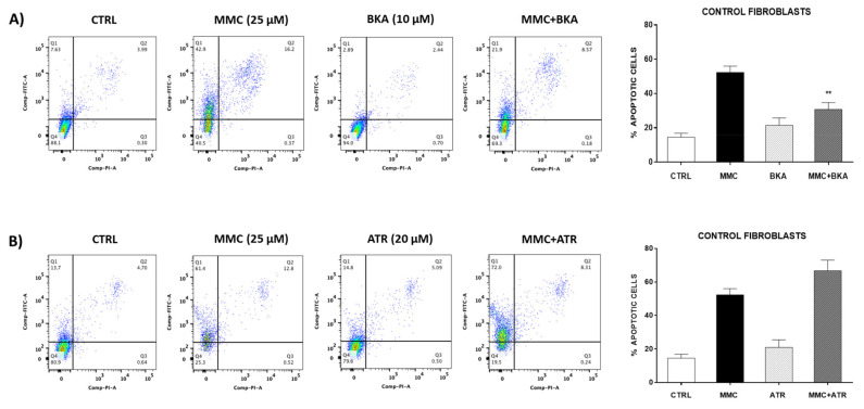
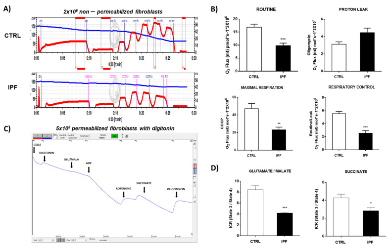
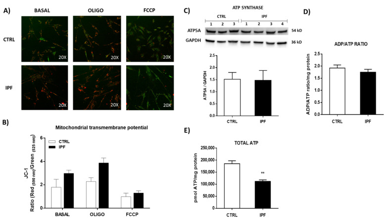
Idiopathic pulmonary fibrosis (IPF) is a devastating disease characterized by increased activation of fibroblasts/myofibroblasts. Previous reports have shown that IPF fibroblasts are resistant to apoptosis, but the mechanisms remain unclear. Since inhibition of the mitochondrial permeability transition pore (mPTP) has been implicated in the resistance to apoptosis, in this study, we analyzed the role of mitochondrial function and the mPTP on the apoptosis resistance of IPF fibroblasts under basal conditions and after mitomycin C-induced apoptosis. We measured the release of cytochrome c, mPTP opening, mitochondrial calcium release, oxygen consumption, mitochondrial membrane potential, ADP/ATP ratio, ATP concentration, and mitochondrial morphology. We found that IPF fibroblasts were resistant to mitomycin C-induced apoptosis and that calcium, a well-established activator of mPTP, is decreased as well as the release of pro-apoptotic proteins such as cytochrome c. Likewise, IPF fibroblasts showed decreased mitochondrial function, while mPTP was less sensitive to ionomycin-induced opening. Although IPF fibroblasts did not present changes in the mitochondrial membrane potential, we found a fragmented mitochondrial network with scarce, thinned, and disordered mitochondria with reduced ATP levels. Our findings demonstrate that IPF fibroblasts are resistant to mitomycin C-induced apoptosis and that altered mPTP opening contributes to this resistance. In addition, IPF fibroblasts show mitochondrial dysfunction evidenced by a decrease in respiratory parameters.

摘要

特发性肺纤维化(IPF)是一种破坏性疾病,其特征在于成纤维细胞/肌成纤维细胞的过度激活。先前的报告表明,IPF 成纤维细胞对细胞凋亡具有抗性,但机制尚不清楚。由于抑制线粒体通透性转换孔(mPTP)与细胞凋亡的抗性有关,因此在这项研究中,我们分析了在基础条件下和丝裂霉素 C 诱导细胞凋亡后,线粒体功能和 mPTP 对 IPF 成纤维细胞抗凋亡的作用。我们测量了细胞色素 c 的释放、mPTP 的开放、线粒体钙释放、耗氧量、线粒体膜电位、ADP/ATP 比、ATP 浓度和线粒体形态。我们发现,IPF 成纤维细胞对丝裂霉素 C 诱导的细胞凋亡具有抗性,钙作为 mPTP 的一种已确立的激活剂,其释放以及细胞色素 c 等促凋亡蛋白的释放减少。同样,IPF 成纤维细胞表现出线粒体功能降低,而 mPTP 对离子霉素诱导的开放不太敏感。尽管 IPF 成纤维细胞的线粒体膜电位没有变化,但我们发现线粒体网络呈碎片化,线粒体稀少、变薄且排列紊乱,ATP 水平降低。我们的研究结果表明,IPF 成纤维细胞对丝裂霉素 C 诱导的细胞凋亡具有抗性,并且改变的 mPTP 开放有助于这种抗性。此外,IPF 成纤维细胞表现出线粒体功能障碍的证据,表现为呼吸参数下降。

https://cdn.ncbi.nlm.nih.gov/pmc/blobs/40ca/8346102/fb41b6b2d239/ijms-22-07870-g010.jpg
https://cdn.ncbi.nlm.nih.gov/pmc/blobs/40ca/8346102/03e30a13f094/ijms-22-07870-g001.jpg
https://cdn.ncbi.nlm.nih.gov/pmc/blobs/40ca/8346102/7980ea8f65d1/ijms-22-07870-g002.jpg
https://cdn.ncbi.nlm.nih.gov/pmc/blobs/40ca/8346102/da7810ac6f8a/ijms-22-07870-g003.jpg
https://cdn.ncbi.nlm.nih.gov/pmc/blobs/40ca/8346102/2e0bbefe1163/ijms-22-07870-g004.jpg
https://cdn.ncbi.nlm.nih.gov/pmc/blobs/40ca/8346102/f5a523a3dd26/ijms-22-07870-g005.jpg
https://cdn.ncbi.nlm.nih.gov/pmc/blobs/40ca/8346102/50d5c836dc2b/ijms-22-07870-g006.jpg
https://cdn.ncbi.nlm.nih.gov/pmc/blobs/40ca/8346102/419ac99c0ab4/ijms-22-07870-g007.jpg
https://cdn.ncbi.nlm.nih.gov/pmc/blobs/40ca/8346102/99ab12730890/ijms-22-07870-g008.jpg
https://cdn.ncbi.nlm.nih.gov/pmc/blobs/40ca/8346102/c3de0496a6ab/ijms-22-07870-g009.jpg
https://cdn.ncbi.nlm.nih.gov/pmc/blobs/40ca/8346102/fb41b6b2d239/ijms-22-07870-g010.jpg

相似文献

1
Mitochondrial Dysfunction and Alterations in Mitochondrial Permeability Transition Pore (mPTP) Contribute to Apoptosis Resistance in Idiopathic Pulmonary Fibrosis Fibroblasts.线粒体功能障碍和线粒体通透性转换孔 (mPTP) 的改变导致特发性肺纤维化成纤维细胞的抗凋亡。
Int J Mol Sci. 2021 Jul 23;22(15):7870. doi: 10.3390/ijms22157870.
2
Mitochondrial permeability transition pore contributes to mitochondrial dysfunction in fibroblasts of patients with sporadic Alzheimer's disease.线粒体通透性转换孔在散发性阿尔茨海默病患者成纤维细胞中线粒体功能障碍中起作用。
Redox Biol. 2018 Oct;19:290-300. doi: 10.1016/j.redox.2018.09.001. Epub 2018 Sep 4.
3
Mitochondrial permeability transition and its regulatory components are implicated in apoptosis of primary cultures of rat proximal tubular cells exposed to lead.线粒体通透性转换及其调节成分与暴露于铅的大鼠近端肾小管细胞原代培养细胞的细胞凋亡有关。
Arch Toxicol. 2016 May;90(5):1193-209. doi: 10.1007/s00204-015-1547-0. Epub 2015 Jun 17.
4
Recent progress in the use of mitochondrial membrane permeability transition pore in mitochondrial dysfunction-related disease therapies.线粒体膜通透性转换孔在与线粒体功能障碍相关疾病治疗中的应用的最新进展。
Mol Cell Biochem. 2021 Jan;476(1):493-506. doi: 10.1007/s11010-020-03926-0. Epub 2020 Sep 30.
5
Amorphous SiO2 nanoparticles promote cardiac dysfunction via the opening of the mitochondrial permeability transition pore in rat heart and human cardiomyocytes.无定形二氧化硅纳米颗粒通过打开大鼠心脏和人心肌细胞中线粒体通透性转换孔促进心脏功能障碍。
Part Fibre Toxicol. 2020 May 7;17(1):15. doi: 10.1186/s12989-020-00346-2.
6
Mitochondrial dysfunction contributes to the senescent phenotype of IPF lung fibroblasts.线粒体功能障碍导致 IPF 肺成纤维细胞的衰老表型。
J Cell Mol Med. 2018 Dec;22(12):5847-5861. doi: 10.1111/jcmm.13855. Epub 2018 Sep 26.
7
Genetic ablation of calcium-independent phospholipase A(2)γ (iPLA(2)γ) attenuates calcium-induced opening of the mitochondrial permeability transition pore and resultant cytochrome c release.钙非依赖型磷脂酶 A2γ(iPLA2γ)基因缺失可减轻钙诱导的线粒体通透性转换孔开放及细胞色素 c 释放。
J Biol Chem. 2012 Aug 24;287(35):29837-50. doi: 10.1074/jbc.M112.373654. Epub 2012 Jul 9.
8
mPTP opening caused by Cdk5 loss is due to increased mitochondrial Ca uptake.Cdk5 缺失导致的 mPTP 开放是由于线粒体钙摄取增加所致。
Oncogene. 2020 Mar;39(13):2797-2806. doi: 10.1038/s41388-020-1188-5. Epub 2020 Feb 5.
9
PGC1α repression in IPF fibroblasts drives a pathologic metabolic, secretory and fibrogenic state.IPF 成纤维细胞中 PGC1α 的抑制导致病理性代谢、分泌和纤维生成状态。
Thorax. 2019 Aug;74(8):749-760. doi: 10.1136/thoraxjnl-2019-213064. Epub 2019 Jun 10.
10
Idiopathic pulmonary fibrosis fibroblasts become resistant to Fas ligand-dependent apoptosis via the alteration of decoy receptor 3.特发性肺纤维化成纤维细胞通过诱饵受体3的改变对Fas配体依赖性凋亡产生抗性。
J Pathol. 2016 Sep;240(1):25-37. doi: 10.1002/path.4749. Epub 2016 Jul 14.

引用本文的文献

1
Intracristal space proteome mapping using super-resolution proximity labeling with isotope-coded probes.使用同位素编码探针的超分辨率邻近标记法进行嵴内空间蛋白质组图谱绘制。
Nat Commun. 2025 Aug 20;16(1):7757. doi: 10.1038/s41467-025-62756-0.
2
PGC-1 alpha regulates mitochondrial biogenesis to promote silica-induced pulmonary fibrosis.过氧化物酶体增殖物激活受体γ辅激活因子1α(PGC-1α)调节线粒体生物发生,以促进二氧化硅诱导的肺纤维化。
Toxicol Res (Camb). 2025 May 23;14(3):tfaf070. doi: 10.1093/toxres/tfaf070. eCollection 2025 Jun.
3
The Mechanism of a Novel Mitochondrial-Targeted Icaritin Derivative in Regulating Apoptosis of BEL-7402 Cells Based on the SIRT3 and CypD-Mediated ROS/p38 MAPK Signaling Pathway.

本文引用的文献

1
Divergent Regulation of Alveolar Type 2 Cell and Fibroblast Apoptosis by Plasminogen Activator Inhibitor 1 in Lung Fibrosis.纤溶酶原激活物抑制剂 1 对肺纤维化中肺泡 2 型细胞和成纤维细胞凋亡的不同调节作用。
Am J Pathol. 2021 Jul;191(7):1227-1239. doi: 10.1016/j.ajpath.2021.04.003. Epub 2021 Apr 20.
2
The Role of Adenine Nucleotide Translocase in the Mitochondrial Permeability Transition.腺嘌呤核苷酸转位酶在线粒体通透性转换中的作用
Cells. 2020 Dec 15;9(12):2686. doi: 10.3390/cells9122686.
3
The Interplay of the Genetic Architecture, Aging, and Environmental Factors in the Pathogenesis of Idiopathic Pulmonary Fibrosis.
基于SIRT3和CypD介导的ROS/p38 MAPK信号通路的新型线粒体靶向淫羊藿素衍生物调控BEL-7402细胞凋亡的机制
Molecules. 2025 Apr 8;30(8):1667. doi: 10.3390/molecules30081667.
4
Dexmedetomidine ameliorates hepatic ischemia reperfusion injury via modulating SIRT3 mediated mitochondrial quality control.右美托咪定通过调节SIRT3介导的线粒体质量控制改善肝脏缺血再灌注损伤。
Sci Rep. 2025 Feb 15;15(1):5630. doi: 10.1038/s41598-025-90069-1.
5
HCMV strain- and cell type-specific alterations in membrane contact sites point to the convergent regulation of organelle remodeling.人巨细胞病毒株和细胞类型特异性在膜接触点的改变指向了细胞器重塑的趋同调控。
J Virol. 2024 Nov 19;98(11):e0109924. doi: 10.1128/jvi.01099-24. Epub 2024 Oct 31.
6
Wild-type S100A3 and S100A13 restore calcium homeostasis and mitigate mitochondrial dysregulation in pulmonary fibrosis patient-derived cells.野生型S100A3和S100A13可恢复钙稳态,并减轻肺纤维化患者来源细胞中的线粒体失调。
Front Cell Dev Biol. 2023 Nov 30;11:1282868. doi: 10.3389/fcell.2023.1282868. eCollection 2023.
7
Mitochondrial Dysfunction in Lung Resident Mesenchymal Stem Cells from Idiopathic Pulmonary Fibrosis Patients.特发性肺纤维化患者肺固有间充质干细胞中线粒体功能障碍。
Cells. 2023 Aug 17;12(16):2084. doi: 10.3390/cells12162084.
8
Treprostinil Reconstitutes Mitochondrial Organisation and Structure in Idiopathic Pulmonary Fibrosis Cells.前列地尔重建特发性肺纤维化细胞的线粒体组织和结构。
Int J Mol Sci. 2023 Jul 29;24(15):12148. doi: 10.3390/ijms241512148.
9
Arsenic-Induced, Mitochondria-Mediated Apoptosis Is Associated with Decreased Peroxisome Proliferator-Activated Receptor γ Coactivator α in Rat Brains.砷诱导的、线粒体介导的细胞凋亡与大鼠脑内过氧化物酶体增殖物激活受体γ共激活因子α的减少有关。
Toxics. 2023 Jul 2;11(7):576. doi: 10.3390/toxics11070576.
10
Mitochondrial Oxidative Metabolism: An Emerging Therapeutic Target to Improve CKD Outcomes.线粒体氧化代谢:改善慢性肾脏病预后的新兴治疗靶点。
Biomedicines. 2023 May 29;11(6):1573. doi: 10.3390/biomedicines11061573.
遗传结构、衰老和环境因素在特发性肺纤维化发病机制中的相互作用。
Am J Respir Cell Mol Biol. 2021 Feb;64(2):163-172. doi: 10.1165/rcmb.2020-0373PS.
4
Cryo-EM structure of the entire mammalian F-type ATP synthase.完整哺乳动物F型ATP合酶的冷冻电镜结构
Nat Struct Mol Biol. 2020 Nov;27(11):1077-1085. doi: 10.1038/s41594-020-0503-8. Epub 2020 Sep 14.
5
Mitochondria dysfunction and metabolic reprogramming as drivers of idiopathic pulmonary fibrosis.线粒体功能障碍和代谢重编程作为特发性肺纤维化的驱动因素。
Redox Biol. 2020 Jun;33:101509. doi: 10.1016/j.redox.2020.101509. Epub 2020 Mar 19.
6
Purified F-ATP synthase forms a Ca-dependent high-conductance channel matching the mitochondrial permeability transition pore.纯化的 F-ATP 合酶形成了一种依赖 Ca2+的高电导通道,与线粒体通透性转换孔相匹配。
Nat Commun. 2019 Sep 25;10(1):4341. doi: 10.1038/s41467-019-12331-1.
7
A Cell's Fate: An Overview of the Molecular Biology and Genetics of Apoptosis.细胞的命运:细胞凋亡的分子生物学和遗传学概述。
Int J Mol Sci. 2019 Aug 24;20(17):4133. doi: 10.3390/ijms20174133.
8
Persistence of the permeability transition pore in human mitochondria devoid of an assembled ATP synthase.人线粒体中缺乏组装的 ATP 合酶时通透性转换孔的持续存在。
Proc Natl Acad Sci U S A. 2019 Jun 25;116(26):12816-12821. doi: 10.1073/pnas.1904005116. Epub 2019 Jun 18.
9
Mitochondrial Permeability Transition: A Molecular Lesion with Multiple Drug Targets.线粒体通透性转换:具有多种药物靶点的分子病变。
Trends Pharmacol Sci. 2019 Jan;40(1):50-70. doi: 10.1016/j.tips.2018.11.004. Epub 2018 Dec 6.
10
Mitochondrial dysfunction contributes to the senescent phenotype of IPF lung fibroblasts.线粒体功能障碍导致 IPF 肺成纤维细胞的衰老表型。
J Cell Mol Med. 2018 Dec;22(12):5847-5861. doi: 10.1111/jcmm.13855. Epub 2018 Sep 26.