• 文献检索
  • 文档翻译
  • 深度研究
  • 学术资讯
  • Suppr Zotero 插件Zotero 插件
  • 邀请有礼
  • 套餐&价格
  • 历史记录
应用&插件
Suppr Zotero 插件Zotero 插件浏览器插件Mac 客户端Windows 客户端微信小程序
定价
高级版会员购买积分包购买API积分包
服务
文献检索文档翻译深度研究API 文档MCP 服务
关于我们
关于 Suppr公司介绍联系我们用户协议隐私条款
关注我们

Suppr 超能文献

核心技术专利:CN118964589B侵权必究
粤ICP备2023148730 号-1Suppr @ 2026

文献检索

告别复杂PubMed语法,用中文像聊天一样搜索,搜遍4000万医学文献。AI智能推荐,让科研检索更轻松。

立即免费搜索

文件翻译

保留排版,准确专业,支持PDF/Word/PPT等文件格式,支持 12+语言互译。

免费翻译文档

深度研究

AI帮你快速写综述,25分钟生成高质量综述,智能提取关键信息,辅助科研写作。

立即免费体验

LSEC 在肝切除术后肝再生和衰竭中的关键作用。

Critical Role of LSEC in Post-Hepatectomy Liver Regeneration and Failure.

机构信息

Laboratory of Hepato-Gastroenterology, Institute of Experimental and Clinical Research, UCLouvain, 1200 Brussels, Belgium.

HPB Surgery Unit, Centre Hospitalier Universitaire UCL Namur, Site Mont-Godinne, 5530 Yvoir, Belgium.

出版信息

Int J Mol Sci. 2021 Jul 28;22(15):8053. doi: 10.3390/ijms22158053.

DOI:10.3390/ijms22158053
PMID:34360818
原文链接:https://pmc.ncbi.nlm.nih.gov/articles/PMC8347197/
Abstract

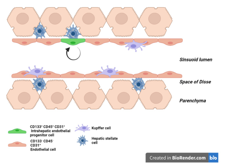
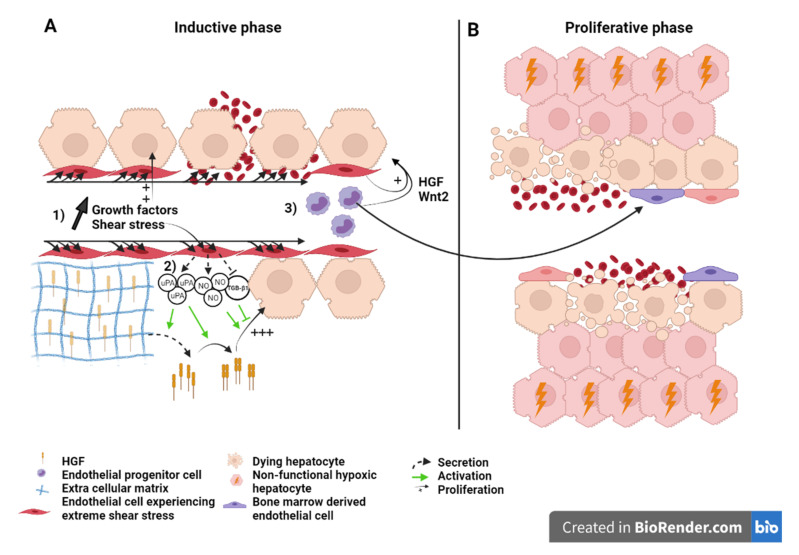
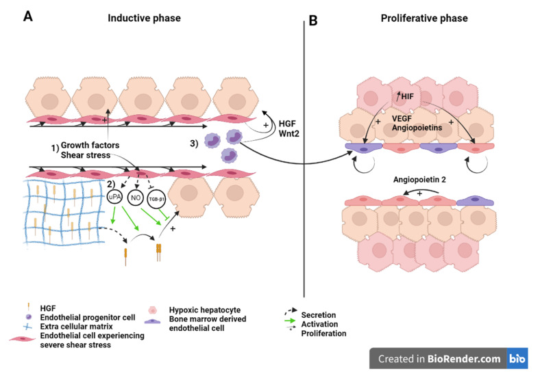
Liver sinusoids are lined by liver sinusoidal endothelial cells (LSEC), which represent approximately 15 to 20% of the liver cells, but only 3% of the total liver volume. LSEC have unique functions, such as fluid filtration, blood vessel tone modulation, blood clotting, inflammatory cell recruitment, and metabolite and hormone trafficking. Different subtypes of liver endothelial cells are also known to control liver zonation and hepatocyte function. Here, we have reviewed the origin of LSEC, the different subtypes identified in the liver, as well as their renewal during homeostasis. The liver has the exceptional ability to regenerate from small remnants. The past decades have seen increasing awareness in the role of non-parenchymal cells in liver regeneration despite not being the most represented population. While a lot of knowledge has emerged, clarification is needed regarding the role of LSEC in sensing shear stress and on their participation in the inductive phase of regeneration by priming the hepatocytes and delivering mitogenic factors. It is also unclear if bone marrow-derived LSEC participate in the proliferative phase of liver regeneration. Similarly, data are scarce as to LSEC having a role in the termination phase of the regeneration process. Here, we review what is known about the interaction between LSEC and other liver cells during the different phases of liver regeneration. We next explain extended hepatectomy and small liver transplantation, which lead to "small for size syndrome" (SFSS), a lethal liver failure. SFSS is linked to endothelial denudation, necrosis, and lobular disturbance. Using the knowledge learned from partial hepatectomy studies on LSEC, we expose several techniques that are, or could be, used to avoid the "small for size syndrome" after extended hepatectomy or small liver transplantation.

摘要

肝窦由肝窦内皮细胞(LSEC)组成,LSEC 约占肝脏细胞的 15%至 20%,但仅占肝脏总体积的 3%。LSEC 具有独特的功能,如液体过滤、血管张力调节、凝血、炎症细胞募集以及代谢物和激素转运。不同类型的肝内皮细胞也被认为控制着肝区带和肝细胞功能。在这里,我们回顾了 LSEC 的起源、在肝脏中鉴定出的不同亚型,以及它们在稳态下的更新。肝脏具有从微小残片中再生的非凡能力。尽管非实质细胞不是最主要的细胞群,但过去几十年来,人们越来越意识到它们在肝脏再生中的作用。虽然已经有了很多的认识,但仍需要澄清 LSEC 在感知切应力中的作用,以及在通过激活肝细胞和传递有丝分裂原因子来启动再生的诱导阶段时,它们在参与再生中的作用。骨髓来源的 LSEC 是否参与肝脏再生的增殖阶段也不清楚。同样,关于 LSEC 在再生过程的终止阶段是否起作用的数据也很少。在这里,我们回顾了 LSEC 在肝脏再生的不同阶段与其他肝脏细胞之间的相互作用。接下来,我们解释了扩大肝切除术和小肝移植导致的“小肝综合征”(SFSS),这是一种致命的肝衰竭。SFSS 与内皮细胞剥脱、坏死和小叶紊乱有关。利用从小鼠部分肝切除研究中获得的关于 LSEC 的知识,我们介绍了几种技术,这些技术目前正在或可能被用于避免在扩大肝切除术或小肝移植后出现“小肝综合征”。

https://cdn.ncbi.nlm.nih.gov/pmc/blobs/5e28/8347197/cc2b6b8f7aaa/ijms-22-08053-g003.jpg
https://cdn.ncbi.nlm.nih.gov/pmc/blobs/5e28/8347197/d5042a87d881/ijms-22-08053-g001.jpg
https://cdn.ncbi.nlm.nih.gov/pmc/blobs/5e28/8347197/493c35b5bde3/ijms-22-08053-g002.jpg
https://cdn.ncbi.nlm.nih.gov/pmc/blobs/5e28/8347197/cc2b6b8f7aaa/ijms-22-08053-g003.jpg
https://cdn.ncbi.nlm.nih.gov/pmc/blobs/5e28/8347197/d5042a87d881/ijms-22-08053-g001.jpg
https://cdn.ncbi.nlm.nih.gov/pmc/blobs/5e28/8347197/493c35b5bde3/ijms-22-08053-g002.jpg
https://cdn.ncbi.nlm.nih.gov/pmc/blobs/5e28/8347197/cc2b6b8f7aaa/ijms-22-08053-g003.jpg

相似文献

1
Critical Role of LSEC in Post-Hepatectomy Liver Regeneration and Failure.LSEC 在肝切除术后肝再生和衰竭中的关键作用。
Int J Mol Sci. 2021 Jul 28;22(15):8053. doi: 10.3390/ijms22158053.
2
Liver sinusoidal endothelial cell progenitor cells promote liver regeneration in rats.肝血窦内皮细胞祖细胞促进大鼠肝脏再生。
J Clin Invest. 2012 Apr;122(4):1567-73. doi: 10.1172/JCI58789. Epub 2012 Mar 12.
3
Inductive angiocrine signals from sinusoidal endothelium are required for liver regeneration.窦状内皮细胞的诱导性血管分泌信号对于肝再生是必需的。
Nature. 2010 Nov 11;468(7321):310-5. doi: 10.1038/nature09493.
4
Lack of endothelial autophagy does not impair liver regeneration after partial hepatectomy in mice.缺乏内皮细胞自噬并不会损害小鼠部分肝切除术后的肝再生。
Liver Int. 2023 Oct;43(10):2302-2308. doi: 10.1111/liv.15677. Epub 2023 Jul 17.
5
Platelet Interactions with Liver Sinusoidal Endothelial Cells and Hepatic Stellate Cells Lead to Hepatocyte Proliferation.血小板与肝窦内皮细胞和肝星状细胞的相互作用导致肝细胞增殖。
Cells. 2020 May 18;9(5):1243. doi: 10.3390/cells9051243.
6
The vascular endothelial growth factor signaling pathway regulates liver sinusoidal endothelial cells during liver regeneration after partial hepatectomy.血管内皮生长因子信号通路在部分肝切除术后肝脏再生过程中调节肝窦内皮细胞。
Expert Rev Gastroenterol Hepatol. 2021 Feb;15(2):139-147. doi: 10.1080/17474124.2020.1815532. Epub 2020 Oct 14.
7
Endothelial cell-derived angiopoietin-2 controls liver regeneration as a spatiotemporal rheostat.内皮细胞衍生的血管生成素-2 作为时空变阻器控制肝脏再生。
Science. 2014 Jan 24;343(6169):416-9. doi: 10.1126/science.1244880.
8
Endothelial GATA4 controls liver fibrosis and regeneration by preventing a pathogenic switch in angiocrine signaling.内皮细胞 GATA4 通过防止血管生成信号的致病转换来控制肝纤维化和再生。
J Hepatol. 2021 Feb;74(2):380-393. doi: 10.1016/j.jhep.2020.08.033. Epub 2020 Sep 9.
9
Human Stem Cells Promote Liver Regeneration After Partial Hepatectomy in BALB/C Nude Mice.人源干细胞促进 BALB/C 裸鼠肝部分切除术后肝再生。
J Surg Res. 2019 Jul;239:191-200. doi: 10.1016/j.jss.2019.02.010. Epub 2019 Mar 4.
10
Angiocrine Hepatocyte Growth Factor Signaling Controls Physiological Organ and Body Size and Dynamic Hepatocyte Proliferation to Prevent Liver Damage during Regeneration.血管生成素肝生长因子信号控制生理器官和身体大小,并控制肝实质细胞的动态增殖,以防止再生过程中的肝损伤。
Am J Pathol. 2020 Feb;190(2):358-371. doi: 10.1016/j.ajpath.2019.10.009. Epub 2019 Nov 27.

引用本文的文献

1
APJ regulates the balance between self-renewal and differentiation of vascular endothelial stem cells.APJ调节血管内皮干细胞自我更新与分化之间的平衡。
Inflamm Regen. 2025 Aug 4;45(1):25. doi: 10.1186/s41232-025-00389-y.
2
Research Progress on the Immune Function of Liver Sinusoidal Endothelial Cells in Sepsis.脓毒症中肝窦内皮细胞免疫功能的研究进展
Cells. 2025 Mar 4;14(5):373. doi: 10.3390/cells14050373.
3
Molecular mechanisms in liver repair and regeneration: from physiology to therapeutics.肝脏修复与再生的分子机制:从生理学到治疗学

本文引用的文献

1
Liver homeostasis is maintained by midlobular zone 2 hepatocytes.肝小叶中区 2 型肝细胞维持肝脏内稳态。
Science. 2021 Feb 26;371(6532). doi: 10.1126/science.abb1625.
2
Liver regeneration: biological and pathological mechanisms and implications.肝脏再生:生物学和病理学机制及其意义。
Nat Rev Gastroenterol Hepatol. 2021 Jan;18(1):40-55. doi: 10.1038/s41575-020-0342-4. Epub 2020 Aug 6.
3
Does Mechanocrine Signaling by Liver Sinusoidal Endothelial Cells Offer New Opportunities for the Development of Anti-fibrotics?肝窦内皮细胞的机械分泌信号传导是否为抗纤维化药物的开发提供了新机遇?
Signal Transduct Target Ther. 2025 Feb 8;10(1):63. doi: 10.1038/s41392-024-02104-8.
4
Circulating proliferative factors versus portal inflow redistribution: mechanistic insights of ALPPS-derived rapid liver regeneration.循环增殖因子与门静脉血流再分布:ALPPS诱导快速肝再生的机制洞察
Front Oncol. 2025 Jan 7;14:1429564. doi: 10.3389/fonc.2024.1429564. eCollection 2024.
5
Human umbilical cord mesenchymal stem cells enhance liver regeneration and decrease collagen content in fibrosis mice after partial hepatectomy by activating Wnt/β-catenin signaling.人脐带间充质干细胞通过激活Wnt/β-连环蛋白信号通路增强肝再生并降低部分肝切除术后纤维化小鼠的胶原含量。
Acta Biochim Biophys Sin (Shanghai). 2024 Dec 23;57(4):604-615. doi: 10.3724/abbs.2024207.
6
Lack of RAMP1 Signaling Suppresses Liver Regeneration and Angiogenesis Following Partial Hepatectomy in Mice.RAMP1 信号缺失抑制小鼠肝部分切除术后的肝再生和血管生成。
In Vivo. 2024 Sep-Oct;38(5):2261-2270. doi: 10.21873/invivo.13691.
7
Gut Microbiota and Liver Regeneration: A Synthesis of Evidence on Structural Changes and Physiological Mechanisms.肠道微生物群与肝脏再生:关于结构变化和生理机制的证据综述
J Clin Exp Hepatol. 2024 Nov-Dec;14(6):101455. doi: 10.1016/j.jceh.2024.101455. Epub 2024 Jun 8.
8
An Endothelial Cell Is Not Simply an Endothelial Cell.内皮细胞并非单纯的内皮细胞。
Stem Cells Dev. 2024 Oct;33(19-20):517-527. doi: 10.1089/scd.2024.0088. Epub 2024 Aug 9.
9
Oit3, a promising hallmark gene for targeting liver sinusoidal endothelial cells.Oit3,一种有前途的肝窦内皮细胞靶向标志物基因。
Signal Transduct Target Ther. 2023 Sep 12;8(1):344. doi: 10.1038/s41392-023-01621-2.
10
Multi-organ Radiomics-Based Prediction of Future Remnant Liver Hypertrophy Following Portal Vein Embolization.多器官影像组学预测门静脉栓塞后残余肝体积的未来增长。
Ann Surg Oncol. 2023 Dec;30(13):7976-7985. doi: 10.1245/s10434-023-14241-5. Epub 2023 Sep 5.
Front Med (Lausanne). 2020 Jan 9;6:312. doi: 10.3389/fmed.2019.00312. eCollection 2019.
4
The role of microbiota in tissue repair and regeneration.微生物群在组织修复和再生中的作用。
J Tissue Eng Regen Med. 2020 Mar;14(3):539-555. doi: 10.1002/term.3009. Epub 2020 Jan 15.
5
Hypoxia protects the liver from Small For Size Syndrome: A lesson learned from the associated liver partition and portal vein ligation for staged hepatectomy (ALPPS) procedure in rats.缺氧可保护肝脏免受小肝综合征的影响:来自大鼠联合肝脏离断和门静脉结扎的分期肝切除术(ALPPS)的经验教训。
Am J Transplant. 2019 Nov;19(11):2979-2990. doi: 10.1111/ajt.15420. Epub 2019 Jun 20.
6
Human Organ-Specific Endothelial Cell Heterogeneity.人类器官特异性内皮细胞异质性
iScience. 2018 Jun 29;4:20-35. doi: 10.1016/j.isci.2018.05.003. Epub 2018 May 9.
7
Paired-cell sequencing enables spatial gene expression mapping of liver endothelial cells.配对细胞测序实现了肝脏内皮细胞的空间基因表达图谱绘制。
Nat Biotechnol. 2018 Nov;36(10):962-970. doi: 10.1038/nbt.4231. Epub 2018 Sep 17.
8
Liver sinusoidal endothelial cells - gatekeepers of hepatic immunity.肝窦内皮细胞 - 肝脏免疫的守门员。
Nat Rev Gastroenterol Hepatol. 2018 Sep;15(9):555-567. doi: 10.1038/s41575-018-0020-y.
9
Structure and Distribution of an Unrecognized Interstitium in Human Tissues.人体组织中未被识别的间质的结构与分布。
Sci Rep. 2018 Mar 27;8(1):4947. doi: 10.1038/s41598-018-23062-6.
10
Characterization of isolated liver sinusoidal endothelial cells for liver bioengineering.分离的肝窦内皮细胞的特征用于肝脏生物工程。
Angiogenesis. 2018 Aug;21(3):581-597. doi: 10.1007/s10456-018-9610-0. Epub 2018 Mar 26.