Department of Ophthalmology, Visual and Anatomical Sciences, School of Medicine, Wayne State University, 540 East Canfield, Detroit, MI 48201, USA.
Department of Pharmacology, School of Medicine, Wayne State University, 540 East Canfield, Detroit, MI 48201, USA.
Int J Mol Sci. 2021 Jul 29;22(15):8130. doi: 10.3390/ijms22158130.
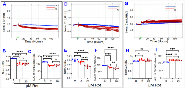
Disruption of retinal pigment epithelial (RPE) barrier integrity is involved in the pathology of several blinding retinal diseases including age-related macular degeneration (AMD) and diabetic retinopathy (DR), but the underlying causes and pathophysiology are not completely well-defined. Mitochondria dysfunction has often been considered as a potential candidate implicated in such a process. In this study, we aimed to dissect the role of different mitochondrial components; specifically, those of oxidative phosphorylation (OxPhos), in maintaining the barrier functionality of RPE. Electric cell-substrate impedance sensing (ECIS) technology was used to collect multi-frequency electrical impedance data to assess in real-time the barrier formation of the RPE cells. For this purpose, the human retinal pigment epithelial cell line-ARPE-19-was used and treated with varying concentrations of specific mitochondrial inhibitors that target different steps in OxPhos: Rotenone for complex I (the largest protein complex in the electron transport chain (ETC)); oligomycin for ATP synthase; and carbonyl cyanide-p-trifluoromethoxyphenyl hydrazone (FCCP) for uncoupling ATP synthesis from the accompanying ETC. Furthermore, data were modeled using the ECIS-Zθ software to investigate in depth the effects of these inhibitors on three separate barrier parameters: cell-cell interactions (R), cell-matrix interactions (α), and the cell membrane capacitance (C). The viability of ARPE-19 cells was determined by lactate dehydrogenase (LDH) Cytotoxicity Assay. The ECIS program's modeling demonstrated that FCCP and thus OxPhos uncoupling disrupt the barrier function in the ARPE-19 cells across all three components of the total resistance (Rb, α, and C) in a dose-dependent manner. On the other hand, oligomycin and thus ATP synthase inhibition mostly affects the ARPE-19 cells' attachment to their substrate evident by a significant decrease in α resistance in a dose-dependent manner, both at the end and throughout the duration of the experiment. On the contrary, rotenone and complex I inhibition mostly affect the ARPE-19 paracellular resistance R in a dose-dependent manner compared to basolateral resistance α or C. Our results clearly demonstrate differential roles for different mitochondrial components in maintaining RPE cell functionality in which uncoupling of OxPhos is a major contributing factor to the disruption barrier function. Such differences can be used in investigating gene expression as well as for screening of selective agents that improve the OxPhos coupling efficiency to be used in the therapeutic approach for treating RPE-related retinal diseases.
视网膜色素上皮 (RPE) 屏障完整性的破坏与几种致盲性视网膜疾病的病理学有关,包括年龄相关性黄斑变性 (AMD) 和糖尿病性视网膜病变 (DR),但其根本原因和病理生理学尚不完全清楚。线粒体功能障碍通常被认为是参与这一过程的一个潜在候选因素。在这项研究中,我们旨在剖析不同线粒体成分的作用;具体来说,那些涉及氧化磷酸化 (OxPhos) 的成分,在维持 RPE 的屏障功能方面的作用。使用电动细胞-基底阻抗感应 (ECIS) 技术收集多频电阻抗数据,实时评估 RPE 细胞的屏障形成情况。为此,使用人视网膜色素上皮细胞系 ARPE-19,并使用不同浓度的特异性线粒体抑制剂处理,这些抑制剂针对 OxPhos 的不同步骤:鱼藤酮针对复合物 I(电子传递链 (ETC) 中最大的蛋白质复合物);寡霉素针对 ATP 合酶;以及羰基氰化物-对三氟甲氧基苯腙 (FCCP) 用于将 ATP 合成与伴随的 ETC 解偶联。此外,使用 ECIS-Zθ 软件对数据进行建模,以深入研究这些抑制剂对三个独立屏障参数的影响:细胞-细胞相互作用 (R)、细胞-基质相互作用 (α) 和细胞膜电容 (C)。通过乳酸脱氢酶 (LDH) 细胞毒性测定法确定 ARPE-19 细胞的活力。ECIS 程序的建模表明,FCCP 因此 OxPhos 解偶联以剂量依赖性方式破坏 ARPE-19 细胞的屏障功能,跨越总电阻 (Rb、α 和 C) 的所有三个组成部分。另一方面,寡霉素因此 ATP 合酶抑制主要影响 ARPE-19 细胞附着在其基质上,这在剂量依赖性方式下通过显著降低 α 电阻来证明,这在实验的结束和整个过程中都是如此。相反,与基底外侧电阻 α 或 C 相比,鱼藤酮和复合物 I 抑制主要以剂量依赖性方式影响 ARPE-19 的细胞旁电阻 R。我们的结果清楚地表明,不同的线粒体成分在维持 RPE 细胞功能方面发挥着不同的作用,其中 OxPhos 的解偶联是破坏屏障功能的主要因素。这些差异可用于研究基因表达以及筛选可提高 OxPhos 偶联效率的选择性药物,以用于治疗与 RPE 相关的视网膜疾病的治疗方法。