Movement Analysis Laboratory, IRCCS Istituto Ortopedico Rizzoli, Via di Barbiano 1/10, Bologna, Italy.
Bentivoglio Orthopaedic Ward, IRCCS Istituto Ortopedico Rizzoli, Bologna, Italy.
Sci Rep. 2021 Aug 9;11(1):16139. doi: 10.1038/s41598-021-95708-x.
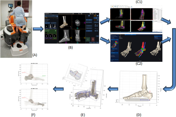
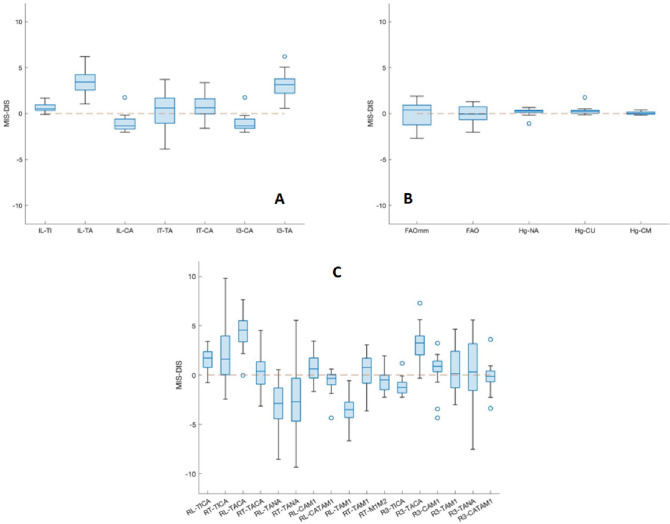
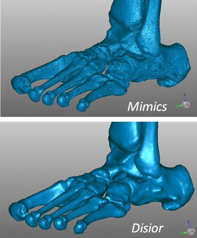
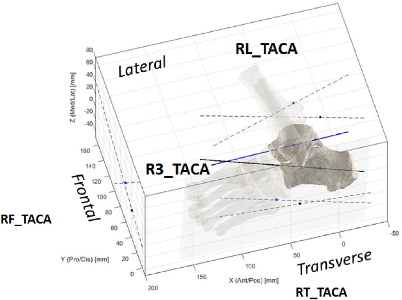
Acquired adult flatfoot is a frequent deformity which implies multiple, complex and combined 3D modifications of the foot skeletal structure. The difficult thorough evaluation of the degree of severity pre-op and the corresponding assessment post-op can now be overcome by cone-beam (CBCT) technology, which can provide access to the 3D skeletal structure in weight-bearing. This study aims to report flatfoot deformities originally in 3D and in weight-bearing, with measurements taken using two different bone segmentation techniques. 21 such patients, with indication for surgical corrections, underwent CBCT (Carestream, US) while standing on one leg. From these scans, 3D models of each bone of the foot were reconstructed by using two different state-of-the-art segmentation tools: a semi-automatic (Mimics Innovation Suite, Materialise, Belgium), and an automatic (Bonelogic Ortho Foot and Ankle, Disior, Finland). From both reconstructed models, Principal Component Analysis was used to define anatomical reference frames, and original foot and ankle angles and other parameters were calculated mostly based on the longitudinal axis of the bones, in anatomical plane projections and in 3D. Both bone model reconstructions revealed a considerable valgus of the calcareous, plantarflexion and internal rotation of the talus, and typical Meary's angles in the lateral and transverse plane projections. The mean difference from these angles between semi-automatic and automatic segmentations was larger than 3.5 degrees for only 3 of the 32 measurements, and a large number of these differences were not statistically significant. CBCT and the present techniques for bone shape reconstruction finally provide a novel and valuable 3D assessment of complex foot deformities in weight-bearing, eliminating previous limitations associated to unloaded feet and bidimensional measures. Corresponding measurements on the bone models from the two segmentation tools compared well. Other more representative measurements can be defined in the future using CBCT and these techniques.
成人获得性扁平足是一种常见的畸形,它意味着足部骨骼结构的多个、复杂和综合的 3D 改变。通过锥形束 CT(CBCT)技术,可以克服术前严重程度的全面评估和术后相应评估的困难,该技术可以提供负重状态下的 3D 骨骼结构。本研究旨在报告原本为 3D 并处于负重状态的扁平足畸形,并使用两种不同的骨骼分割技术进行测量。21 名有手术矫正指征的此类患者在单腿站立时接受了 CBCT(美国 Carestream)检查。从这些扫描中,使用两种不同的最先进的分割工具(比利时 Materialise 的 Mimics Innovation Suite 和芬兰 Disior 的 Bonelogic Ortho Foot and Ankle)重建了每个足部骨骼的 3D 模型。从这两个重建模型中,使用主成分分析来定义解剖参考框架,并根据骨骼的纵轴在解剖平面投影和 3D 中计算原始足部和踝关节角度和其他参数。这两种骨骼模型重建都显示出距骨的明显外旋、跖屈和内旋,以及横向和横向平面投影中典型的 Meary 角。半自动和自动分割之间这些角度的平均差异大于 3.5 度的仅为 32 个测量值中的 3 个,而且大量这些差异在统计学上并不显著。CBCT 和目前的骨骼形状重建技术最终提供了一种新的、有价值的负重状态下复杂足部畸形的 3D 评估方法,消除了以前与非负重和二维测量相关的局限性。两种分割工具的骨骼模型上的相应测量值非常吻合。未来可以使用 CBCT 和这些技术定义其他更具代表性的测量值。