• 文献检索
  • 文档翻译
  • 深度研究
  • 学术资讯
  • Suppr Zotero 插件Zotero 插件
  • 邀请有礼
  • 套餐&价格
  • 历史记录
应用&插件
Suppr Zotero 插件Zotero 插件浏览器插件Mac 客户端Windows 客户端微信小程序
定价
高级版会员购买积分包购买API积分包
服务
文献检索文档翻译深度研究API 文档MCP 服务
关于我们
关于 Suppr公司介绍联系我们用户协议隐私条款
关注我们

Suppr 超能文献

核心技术专利:CN118964589B侵权必究
粤ICP备2023148730 号-1Suppr @ 2026

文献检索

告别复杂PubMed语法,用中文像聊天一样搜索,搜遍4000万医学文献。AI智能推荐,让科研检索更轻松。

立即免费搜索

文件翻译

保留排版,准确专业,支持PDF/Word/PPT等文件格式,支持 12+语言互译。

免费翻译文档

深度研究

AI帮你快速写综述,25分钟生成高质量综述,智能提取关键信息,辅助科研写作。

立即免费体验

临床人群负重 CT 测量后足对线角度的 3D 测量技术。

3D measurement techniques for the hindfoot alignment angle from weight-bearing CT in a clinical population.

机构信息

Movement Analysis Laboratory, IRCCS Istituto Ortopedico Rizzoli, Via di Barbiano 1/10, Bologna, Italy.

Bentivoglio Orthopaedic Ward, IRCCS Istituto Ortopedico Rizzoli, Bologna, Italy.

出版信息

Sci Rep. 2022 Oct 7;12(1):16900. doi: 10.1038/s41598-022-21440-9.

DOI:10.1038/s41598-022-21440-9
PMID:36207422
原文链接:https://pmc.ncbi.nlm.nih.gov/articles/PMC9546907/
Abstract

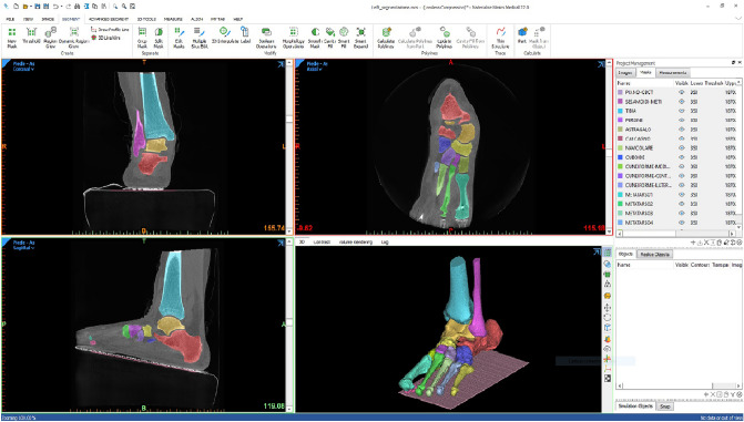
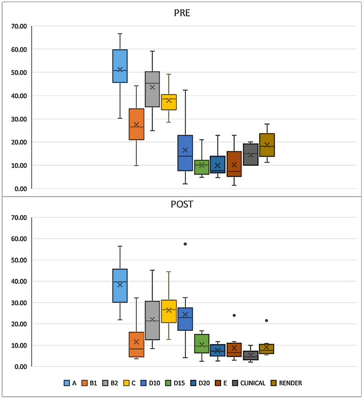
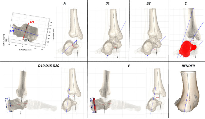
Cone-beam CT (CBCT) scans now enable accurate measurements on foot skeletal structures with the advantage of observing these in 3D and in weight-bearing. Among the most common skeletal deformities, the varus/valgus of the hindfoot is the most complex to be represented, and a number of measure proposals have been published. This study aims to analyze and to compare these measurements from CBCT scans in a real clinical population with large such deformity. Ten patients with severe acquired adult flatfoot and indication for surgery underwent CBCT scans (Carestream, USA) while standing on that leg, before and after surgical correction. Corresponding 3D shape of each bone of the distal shank and hindfoot were defined (Materialise, Belgium). Six different techniques from the literature were used to calculate the varus/valgus deformity, i.e. the inclination of the hindfoot in the frontal plane of the shank. Standard clinical measurements by goniometers were taken for comparison. According to these techniques, and starting from a careful 3D reconstruction of the relevant foot skeletal structures, a large spectrum of measurements was found to represent the same hindfoot alignment angle. Most of them were very different from the traditional clinical measures. The assessment of the pre-operative valgus deformity and of the corresponding post-operative correction varied considerably. CBCT finally allows 3D assessment of foot deformities in weight-bearing. Measurements from the different available techniques do not compare well, as they are based on very different approaches. It is recommended to be aware of the anatomical and functional concepts behind these techniques before clinical and surgical conclusions.

摘要

锥形束 CT(CBCT)扫描现在可以通过观察三维和负重状态下的足部骨骼结构,实现对足部骨骼结构的精确测量。在最常见的骨骼畸形中,后足的内翻/外翻畸形最为复杂,已有多种测量方法被提出。本研究旨在分析和比较这些在存在严重获得性成人扁平足并需要手术的临床实际患者的 CBCT 扫描中的测量值,并对存在此类严重畸形的患者进行研究。10 例患有严重获得性成人扁平足且需要手术的患者在手术矫正前后,单腿站立时接受了 CBCT 扫描(美国 Carestream)。对每个跟骨和后足的远端骨骼的相应 3D 形状进行了定义(比利时 Materialise)。使用文献中的 6 种不同技术来计算内翻/外翻畸形,即跟骨在小腿的额状面的倾斜度。同时还进行了传统的测角计临床测量作为比较。根据这些技术,并且从对相关足部骨骼结构的仔细 3D 重建开始,发现了大量的测量值来代表相同的后足对线角度。其中大多数与传统的临床测量值都有很大的不同。术前外翻畸形的评估和相应的术后矫正效果存在较大差异。CBCT 最终允许在负重状态下对足部畸形进行 3D 评估。来自不同可用技术的测量值之间的比较效果并不理想,因为它们基于非常不同的方法。建议在进行临床和手术结论之前,了解这些技术背后的解剖学和功能概念。

https://cdn.ncbi.nlm.nih.gov/pmc/blobs/c8bd/9546907/da9e843fc132/41598_2022_21440_Fig3_HTML.jpg
https://cdn.ncbi.nlm.nih.gov/pmc/blobs/c8bd/9546907/ba7f18f68823/41598_2022_21440_Fig1_HTML.jpg
https://cdn.ncbi.nlm.nih.gov/pmc/blobs/c8bd/9546907/705ae2993096/41598_2022_21440_Fig2_HTML.jpg
https://cdn.ncbi.nlm.nih.gov/pmc/blobs/c8bd/9546907/da9e843fc132/41598_2022_21440_Fig3_HTML.jpg
https://cdn.ncbi.nlm.nih.gov/pmc/blobs/c8bd/9546907/ba7f18f68823/41598_2022_21440_Fig1_HTML.jpg
https://cdn.ncbi.nlm.nih.gov/pmc/blobs/c8bd/9546907/705ae2993096/41598_2022_21440_Fig2_HTML.jpg
https://cdn.ncbi.nlm.nih.gov/pmc/blobs/c8bd/9546907/da9e843fc132/41598_2022_21440_Fig3_HTML.jpg

相似文献

1
3D measurement techniques for the hindfoot alignment angle from weight-bearing CT in a clinical population.临床人群负重 CT 测量后足对线角度的 3D 测量技术。
Sci Rep. 2022 Oct 7;12(1):16900. doi: 10.1038/s41598-022-21440-9.
2
Hindfoot alignment of adult acquired flatfoot deformity: A comparison of clinical assessment and weightbearing cone beam CT examinations.成人获得性平足畸形的后足对线:临床评估与负重锥形束 CT 检查的比较。
Foot Ankle Surg. 2019 Dec;25(6):790-797. doi: 10.1016/j.fas.2018.10.008. Epub 2018 Nov 5.
3
Is Lower-limb Alignment Associated with Hindfoot Deformity in the Coronal Plane? A Weightbearing CT Analysis.下肢对线与冠状面后足畸形有关吗?负重 CT 分析。
Clin Orthop Relat Res. 2020 Jan;478(1):154-168. doi: 10.1097/CORR.0000000000001067.
4
Angular and linear measurements of adult flexible flatfoot via weight-bearing CT scans and 3D bone reconstruction tools.成人柔性平足的 CT 扫描负重位下的角度和线性测量及 3D 骨骼重建工具。
Sci Rep. 2021 Aug 9;11(1):16139. doi: 10.1038/s41598-021-95708-x.
5
Correlation of Clinical Evaluation and Radiographic Hindfoot Alignment in Stage II Adult-Acquired Flatfoot Deformity.II期成人获得性平足畸形的临床评估与影像学后足对线的相关性
Foot Ankle Int. 2018 Jul;39(7):771-779. doi: 10.1177/1071100718762113. Epub 2018 Mar 28.
6
Morphological changes in flatfoot: a 3D analysis using weight-bearing CT scans.扁平足的形态变化:使用负重 CT 扫描的 3D 分析。
BMC Med Imaging. 2024 Aug 19;24(1):219. doi: 10.1186/s12880-024-01396-0.
7
Weight-bearing radiographs and cone-beam computed tomography examinations in adult acquired flatfoot deformity.成人获得性扁平足畸形的负重位 X 线片和锥形束 CT 检查。
Foot Ankle Surg. 2021 Feb;27(2):201-206. doi: 10.1016/j.fas.2020.04.011. Epub 2020 Apr 30.
8
Measuring hindfoot alignment in weight bearing CT: A novel clinical relevant measurement method.在负重CT中测量后足对线:一种新的临床相关测量方法。
Foot Ankle Surg. 2016 Dec;22(4):233-238. doi: 10.1016/j.fas.2015.10.002. Epub 2015 Oct 30.
9
Tripod index: a new radiographic parameter assessing foot alignment.三脚架指数:一种新的评估足弓对线的影像学参数。
Foot Ankle Int. 2013 Oct;34(10):1411-20. doi: 10.1177/1071100713488761. Epub 2013 May 8.
10
Three-dimensional displacement after a medializing calcaneal osteotomy in relation to the osteotomy angle and hindfoot alignment.距下骨切开术后的三维位移与骨切开角度和后足对线的关系。
Foot Ankle Surg. 2020 Jan;26(1):78-84. doi: 10.1016/j.fas.2018.11.015. Epub 2018 Dec 7.

引用本文的文献

1
Trends in the Use of Weightbearing Computed Tomography.负重计算机断层扫描的使用趋势
J Clin Med. 2024 Sep 18;13(18):5519. doi: 10.3390/jcm13185519.
2
Implementing automated 3D measurements to quantify reference values and side-to-side differences in the ankle syndesmosis.实现踝关节联合部的自动 3D 测量,以量化参考值和左右侧的差异。
Sci Rep. 2023 Aug 23;13(1):13774. doi: 10.1038/s41598-023-40599-3.

本文引用的文献

1
Republication of "How Accurate Is Clinical Evaluation in Hindfoot Coronal Alignment?".《后足冠状面排列的临床评估有多准确?》再版
Foot Ankle Orthop. 2023 Jul 23;8(3):24730114231188111. doi: 10.1177/24730114231188111. eCollection 2023 Jul.
2
Intra- and Interobserver Reliability of the New Classification System of Progressive Collapsing Foot Deformity.新型渐进性塌陷足畸形分类系统的观察者内和观察者间可靠性。
Foot Ankle Int. 2022 Apr;43(4):582-589. doi: 10.1177/10711007211058154. Epub 2021 Dec 1.
3
Imaging of progressive collapsing foot deformity with emphasis on the role of weightbearing cone beam CT.
进行性足塌陷畸形的影像学表现,重点关注负重锥形束CT的作用。
Skeletal Radiol. 2022 Jun;51(6):1127-1141. doi: 10.1007/s00256-021-03942-1. Epub 2021 Oct 25.
4
Angular and linear measurements of adult flexible flatfoot via weight-bearing CT scans and 3D bone reconstruction tools.成人柔性平足的 CT 扫描负重位下的角度和线性测量及 3D 骨骼重建工具。
Sci Rep. 2021 Aug 9;11(1):16139. doi: 10.1038/s41598-021-95708-x.
5
Weight-bearing cone-beam computed tomography in the foot and ankle specialty: where we are and where we are going - an update.足踝专科中的负重锥形束计算机断层扫描:我们的现状与未来发展——最新进展
Radiol Bras. 2021 May-Jun;54(3):177-184. doi: 10.1590/0100-3984.2020.0048.
6
Measuring standing hindfoot alignment: reliability of different approaches in conventional x-ray and cone-beam CT.测量站立位后足对线:传统 X 射线和锥形束 CT 中不同方法的可靠性。
Arch Orthop Trauma Surg. 2022 Nov;142(11):3035-3043. doi: 10.1007/s00402-021-03904-1. Epub 2021 Apr 22.
7
Weight-bearing CT in foot and ankle pathology.足部和踝关节病变的负重 CT
Orthop Traumatol Surg Res. 2021 Feb;107(1S):102772. doi: 10.1016/j.otsr.2020.102772. Epub 2020 Dec 13.
8
[Etiology, pathogenesis, clinical features, diagnostics and conservative treatment of adult flatfoot].[成人扁平足的病因、发病机制、临床特征、诊断及保守治疗]
Orthopade. 2020 Nov;49(11):942-953. doi: 10.1007/s00132-020-03995-5.
9
Classification and Nomenclature: Progressive Collapsing Foot Deformity.分类和命名:进行性塌陷足畸形。
Foot Ankle Int. 2020 Oct;41(10):1271-1276. doi: 10.1177/1071100720950722. Epub 2020 Aug 28.
10
Foot and ankle measurements on cone beam weightbearing computed tomography.足和踝关节在锥形束负重 CT 中的测量。
J Biol Regul Homeost Agents. 2020 May-Jun;34(3 Suppl. 2):23-32. ADVANCES IN MUSCULOSKELETAL DISEASES AND INFECTIONS - SOTIMI 2019.