Division of Vascular and Endovascular Surgery, Department of Surgery, Beth Israel Deaconess Medical Center, Harvard Medical School, Boston, Massachusetts, USA.
Division of Vascular Surgery and Endovascular Therapy, Department of Surgery, Yale University School of Medicine, New Haven, Connecticut, USA.
Physiol Rep. 2021 Aug;9(16):e15008. doi: 10.14814/phy2.15008.
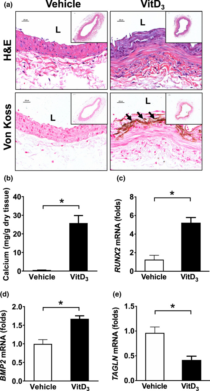
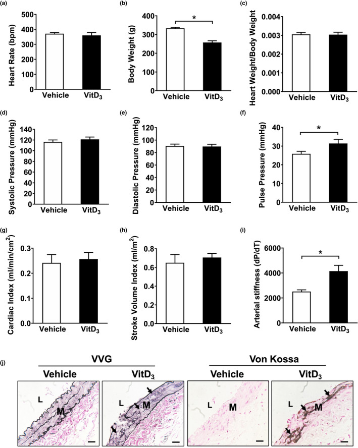
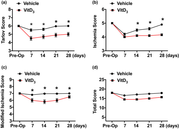
Medial artery calcification results from deposition of calcium hydroxyapatite crystals on elastin layers, and osteogenic changes in vascular smooth muscle cells. It is highly prevalent in patients with chronic kidney disease, diabetes, and peripheral artery disease (PAD), and when identified in lower extremity vessels, it is associated with increased amputation rates. This study aims to evaluate the effects of medial calcification on perfusion and functional recovery after hindlimb ischemia in rats. Medial artery calcification and acute limb ischemia were induced by vitamin D (VitD ) injection and femoral artery ligation in rats. VitD injection robustly induced calcification in the medial layer of femoral arteries in vivo. Laser Doppler perfusion imaging revealed that perfusion decreased and then partially recovered after hindlimb ischemia in vehicle-injected rats. In contrast, VitD -injected rats showed markedly impaired recovery of perfusion following limb ischemia. Accordingly, rats with medial calcification showed worse ischemia scores and delayed functional recovery compared with controls. Immunohistochemical and histological staining did not show differences in capillary density or muscle morphology between VitD - and vehicle-injected rats at 28 days after femoral artery ligation. The evaluation of cardiac and hemodynamic parameters showed that arterial stiffness was increased while cardiac function was preserved in VitD -injected rats. These findings suggest that medial calcification may contribute to impaired perfusion in PAD by altering vascular compliance, however, the specific mechanisms remain poorly understood. Reducing or slowing the progression of arterial calcification in patients with PAD may improve clinical outcomes.
中层动脉钙化是由于钙羟磷灰石晶体在弹力层上的沉积和血管平滑肌细胞的成骨样变化所致。它在慢性肾脏病、糖尿病和外周动脉疾病(PAD)患者中非常普遍,当在下肢血管中发现时,与截肢率增加相关。本研究旨在评估中层钙化对大鼠后肢缺血后灌注和功能恢复的影响。通过维生素 D(VitD)注射和股动脉结扎在大鼠中诱导中层钙化和急性肢体缺血。VitD 注射在体内强烈诱导股动脉中层钙化。激光多普勒灌注成像显示,在注射载体的大鼠后肢缺血后,灌注减少,然后部分恢复。相比之下,VitD 注射的大鼠在肢体缺血后显示出灌注恢复明显受损。因此,与对照组相比,中层钙化的大鼠表现出更差的缺血评分和延迟的功能恢复。免疫组织化学和组织学染色显示,在股动脉结扎后 28 天,VitD 注射和载体注射的大鼠之间毛细血管密度或肌肉形态没有差异。对心脏和血流动力学参数的评估表明,VitD 注射的大鼠动脉僵硬增加,而心脏功能保持不变。这些发现表明,中层钙化可能通过改变血管顺应性导致 PAD 中的灌注受损,然而,具体机制仍知之甚少。减少或减缓 PAD 患者的动脉钙化进展可能改善临床结局。