Moon Seo Yun, Lee Heejin, Kim Seoree, Hong Ji Hyung, Chun Sang Hoon, Lee Hee Yeon, Kang Keunsoo, Kim Ho Shik, Won Hye Sung, Ko Yoon Ho
Department of Biomedicine & Health Sciences, College of Medicine, The Catholic University of Korea, Seoul, Republic of Korea.
Cancer Research Institute, College of Medicine, The Catholic University of Korea, Seoul, Republic of Korea.
BMC Cancer. 2021 Aug 18;21(1):931. doi: 10.1186/s12885-021-08641-7.
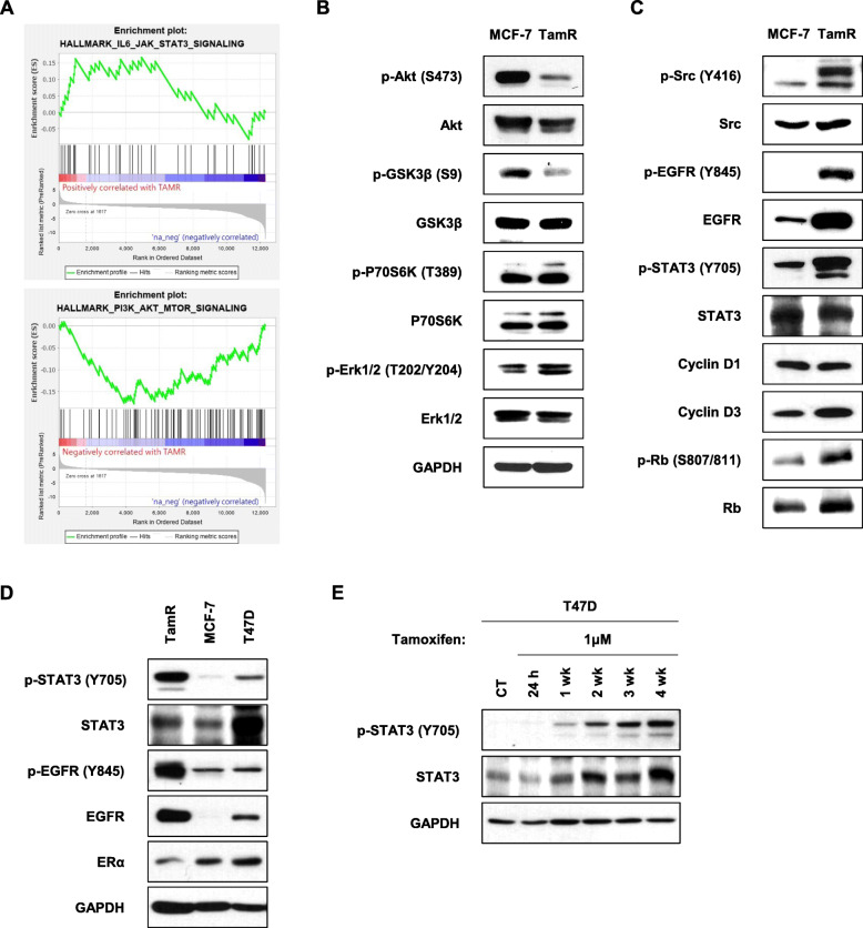
The mechanisms of endocrine resistance are complex, and deregulation of several oncogenic signalling pathways has been proposed. We aimed to investigate the role of the EGFR and Src-mediated STAT3 signalling pathway in tamoxifen-resistant breast cancer cells.
The ER-positive luminal breast cancer cell lines, MCF-7 and T47D, were used. We have established an MCF-7-derived tamoxifen-resistant cell line (TamR) by long-term culture of MCF-7 cells with 4-hydroxytamoxifen. Cell viability was determined using an MTT assay, and protein expression levels were determined using western blot. Cell cycle and annexin V staining were analysed using flow cytometry.
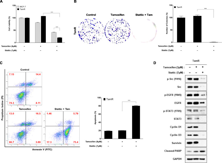
TamR cells showed decreased expression of estrogen receptor and increased expression of EGFR. TamR cells showed an acceleration of the G1 to S phase transition. The protein expression levels of phosphorylated Src, EGFR (Y845), and STAT3 was increased in TamR cells, while phosphorylated Akt was decreased. The expression of p-STAT3 was enhanced according to exposure time of tamoxifen in T47D cells, suggesting that activation of STAT3 can cause tamoxifen resistance in ER-positive breast cancer cells. Both dasatinib (Src inhibitor) and stattic (STAT3 inhibitor) inhibited cell proliferation and induced apoptosis in TamR cells. However, stattic showed a much stronger effect than dasatinib. Knockdown of STAT3 expression by siRNA had no effect on sensitivity to tamoxifen in MCF-7 cells, while that enhanced sensitivity to tamoxifen in TamR cells. There was not a significant synergistic effect of dasatinib and stattic on cell survival. TamR cells have low nuclear p21(Cip1) expression compared to MCF-7 cells and inhibition of STAT3 increased the expression of nuclear p21(Cip1) in TamR cells.
The EGFR and Src-mediated STAT3 signalling pathway is activated in TamR cells, and inhibition of STAT3 may be a potential target in tamoxifen-resistant breast cancer. An increase in nuclear p21(Cip1) may be a key step in STAT3 inhibitor-induced cell death in TamR cells.
内分泌耐药机制复杂,有人提出多种致癌信号通路失调与之相关。我们旨在研究表皮生长因子受体(EGFR)和Src介导的信号转导与转录激活因子3(STAT3)信号通路在耐他莫昔芬乳腺癌细胞中的作用。
使用雌激素受体(ER)阳性的管腔型乳腺癌细胞系MCF-7和T47D。通过用4-羟基他莫昔芬长期培养MCF-7细胞,建立了一株源自MCF-7的耐他莫昔芬细胞系(TamR)。采用MTT法测定细胞活力,用蛋白质免疫印迹法测定蛋白质表达水平。使用流式细胞术分析细胞周期和膜联蛋白V染色情况。
TamR细胞雌激素受体表达降低,EGFR表达增加。TamR细胞从G1期到S期的转变加速。TamR细胞中磷酸化Src、EGFR(Y845)和STAT3的蛋白质表达水平升高,而磷酸化Akt降低。在T47D细胞中,p-STAT3的表达随着他莫昔芬暴露时间的延长而增强,这表明STAT3的激活可导致ER阳性乳腺癌细胞产生他莫昔芬耐药。达沙替尼(Src抑制剂)和Stattic(STAT3抑制剂)均抑制TamR细胞增殖并诱导其凋亡。然而,Stattic的作用比达沙替尼强得多。通过小干扰RNA(siRNA)敲低STAT3表达对MCF-7细胞对他莫昔芬的敏感性没有影响,而增强了TamR细胞对他莫昔芬的敏感性。达沙替尼和Stattic对细胞存活没有显著的协同作用。与MCF-7细胞相比,TamR细胞中核p21(Cip1)表达较低,抑制STAT3可增加TamR细胞中核p21(Cip1)的表达。
EGFR和Src介导的STAT3信号通路在TamR细胞中被激活,抑制STAT3可能是耐他莫昔芬乳腺癌的一个潜在靶点。核p21(Cip1)的增加可能是STAT3抑制剂诱导TamR细胞死亡的关键步骤。