• 文献检索
  • 文档翻译
  • 深度研究
  • 学术资讯
  • Suppr Zotero 插件Zotero 插件
  • 邀请有礼
  • 套餐&价格
  • 历史记录
应用&插件
Suppr Zotero 插件Zotero 插件浏览器插件Mac 客户端Windows 客户端微信小程序
定价
高级版会员购买积分包购买API积分包
服务
文献检索文档翻译深度研究API 文档MCP 服务
关于我们
关于 Suppr公司介绍联系我们用户协议隐私条款
关注我们

Suppr 超能文献

核心技术专利:CN118964589B侵权必究
粤ICP备2023148730 号-1Suppr @ 2026

文献检索

告别复杂PubMed语法,用中文像聊天一样搜索,搜遍4000万医学文献。AI智能推荐,让科研检索更轻松。

立即免费搜索

文件翻译

保留排版,准确专业,支持PDF/Word/PPT等文件格式,支持 12+语言互译。

免费翻译文档

深度研究

AI帮你快速写综述,25分钟生成高质量综述,智能提取关键信息,辅助科研写作。

立即免费体验

一种基于对比增强CT的分类树模型,用于对中国临床人群中的实性肺肿瘤恶性程度进行分类。

A contrast-enhanced-CT-based classification tree model for classifying malignancy of solid lung tumors in a Chinese clinical population.

作者信息

Cui Xiaonan, Heuvelmans Marjolein A, Sidorenkov Grigory, Zhao Yingru, Fan Shuxuan, Groen Harry J M, Dorrius Monique D, Oudkerk Matthijs, de Bock Geertruida H, Vliegenthart Rozemarijn, Ye Zhaoxiang

机构信息

Department of Radiology, Key Laboratory of Cancer Prevention and Therapy, Tianjin Medical University Cancer Institute and Hospital, National Clinical Research Centre of Cancer, Tianjin, China.

Department of Radiology, University Medical Center Groningen, University of Groningen, Groningen, The Netherlands.

出版信息

J Thorac Dis. 2021 Jul;13(7):4407-4417. doi: 10.21037/jtd-21-588.

DOI:10.21037/jtd-21-588
PMID:34422367
原文链接:https://pmc.ncbi.nlm.nih.gov/articles/PMC8339765/
Abstract

BACKGROUND

To develop and validate a contrast-enhanced CT based classification tree model for classifying solid lung tumors in clinical patients into malignant or benign.

METHODS

Between January 2015 and October 2017, 827 pathologically confirmed solid lung tumors (487 malignant, 340 benign; median size, 27.0 mm, IQR 18.0-39.0 mm) from 827 patients from a dedicated Chinese cancer hospital were identified. Nodules were divided randomly into two groups, a training group (575 cases) and a testing group (252 cases). CT characteristics were collected by two radiologists, and analyzed using a classification and regression tree (CART) model. For validation, we used the decision analysis threshold to evaluate the classification performance of the CART model and radiologist's diagnosis (benign; malignant) in the testing group.

RESULTS

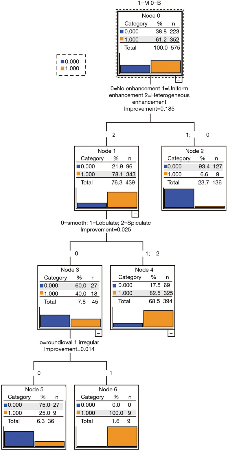
Three out of 19 characteristics [margin (smooth; slightly lobulated/lobulated/spiculated), and shape (round/oval; irregular), subjective enhancement (no/uniform enhancement; heterogeneous enhancement)] were automatically generated by the CART model for classifying solid lung tumors. The sensitivity, specificity, PPV, NPV, and diagnostic accuracy of the CART model is 98.5%, 58.1%, 80.6%, 98.6%, 79.8%, and 90.4%, 54.7%, 82.4% 98.5%, 74.2% for the radiologist's diagnosis by using three-threshold decision analysis.

CONCLUSIONS

Tumor margin and shape, and subjective tumor enhancement were the most important CT characteristics in the CART model for classifying solid lung tumors as malignant. The CART model had higher discriminatory power than radiologist's diagnosis. The CART model could help radiologists making recommendations regarding follow-up or surgery in clinical patients with a solid lung tumor.

摘要

背景

开发并验证一种基于对比增强CT的分类树模型,用于将临床患者的实性肺肿瘤分为恶性或良性。

方法

2015年1月至2017年10月期间,从一家专门的中国癌症医院的827例患者中确定了827个经病理证实的实性肺肿瘤(487个恶性,340个良性;中位大小27.0mm,IQR 18.0 - 39.0mm)。结节被随机分为两组,训练组(575例)和测试组(252例)。由两名放射科医生收集CT特征,并使用分类回归树(CART)模型进行分析。为进行验证,我们使用决策分析阈值来评估CART模型和放射科医生在测试组中的诊断(良性;恶性)分类性能。

结果

CART模型自动生成了19个特征中的3个[边缘(光滑;轻度分叶/分叶状/毛刺状)、形状(圆形/椭圆形;不规则)、主观强化(无/均匀强化;不均匀强化)]用于实性肺肿瘤的分类。使用三阈值决策分析时,CART模型的敏感性、特异性、阳性预测值、阴性预测值和诊断准确性分别为98.5%、58.1%、80.6%、98.6%、79.8%,放射科医生诊断的分别为90.4%、54.7%、82.4%、98.5%、74.2%。

结论

肿瘤边缘和形状以及主观肿瘤强化是CART模型中将实性肺肿瘤分类为恶性的最重要CT特征。CART模型比放射科医生的诊断具有更高的鉴别力。CART模型可以帮助放射科医生对患有实性肺肿瘤的临床患者提出随访或手术建议。

https://cdn.ncbi.nlm.nih.gov/pmc/blobs/c1fc/8339765/751aac36fec2/jtd-13-07-4407-f2.jpg
https://cdn.ncbi.nlm.nih.gov/pmc/blobs/c1fc/8339765/ba30333fdf0b/jtd-13-07-4407-f1.jpg
https://cdn.ncbi.nlm.nih.gov/pmc/blobs/c1fc/8339765/751aac36fec2/jtd-13-07-4407-f2.jpg
https://cdn.ncbi.nlm.nih.gov/pmc/blobs/c1fc/8339765/ba30333fdf0b/jtd-13-07-4407-f1.jpg
https://cdn.ncbi.nlm.nih.gov/pmc/blobs/c1fc/8339765/751aac36fec2/jtd-13-07-4407-f2.jpg

相似文献

1
A contrast-enhanced-CT-based classification tree model for classifying malignancy of solid lung tumors in a Chinese clinical population.一种基于对比增强CT的分类树模型,用于对中国临床人群中的实性肺肿瘤恶性程度进行分类。
J Thorac Dis. 2021 Jul;13(7):4407-4417. doi: 10.21037/jtd-21-588.
2
Limited value of shape, margin and CT density in the discrimination between benign and malignant screen detected solid pulmonary nodules of the NELSON trial.在NELSON试验中,形态、边缘和CT密度在鉴别筛查发现的实性肺结节良恶性方面价值有限。
Eur J Radiol. 2008 Nov;68(2):347-52. doi: 10.1016/j.ejrad.2007.08.027. Epub 2007 Oct 24.
3
Role of baseline nodule density and changes in density and nodule features in the discrimination between benign and malignant solid indeterminate pulmonary nodules.基线结节密度以及密度和结节特征变化在鉴别实性不确定肺结节良恶性中的作用
Eur J Radiol. 2009 Jun;70(3):492-8. doi: 10.1016/j.ejrad.2008.02.022. Epub 2008 Apr 15.
4
A Subsolid Nodules Imaging Reporting System (SSN-IRS) for Classifying 3 Subtypes of Pulmonary Adenocarcinoma.一种用于对肺腺癌三种亚型进行分类的亚实性结节影像报告系统(SSN-IRS)。
Clin Lung Cancer. 2020 Jul;21(4):314-325.e4. doi: 10.1016/j.cllc.2020.01.014. Epub 2020 Feb 6.
5
Comparison of Veterans Affairs, Mayo, Brock classification models and radiologist diagnosis for classifying the malignancy of pulmonary nodules in Chinese clinical population.退伍军人事务部、梅奥诊所、布罗克分类模型与放射科医生诊断在中国临床人群中对肺结节恶性程度分类的比较。
Transl Lung Cancer Res. 2019 Oct;8(5):605-613. doi: 10.21037/tlcr.2019.09.17.
6
Deep learning for classifying fibrotic lung disease on high-resolution computed tomography: a case-cohort study.深度学习在高分辨率 CT 上对肺纤维化疾病的分类:一项病例队列研究。
Lancet Respir Med. 2018 Nov;6(11):837-845. doi: 10.1016/S2213-2600(18)30286-8. Epub 2018 Sep 16.
7
An MRI-based decision tree to distinguish lipomas and lipoma variants from well-differentiated liposarcoma of the extremity and superficial trunk: Classification and regression tree (CART) analysis.基于 MRI 的决策树鉴别肢体和浅表躯干的良、恶性脂肪肉瘤及脂肪肉瘤变异型:分类回归树(CART)分析。
Eur J Radiol. 2020 Jun;127:109012. doi: 10.1016/j.ejrad.2020.109012. Epub 2020 Apr 18.
8
[Three Dimensional Volumetric Analysis of Solid Pulmonary Nodules on Chest CT: 
Cancer Risk Assessment].[胸部CT上实性肺结节的三维容积分析:癌症风险评估]
Zhongguo Fei Ai Za Zhi. 2016 May 20;19(5):279-85. doi: 10.3779/j.issn.1009-3419.2016.05.05.
9
High-resolution CT image analysis based on 3D convolutional neural network can enhance the classification performance of radiologists in classifying pulmonary non-solid nodules.基于 3D 卷积神经网络的高分辨率 CT 图像分析可以提高放射科医生对肺非实性结节分类的分类性能。
Eur J Radiol. 2021 Aug;141:109810. doi: 10.1016/j.ejrad.2021.109810. Epub 2021 Jun 3.
10
Whole-Lesion Computed Tomography-Based Entropy Parameters for the Differentiation of Minimally Invasive and Invasive Adenocarcinomas Appearing as Pulmonary Subsolid Nodules.基于全病灶计算机断层扫描的熵参数在鉴别表现为肺磨玻璃结节的微浸润性腺癌和浸润性腺癌中的应用
J Comput Assist Tomogr. 2019 Sep/Oct;43(5):817-824. doi: 10.1097/RCT.0000000000000889.

引用本文的文献

1
The added value of quantitative contrast-enhanced CT parameters in distinguishing malignant from benign solid pulmonary nodules.定量对比增强CT参数在鉴别肺实性结节良恶性中的附加值。
BMC Med Imaging. 2025 Jul 24;25(1):294. doi: 10.1186/s12880-025-01832-9.
2
Integrated Machine Learning Decision Tree Model for Risk Evaluation in Patients with Non-Valvular Atrial Fibrillation When Taking Different Doses of Dabigatran.基于整合机器学习决策树模型评估非瓣膜性心房颤动患者应用不同剂量达比加群的风险。
Int J Environ Res Public Health. 2023 Jan 29;20(3):2359. doi: 10.3390/ijerph20032359.

本文引用的文献

1
Clinical characteristics and work-up of small to intermediate-sized pulmonary nodules in a Chinese dedicated cancer hospital.中国癌症专科医院中小至中等大小肺结节的临床特征和检查。
Cancer Biol Med. 2020 Feb 15;17(1):199-207. doi: 10.20892/j.issn.2095-3941.2019.0028.
2
A Subsolid Nodules Imaging Reporting System (SSN-IRS) for Classifying 3 Subtypes of Pulmonary Adenocarcinoma.一种用于对肺腺癌三种亚型进行分类的亚实性结节影像报告系统(SSN-IRS)。
Clin Lung Cancer. 2020 Jul;21(4):314-325.e4. doi: 10.1016/j.cllc.2020.01.014. Epub 2020 Feb 6.
3
Comparison of Veterans Affairs, Mayo, Brock classification models and radiologist diagnosis for classifying the malignancy of pulmonary nodules in Chinese clinical population.
退伍军人事务部、梅奥诊所、布罗克分类模型与放射科医生诊断在中国临床人群中对肺结节恶性程度分类的比较。
Transl Lung Cancer Res. 2019 Oct;8(5):605-613. doi: 10.21037/tlcr.2019.09.17.
4
Comparing attenuations of malignant and benign solitary pulmonary nodule using semi-automated region of interest selection on contrast-enhanced CT.在对比增强CT上使用半自动感兴趣区选择来比较恶性和良性孤立性肺结节的衰减值。
J Thorac Dis. 2019 Jun;11(6):2392-2401. doi: 10.21037/jtd.2019.05.56.
5
MRI-based decision tree model for diagnosis of biliary atresia.基于 MRI 的先天性胆道闭锁诊断决策树模型。
Eur Radiol. 2018 Aug;28(8):3422-3431. doi: 10.1007/s00330-018-5327-0. Epub 2018 Feb 23.
6
European position statement on lung cancer screening.欧洲肺癌筛查立场声明。
Lancet Oncol. 2017 Dec;18(12):e754-e766. doi: 10.1016/S1470-2045(17)30861-6.
7
Clinical Equipoise and Shared Decision-making in Pulmonary Nodule Management. A Survey of American Thoracic Society Clinicians.肺部结节管理中的临床均衡与共同决策。美国胸科学会临床医生调查。
Ann Am Thorac Soc. 2017 Jun;14(6):968-975. doi: 10.1513/AnnalsATS.201609-727OC.
8
Guidelines for Management of Incidental Pulmonary Nodules Detected on CT Images: From the Fleischner Society 2017.CT 图像上偶然发现的肺结节管理指南:来自 2017 年 Fleischner 学会。
Radiology. 2017 Jul;284(1):228-243. doi: 10.1148/radiol.2017161659. Epub 2017 Feb 23.
9
Physician Assessment of Pretest Probability of Malignancy and Adherence With Guidelines for Pulmonary Nodule Evaluation.医生对恶性肿瘤的预测试概率评估以及对肺结节评估指南的遵循情况
Chest. 2017 Aug;152(2):263-270. doi: 10.1016/j.chest.2017.01.018. Epub 2017 Jan 20.
10
[China National Guideline of Classification, Diagnosis and Treatment for Lung Nodules (2016 Version)].《中国肺结节诊治中国专家共识(2016年版)》
Zhongguo Fei Ai Za Zhi. 2016 Dec 20;19(12):793-798. doi: 10.3779/j.issn.1009-3419.2016.12.12.