School of Pharmacy and Bioengineering, Keele University, Staffordshire ST5 5BG, UK.
Robert Jones and Agnes Hunt Orthopaedic Hospital, Shropshire SY10 7AG, UK.
Cells. 2021 Jul 27;10(8):1903. doi: 10.3390/cells10081903.
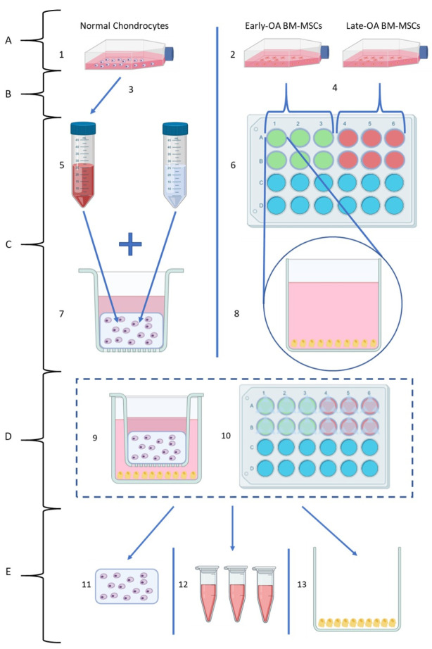
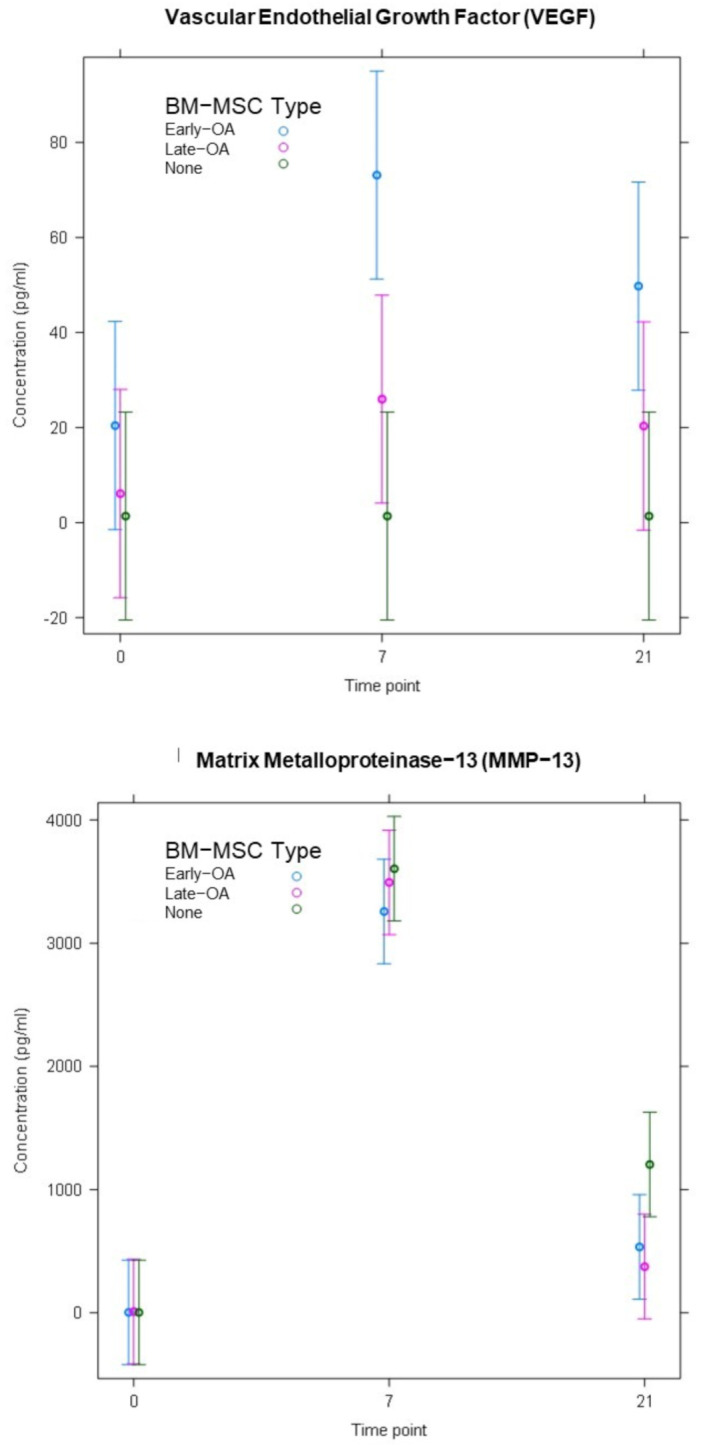
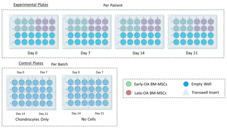
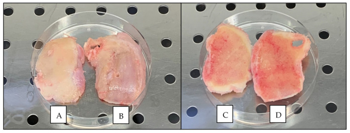
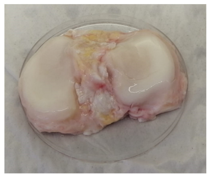
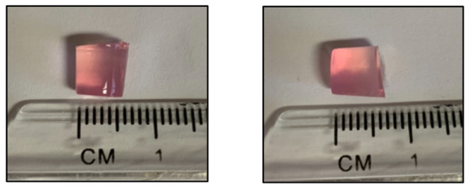
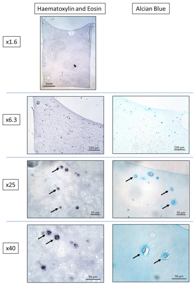
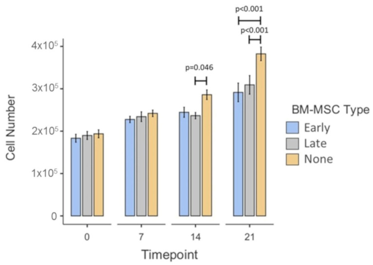
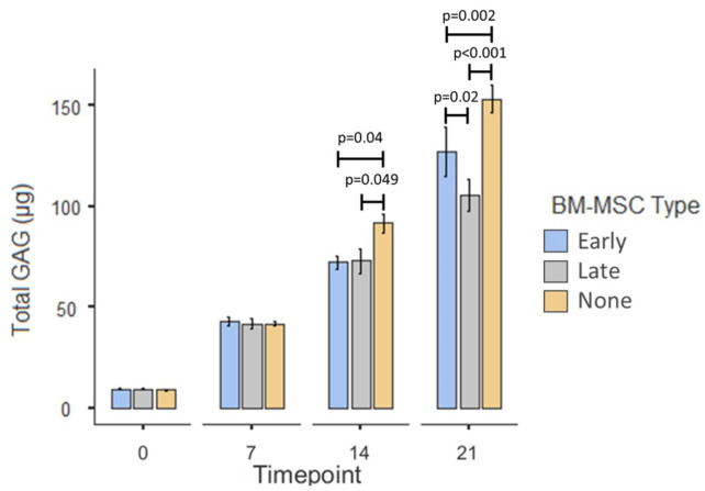
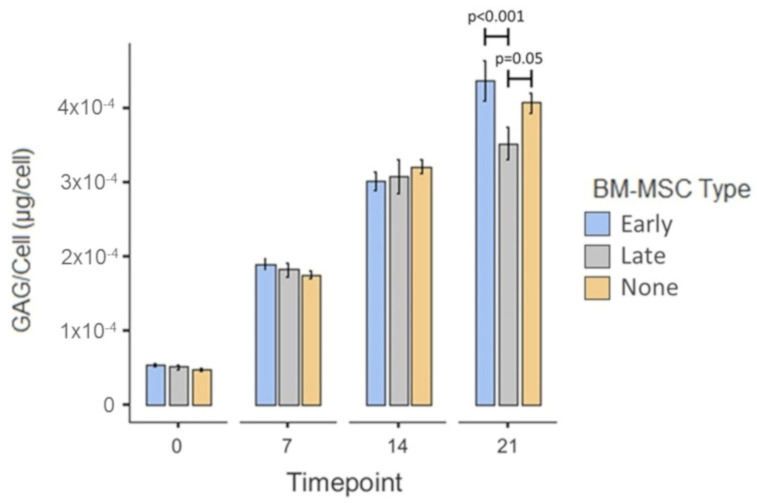
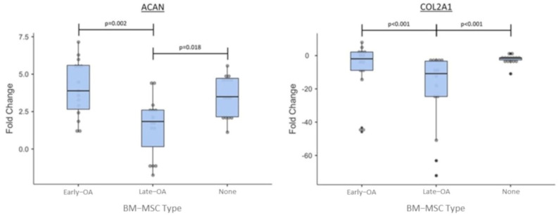
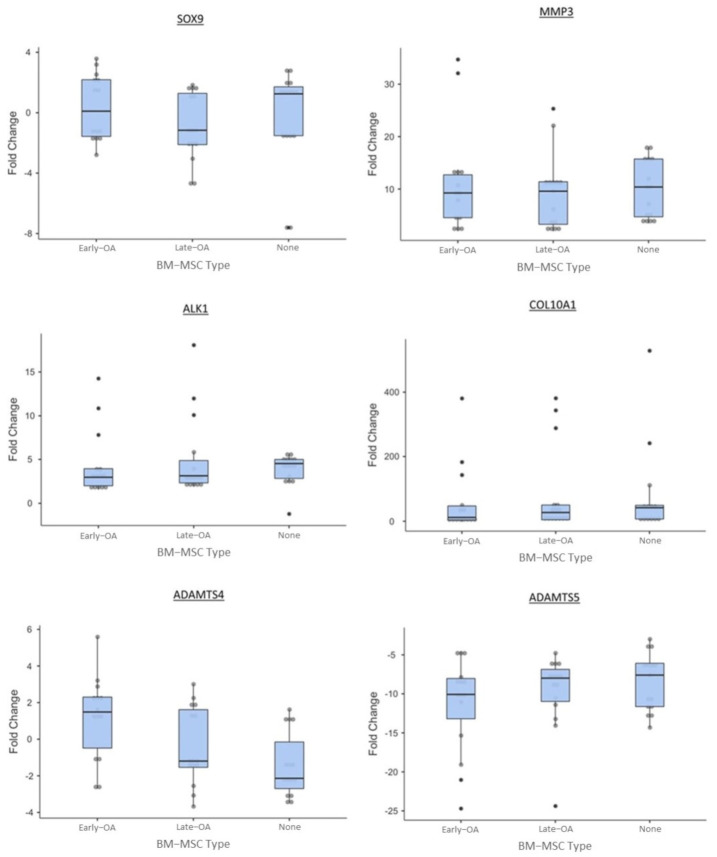
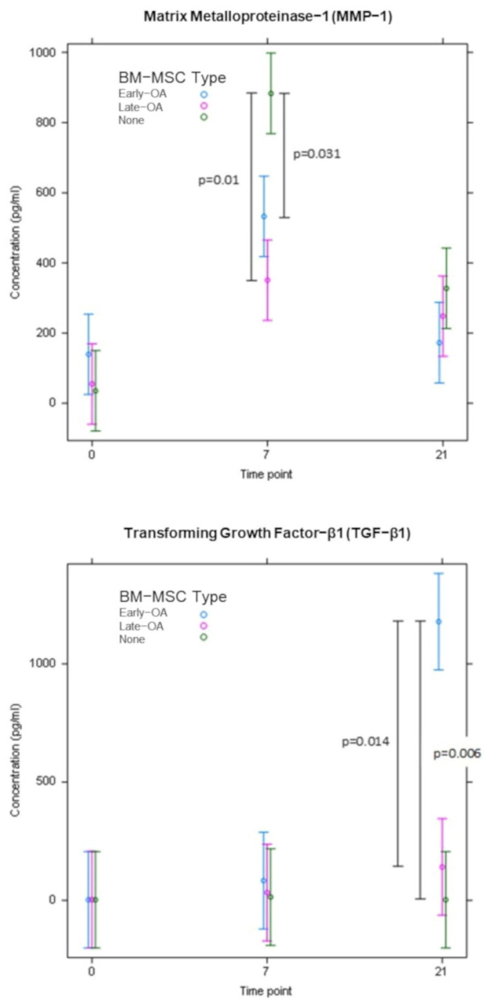
Chondrocyte-based cartilage repair strategies, such as articular chondrocyte implantation, are widely used, but few studies addressed the communication between native subchondral bone cells and the transplanted chondrocytes. An indirect co-culture model was developed, representing a chondrocyte/scaffold-construct repair of a cartilage defect adjoining bone, where the bone could have varying degrees of degeneration. Human BM-MSCs were isolated from two areas of subchondral bone in each of five osteochondral tissue specimens from five patients undergoing knee arthroplasty. These two areas underlaid the macroscopically and histologically best and worst cartilage, representing early and late-stage OA, respectively. BM-MSCs were co-cultured with normal chondrocytes suspended in agarose, with the two cell types separated by a porous membrane. After 0, 7, 14 and 21 days, chondrocyte-agarose scaffolds were assessed by gene expression and biochemical analyses, and the abundance of selected proteins in conditioned media was assessed by ELISA. Co-culture with late-OA BM-MSCs resulted in a reduction in GAG deposition and a decreased expression of genes encoding matrix-specific proteins ( and ), compared to culturing with early OA BM-MSCs. The concentration of TGF-β1 was significantly higher in the early OA conditioned media. The results of this study have clinical implications for cartilage repair, suggesting that the health of the subchondral bone may influence the outcomes of chondrocyte-based repair strategies.
基于软骨细胞的软骨修复策略,如关节软骨细胞植入,被广泛应用,但很少有研究涉及到固有软骨下骨细胞与移植的软骨细胞之间的通讯。本研究建立了一种间接共培养模型,代表了软骨缺损毗邻骨的软骨细胞/支架构建修复,其中骨可能有不同程度的退化。从 5 名接受膝关节置换术的患者的 5 个骨软骨组织标本的两个软骨下骨区域中分离出人 BM-MSCs。这两个区域分别位于宏观和组织学上最好和最差的软骨下,分别代表 OA 的早期和晚期。将 BM-MSCs 与悬浮在琼脂糖中的正常软骨细胞共培养,两种细胞类型被多孔膜隔开。在 0、7、14 和 21 天后,通过基因表达和生化分析评估软骨细胞-琼脂糖支架,通过 ELISA 评估条件培养基中选定蛋白质的丰度。与培养早期 OA BM-MSCs 相比,与晚期 OA BM-MSCs 共培养导致 GAG 沉积减少,编码基质特异性蛋白(和)的基因表达降低。早期 OA 条件培养基中 TGF-β1 的浓度明显更高。这项研究的结果对软骨修复具有临床意义,表明软骨下骨的健康状况可能影响基于软骨细胞的修复策略的结果。