• 文献检索
  • 文档翻译
  • 深度研究
  • 学术资讯
  • Suppr Zotero 插件Zotero 插件
  • 邀请有礼
  • 套餐&价格
  • 历史记录
应用&插件
Suppr Zotero 插件Zotero 插件浏览器插件Mac 客户端Windows 客户端微信小程序
定价
高级版会员购买积分包购买API积分包
服务
文献检索文档翻译深度研究API 文档MCP 服务
关于我们
关于 Suppr公司介绍联系我们用户协议隐私条款
关注我们

Suppr 超能文献

核心技术专利:CN118964589B侵权必究
粤ICP备2023148730 号-1Suppr @ 2026

文献检索

告别复杂PubMed语法,用中文像聊天一样搜索,搜遍4000万医学文献。AI智能推荐,让科研检索更轻松。

立即免费搜索

文件翻译

保留排版,准确专业,支持PDF/Word/PPT等文件格式,支持 12+语言互译。

免费翻译文档

深度研究

AI帮你快速写综述,25分钟生成高质量综述,智能提取关键信息,辅助科研写作。

立即免费体验

亨氏单位在孤立性蝶窦病变评估中的价值

Value of Hounsfield Units in the Evaluation of Isolated Sphenoid Sinus Lesions.

作者信息

Tunç Orhan, Yazıcı Alper, Aytaç İsmail, Tümüklü Koray, Akşamoğlu Melih

机构信息

Gaziantep University, Gaziantep, Turkey.

Sanko University, Gaziantep, Turkey.

出版信息

Allergy Rhinol (Providence). 2021 Aug 23;12:21526567211032560. doi: 10.1177/21526567211032560. eCollection 2021 Jan-Dec.

DOI:10.1177/21526567211032560
PMID:34457372
原文链接:https://pmc.ncbi.nlm.nih.gov/articles/PMC8387604/
Abstract

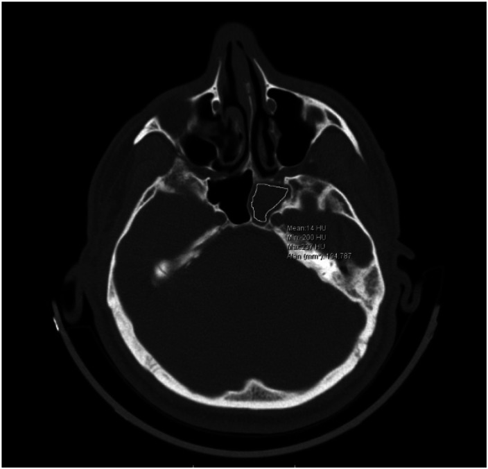
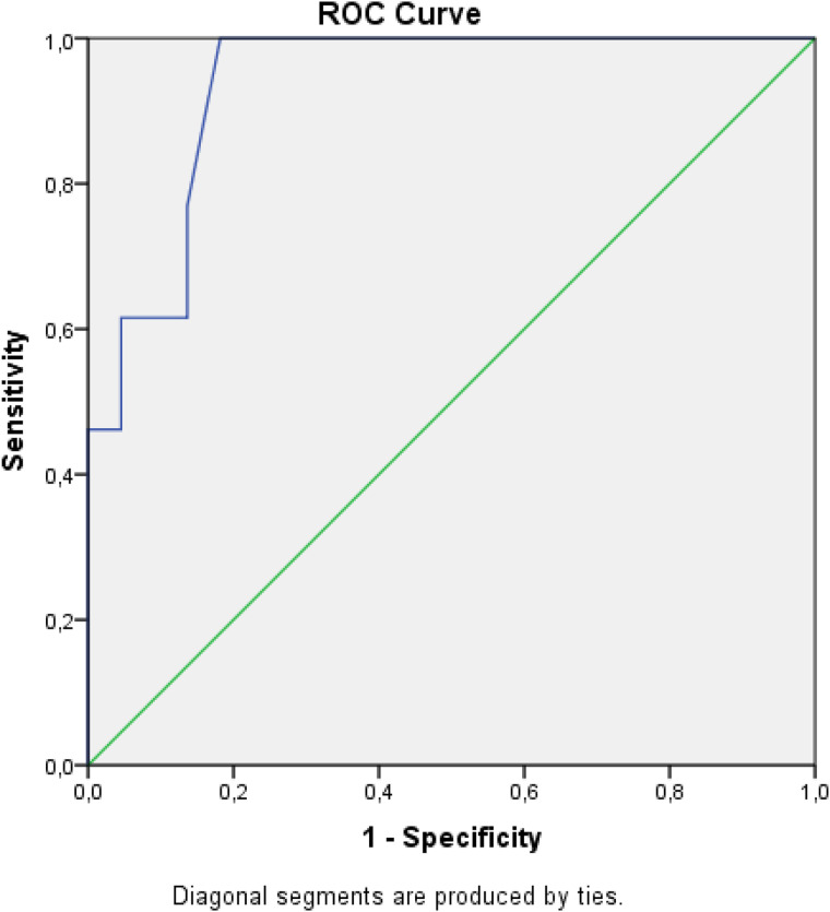
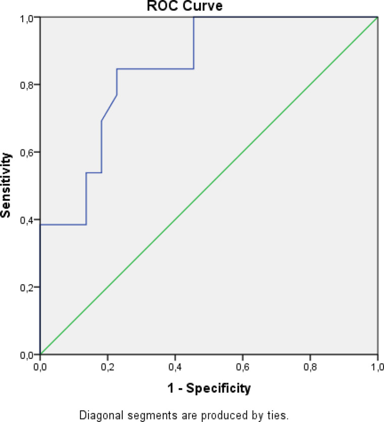
Radiologic findings of fungal sinus disease are generally opacification in paranasal computed tomography (CT) images. The Hounsfield unit (HU) is a standardized objective unit that is also suitable for measuring remodeling and opacifications on CT scans of bone sections of patients with chronic rhinosinusitis. We hypothesized that HU values could provide valuable information in isolated sphenoid sinus lesions before surgery. Between 2012 and 2019, 35 patients underwent functional endoscopic sinus surgery for sphenoid sinus lesions. Tissues obtained from the sphenoid sinus were divided into two groups, fungal and nonfungal, according to the findings of histopathologic examinations. HU values were measured in sphenoid sinus sections on paranasal CT scans of these two groups. Differences in mean and maximum HU values between the two groups were statistically significant (<.05). The maximum HU values calculated from the sphenoid sinus were 435.08 and 196.23 (≤.05) in the fungal group and nonfungal group, respectively. The mean HU values calculated from the sphenoid sinus were 64.31 and 29 (≤.05) in the fungal and nonfungal groups, respectively. At the maximum cutoff value of 241, the sensitivity and specificity of the HU maximum were 84.6% and 77.3%, respectively. At the mean cutoff value of 41.5, the sensitivity and specificity of the HU mean were 76.9% and 86.4%, respectively. HU is an objective value used in radiographic density measurement. The HU values were higher in fungal lesions than in nonfungal inflammations, and they are useful in preoperative measurement.

摘要

真菌性鼻窦疾病的放射学表现通常是鼻窦计算机断层扫描(CT)图像中的混浊。亨氏单位(HU)是一个标准化的客观单位,也适用于测量慢性鼻窦炎患者骨切片CT扫描上的骨质重塑和混浊情况。我们假设HU值可以在术前为孤立性蝶窦病变提供有价值的信息。2012年至2019年期间,35例患者因蝶窦病变接受了功能性鼻内镜鼻窦手术。根据组织病理学检查结果,将从蝶窦获取的组织分为真菌性和非真菌性两组。在这两组患者的鼻窦CT扫描的蝶窦切片中测量HU值。两组之间平均和最大HU值的差异具有统计学意义(<.05)。真菌组和非真菌组蝶窦计算出的最大HU值分别为435.08和196. .23(≤.05)。真菌组和非真菌组蝶窦计算出的平均HU值分别为64.31和29(≤.05)。在最大临界值为241时,HU最大值的敏感性和特异性分别为84.6%和77.3%。在平均临界值为41.5时,HU平均值的敏感性和特异性分别为76.9%和86.4%。HU是用于放射密度测量的客观值。真菌性病变中的HU值高于非真菌性炎症,并且它们在术前测量中很有用。

https://cdn.ncbi.nlm.nih.gov/pmc/blobs/cf7f/8387604/e9aad4a5977b/10.1177_21526567211032560-fig3.jpg
https://cdn.ncbi.nlm.nih.gov/pmc/blobs/cf7f/8387604/73125d46ab98/10.1177_21526567211032560-fig1.jpg
https://cdn.ncbi.nlm.nih.gov/pmc/blobs/cf7f/8387604/8aef1c6f1f91/10.1177_21526567211032560-fig2.jpg
https://cdn.ncbi.nlm.nih.gov/pmc/blobs/cf7f/8387604/e9aad4a5977b/10.1177_21526567211032560-fig3.jpg
https://cdn.ncbi.nlm.nih.gov/pmc/blobs/cf7f/8387604/73125d46ab98/10.1177_21526567211032560-fig1.jpg
https://cdn.ncbi.nlm.nih.gov/pmc/blobs/cf7f/8387604/8aef1c6f1f91/10.1177_21526567211032560-fig2.jpg
https://cdn.ncbi.nlm.nih.gov/pmc/blobs/cf7f/8387604/e9aad4a5977b/10.1177_21526567211032560-fig3.jpg

相似文献

1
Value of Hounsfield Units in the Evaluation of Isolated Sphenoid Sinus Lesions.亨氏单位在孤立性蝶窦病变评估中的价值
Allergy Rhinol (Providence). 2021 Aug 23;12:21526567211032560. doi: 10.1177/21526567211032560. eCollection 2021 Jan-Dec.
2
Objective radiographic density measurements of sinus opacities are not strong predictors of noninvasive fungal disease.鼻窦混浊的客观影像学密度测量并非无创性真菌病的有力预测指标。
Am J Rhinol Allergy. 2014 Nov-Dec;28(6):483-6. doi: 10.2500/ajra.2014.28.4104.
3
Value of MRI signal intensity in evaluation of allergic fungal rhinosinusitis compared with CT Hounsfield units: Retrospective study.MRI 信号强度评估变应性真菌性鼻-鼻窦炎与 CT 亨氏单位的价值:回顾性研究。
Medicine (Baltimore). 2024 Jul 12;103(28):e38951. doi: 10.1097/MD.0000000000038951.
4
Sensitivity and specificity of computed tomography and magnetic resonance imaging in the diagnosis of isolated sphenoid sinus diseases.计算机断层扫描和磁共振成像在诊断孤立性蝶窦疾病中的敏感性和特异性。
Laryngoscope. 2011 Jul;121(7):1584-9. doi: 10.1002/lary.21828. Epub 2011 Jun 9.
5
Evaluating bone remodeling by measuring Hounsfield units in a rabbit model of rhinosinusitis: is it superior to measuring bone thickness?评估兔鼻窦炎模型中通过测量亨氏单位进行骨重建:优于测量骨厚度吗?
Int Forum Allergy Rhinol. 2018 Nov;8(11):1342-1348. doi: 10.1002/alr.22205. Epub 2018 Sep 20.
6
Chronic rhinosinusitis symptoms and computed tomography staging: improved correlation by incorporating radiographic density.慢性鼻-鼻窦炎症状与计算机断层扫描分期:通过引入放射密度提高相关性。
Int Forum Allergy Rhinol. 2012 Sep-Oct;2(5):386-91. doi: 10.1002/alr.21042. Epub 2012 Apr 30.
7
Endoscopic Treatment of Isolated Sphenoid Sinus Disease in Children.儿童孤立性蝶窦疾病的内镜治疗
Ear Nose Throat J. 2019 Aug;98(7):425-430. doi: 10.1177/0145561319841227. Epub 2019 Apr 23.
8
Predictive Significance of Radiographic Density of Sinus Opacity and Bone Thickness in Unilateral Maxillary Sinus Mycetoma.单侧上颌窦真菌球中鼻窦混浊的影像学密度及骨厚度的预测意义
ORL J Otorhinolaryngol Relat Spec. 2019;81(2-3):111-120. doi: 10.1159/000496829. Epub 2019 Jun 25.
9
Diagnostic Accuracy of Hounsfield Unit Value and Hounsfield Unit to Hematocrit Ratio in Predicting Cerebral Venous Sinus Thrombosis: A Retrospective Case-Control Study.亨氏单位值及亨氏单位与血细胞比容比值在预测脑静脉窦血栓形成中的诊断准确性:一项回顾性病例对照研究
Cureus. 2024 Apr 3;16(4):e57567. doi: 10.7759/cureus.57567. eCollection 2024 Apr.
10
Can Volumetric and Morphological Variants of Sphenoid Sinuses Influence Sinuses Opacification?蝶窦的容积和形态变异会影响鼻窦的混浊吗?
J Craniofac Surg. 2018 Nov;29(8):2344-2347. doi: 10.1097/SCS.0000000000004797.

引用本文的文献

1
Using the Hounsfield Units Derived From Computed Tomography (CT) Scans to Differentiate Between the Subtypes of Allergic Fungal Rhinosinusitis: A Retrospective Study.利用计算机断层扫描(CT)扫描得出的亨氏单位鉴别变应性真菌性鼻-鼻窦炎的亚型:一项回顾性研究
Cureus. 2025 Mar 6;17(3):e80151. doi: 10.7759/cureus.80151. eCollection 2025 Mar.

本文引用的文献

1
MR and CT imaging features of sino-nasal organized hematomas.鼻窦组织性血肿的磁共振成像(MR)和计算机断层扫描(CT)影像特征
Oral Radiol. 2021 Apr;37(2):297-304. doi: 10.1007/s11282-020-00450-1. Epub 2020 May 27.
2
Hounsfield Unit Value of Interpeduncular Cistern Hematomas Can Predict Symptomatic Vasospasm.脚间池血肿的 Hounsfield 单位值可预测症状性血管痉挛。
Stroke. 2020 Jan;51(1):143-148. doi: 10.1161/STROKEAHA.119.026962. Epub 2019 Nov 7.
3
A novel use of attenuation value (Hounsfield unit) in non-contrast CT: diagnosis of pyonephrosis in obstructed systems.
非增强CT中衰减值(亨氏单位)的一种新用途:梗阻性系统中脓性肾盂炎的诊断。
Int Urol Nephrol. 2020 Jan;52(1):9-14. doi: 10.1007/s11255-019-02283-2. Epub 2019 Sep 20.
4
Osteitis in Chronic Rhinosinusitis.慢性鼻-鼻窦炎中的骨炎。
Curr Allergy Asthma Rep. 2019 Mar 14;19(5):24. doi: 10.1007/s11882-019-0855-5.
5
Isolated sphenoid sinus opacifications: a systematic review and meta-analysis.孤立性蝶窦混浊:系统评价和荟萃分析。
Int Forum Allergy Rhinol. 2017 Dec;7(12):1201-1206. doi: 10.1002/alr.22023. Epub 2017 Oct 12.
6
Isolated sphenoid sinus fungus ball: a retrospective study conducted at a tertiary care referral center in Korea.孤立性蝶窦真菌球:在韩国一家三级医疗转诊中心进行的一项回顾性研究。
Eur Arch Otorhinolaryngol. 2017 Jun;274(6):2453-2459. doi: 10.1007/s00405-017-4468-0. Epub 2017 Mar 1.
7
Computed tomography and histopathological evaluation of osteitis in rabbit models with rhinosinusitis.兔鼻窦炎模型中骨炎的计算机断层扫描及组织病理学评估
Acta Otolaryngol. 2017 May;137(5):534-540. doi: 10.1080/00016489.2016.1252060. Epub 2016 Nov 11.
8
The radiologic evaluation of osteitis type and formation in chronic rhinosinusitis with and without nasal polyposis.伴或不伴鼻息肉的慢性鼻窦炎中骨炎类型及形成的影像学评估
Am J Rhinol Allergy. 2015 Nov-Dec;29(6):e201-4. doi: 10.2500/ajra.2015.29.4240.
9
Potential correlations of dentogenic factors to the development of clinically verified fungus balls: A retrospective computed tomography-based analysis.牙源性因素与经临床验证的真菌球形成之间的潜在相关性:一项基于计算机断层扫描的回顾性分析。
Laryngoscope. 2016 Jan;126(1):39-43. doi: 10.1002/lary.25416. Epub 2015 Jul 21.
10
Maxillary fungus ball: zinc-oxide endodontic materials as a risk factor.上颌真菌球:氧化锌牙髓材料作为一个危险因素
Acta Otorhinolaryngol Ital. 2015 Apr;35(2):93-6.