• 文献检索
  • 文档翻译
  • 深度研究
  • 学术资讯
  • Suppr Zotero 插件Zotero 插件
  • 邀请有礼
  • 套餐&价格
  • 历史记录
应用&插件
Suppr Zotero 插件Zotero 插件浏览器插件Mac 客户端Windows 客户端微信小程序
定价
高级版会员购买积分包购买API积分包
服务
文献检索文档翻译深度研究API 文档MCP 服务
关于我们
关于 Suppr公司介绍联系我们用户协议隐私条款
关注我们

Suppr 超能文献

核心技术专利:CN118964589B侵权必究
粤ICP备2023148730 号-1Suppr @ 2026

文献检索

告别复杂PubMed语法,用中文像聊天一样搜索,搜遍4000万医学文献。AI智能推荐,让科研检索更轻松。

立即免费搜索

文件翻译

保留排版,准确专业,支持PDF/Word/PPT等文件格式,支持 12+语言互译。

免费翻译文档

深度研究

AI帮你快速写综述,25分钟生成高质量综述,智能提取关键信息,辅助科研写作。

立即免费体验

乳腺磁共振成像在新诊断乳腺癌患者腋窝淋巴结分期中的应用

Performance of breast magnetic resonance imaging in axillary nodal staging in newly diagnosed breast cancer patients.

作者信息

Chayakulkheeree Jatuporn, Pungrassami Dirapit, Prueksadee Jenjeera

机构信息

Division of Diagnostic Radiology, Department of Radiology, Faculty of Medicine Chulalongkorn University, Thailand.

出版信息

Pol J Radiol. 2019 Oct 18;84:e413-e418. doi: 10.5114/pjr.2019.89690. eCollection 2019.

DOI:10.5114/pjr.2019.89690
PMID:31969959
原文链接:https://pmc.ncbi.nlm.nih.gov/articles/PMC6964355/
Abstract

PURPOSE

To determine the diagnostic value of breast magnetic resonance imaging (MRI) in detecting axillary metastatic node in newly diagnosed breast cancer, we assessed the sensitivity, specificity, positive predictive value (PPV), and negative predictive value (NPV) of breast MRI.

MATERIAL AND METHODS

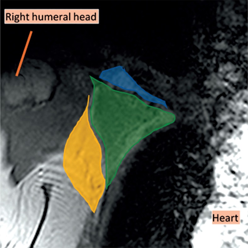
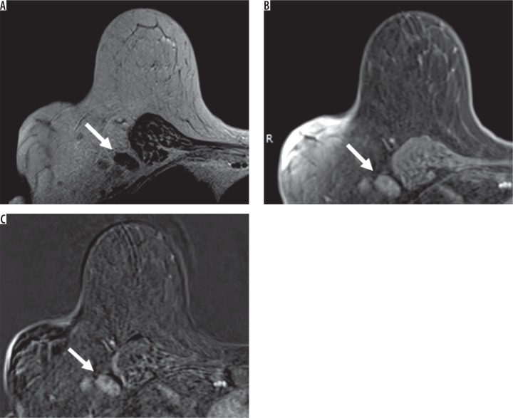
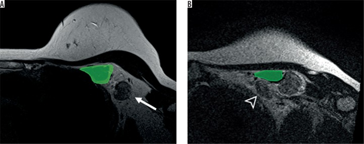
Data were collected retrospectively from January 2011 to December 2018. Preoperative breast MRI of women with newly diagnosed breast cancer were reviewed to evaluate the axillary nodal staging by using the morphological characteristic of metastatic axillary lymph node. Statistical analysis was performed to assess the performance of breast MRI in diagnosing metastatic axillary lymph nodes based on the pathological result.

RESULTS

A total of 131 women with breast cancer were analysed. Two hundred and twenty-seven axillary lymph nodes from preoperative breast MRIs were considered to be metastasis. 65.65% (86 patients) of the breast MRI results matched with the pathological results: 37 patients with N0 stage, 39 patients with N1 stage, eight patients with N2 stage, and two patients with N3 stage. Sensitivity of breast MRI for axillary nodal staging was 98.5% (95% CI: 92-100%), and the negative predictive value was 96.4% (86.2-99.9%). Specificity of breast MRI for axillary nodal staging was 57.8% (44.8-70.1%) and the positive predictive value was 71% (60.6-79.9%).

CONCLUSIONS

Our study showed that the breast MRI had a high sensitivity (98.5%) and high NPV (96.4%) in detecting metastatic axillary lymph nodes, but its specificity was only fair (57.8%). Overestimation and underestimation of the MRI in N staging were also found in 20.61% and 12.98% of cases, respectively.

摘要

目的

为了确定乳腺磁共振成像(MRI)在检测新诊断乳腺癌腋窝转移淋巴结方面的诊断价值,我们评估了乳腺MRI的敏感性、特异性、阳性预测值(PPV)和阴性预测值(NPV)。

材料与方法

回顾性收集2011年1月至2018年12月的数据。对新诊断乳腺癌女性的术前乳腺MRI进行回顾,通过转移性腋窝淋巴结的形态特征评估腋窝淋巴结分期。基于病理结果进行统计分析,以评估乳腺MRI在诊断转移性腋窝淋巴结方面的表现。

结果

共分析了131例乳腺癌女性。术前乳腺MRI检查出的227个腋窝淋巴结被认为有转移。65.65%(86例患者)的乳腺MRI结果与病理结果相符:37例N0期患者,39例N1期患者,8例N2期患者,2例N3期患者。乳腺MRI对腋窝淋巴结分期的敏感性为98.5%(95%CI:92 - 100%),阴性预测值为96.4%(86.2 - 99.9%)。乳腺MRI对腋窝淋巴结分期的特异性为57.8%(44.8 - 70.1%),阳性预测值为71%(60.6 - 79.9%)。

结论

我们的研究表明,乳腺MRI在检测转移性腋窝淋巴结方面具有高敏感性(98.5%)和高阴性预测值(96.4%),但其特异性仅为一般水平(57.8%)。在20.61%和12.98%的病例中,还分别发现MRI在N分期中存在高估和低估的情况。

https://cdn.ncbi.nlm.nih.gov/pmc/blobs/3164/6964355/2570697e0a49/PJR-84-38577-g003.jpg
https://cdn.ncbi.nlm.nih.gov/pmc/blobs/3164/6964355/a887a03a5940/PJR-84-38577-g001.jpg
https://cdn.ncbi.nlm.nih.gov/pmc/blobs/3164/6964355/6540d33674f6/PJR-84-38577-g002.jpg
https://cdn.ncbi.nlm.nih.gov/pmc/blobs/3164/6964355/2570697e0a49/PJR-84-38577-g003.jpg
https://cdn.ncbi.nlm.nih.gov/pmc/blobs/3164/6964355/a887a03a5940/PJR-84-38577-g001.jpg
https://cdn.ncbi.nlm.nih.gov/pmc/blobs/3164/6964355/6540d33674f6/PJR-84-38577-g002.jpg
https://cdn.ncbi.nlm.nih.gov/pmc/blobs/3164/6964355/2570697e0a49/PJR-84-38577-g003.jpg

相似文献

1
Performance of breast magnetic resonance imaging in axillary nodal staging in newly diagnosed breast cancer patients.乳腺磁共振成像在新诊断乳腺癌患者腋窝淋巴结分期中的应用
Pol J Radiol. 2019 Oct 18;84:e413-e418. doi: 10.5114/pjr.2019.89690. eCollection 2019.
2
Noninvasive nodal staging in patients with breast cancer using gadofosveset-enhanced magnetic resonance imaging: a feasibility study.使用钆塞酸二钠增强磁共振成像对乳腺癌患者进行非侵入性淋巴结分期的可行性研究。
Invest Radiol. 2013 Mar;48(3):134-9. doi: 10.1097/RLI.0b013e318277f056.
3
Added value of dedicated axillary hybrid 18F-FDG PET/MRI for improved axillary nodal staging in clinically node-positive breast cancer patients: a feasibility study.专门的腋窝杂交 18F-FDG PET/MRI 对临床淋巴结阳性乳腺癌患者腋窝淋巴结分期的增值作用:一项可行性研究。
Eur J Nucl Med Mol Imaging. 2018 Feb;45(2):179-186. doi: 10.1007/s00259-017-3823-0. Epub 2017 Sep 14.
4
[Diagnostic accuracy of 3.0T high-resolution MRI for assessment mesorectal lymph node metastases in patients with rectal cancer].[3.0T高分辨率磁共振成像评估直肠癌患者中直肠系膜淋巴结转移的诊断准确性]
Zhonghua Wei Chang Wai Ke Za Zhi. 2018 Jul 25;21(7):786-792.
5
Routine use of standard breast MRI compared to axillary ultrasound for differentiating between no, limited and advanced axillary nodal disease in newly diagnosed breast cancer patients.在新诊断的乳腺癌患者中,将标准乳腺磁共振成像(MRI)与腋窝超声的常规使用进行比较,以区分腋窝淋巴结疾病为无、局限或进展期。
Eur J Radiol. 2016 Dec;85(12):2288-2294. doi: 10.1016/j.ejrad.2016.10.030. Epub 2016 Oct 28.
6
Assessment of multifocality and axillary nodal involvement in early-stage breast cancer patients using 18F-FDG PET/CT compared to contrast-enhanced and diffusion-weighted magnetic resonance imaging and sentinel node biopsy.与对比增强磁共振成像、扩散加权磁共振成像及前哨淋巴结活检相比,利用18F-FDG PET/CT评估早期乳腺癌患者的多灶性及腋窝淋巴结受累情况。
Acta Radiol. 2015 Aug;56(8):917-23. doi: 10.1177/0284185114539786. Epub 2014 Jul 10.
7
Reliability of predicting low-burden (≤ 2) positive axillary lymph nodes indicating sentinel lymph node biopsy in primary operable breast cancer - a retrospective comparative study with PET/CT and breast MRI.预测低负担(≤2)阳性腋窝淋巴结以指示原发性可手术乳腺癌前哨淋巴结活检的可靠性——一项 PET/CT 和乳腺 MRI 的回顾性对比研究。
World J Surg Oncol. 2024 Jan 6;22(1):12. doi: 10.1186/s12957-023-03297-y.
8
Diagnostic performance of axillary ultrasound and standard breast MRI for differentiation between limited and advanced axillary nodal disease in clinically node-positive breast cancer patients.腋窝超声和标准乳腺 MRI 对临床淋巴结阳性乳腺癌患者腋窝局限性和进展性淋巴结疾病的鉴别诊断性能。
Sci Rep. 2019 Nov 25;9(1):17476. doi: 10.1038/s41598-019-54017-0.
9
Role of diffusion-weighted MRI: predicting axillary lymph node metastases in breast cancer.扩散加权磁共振成像的作用:预测乳腺癌腋窝淋巴结转移
Acta Radiol. 2014 Oct;55(8):909-16. doi: 10.1177/0284185113509094. Epub 2013 Nov 14.
10
Preoperative axillary lymph node evaluation in breast cancer patients by breast magnetic resonance imaging (MRI): Can breast MRI exclude advanced nodal disease?乳腺癌患者术前通过乳腺磁共振成像(MRI)评估腋窝淋巴结:乳腺MRI能否排除晚期淋巴结疾病?
Eur Radiol. 2016 Nov;26(11):3865-3873. doi: 10.1007/s00330-016-4235-4. Epub 2016 Feb 2.

引用本文的文献

1
Deep learning-based breast MRI for predicting axillary lymph node metastasis: a systematic review and meta-analysis.基于深度学习的乳腺磁共振成像预测腋窝淋巴结转移:一项系统评价与荟萃分析
Cancer Imaging. 2025 Mar 31;25(1):44. doi: 10.1186/s40644-025-00863-3.
2
Diagnostic Usefulness of Diffusion-Weighted MRI for Axillary Lymph Node Evaluation in Patients with Breast Cancer.扩散加权磁共振成像在乳腺癌患者腋窝淋巴结评估中的诊断价值
Diagnostics (Basel). 2023 Jan 31;13(3):513. doi: 10.3390/diagnostics13030513.
3
Beyond N Staging in Breast Cancer: Importance of MRI and Ultrasound-based Imaging.

本文引用的文献

1
Diagnostic accuracy of metastatic axillary lymph nodes in breast MRI.乳腺MRI中腋窝转移性淋巴结的诊断准确性
Springerplus. 2016 Jun 16;5(1):735. doi: 10.1186/s40064-016-2419-7. eCollection 2016.
2
The value of T2* in differentiating metastatic from benign axillary lymph nodes in patients with breast cancer--a preliminary in vivo study.在乳腺癌患者中,T2* 值在鉴别转移性与良性腋窝淋巴结中的价值——一项初步的体内研究。
PLoS One. 2014 Jan 16;9(1):e84038. doi: 10.1371/journal.pone.0084038. eCollection 2014.
3
Axillary staging of breast cancer: what the radiologist should know.
乳腺癌N分期之外:基于MRI和超声成像的重要性
Cancers (Basel). 2022 Aug 31;14(17):4270. doi: 10.3390/cancers14174270.
4
Diffusion tensor imaging and diffusion-weighted imaging on axillary lymph node status in breast cancer patients.乳腺癌患者腋窝淋巴结状态的弥散张量成像和弥散加权成像。
Diagn Interv Radiol. 2022 Jul;28(4):329-336. doi: 10.5152/dir.2022.21460.
5
Diagnostic Value of Axillary Ultrasound, MRI, and F-FDG-PET/ CT in Determining Axillary Lymph Node Status in Breast Cancer Patients.腋窝超声、MRI及F-FDG-PET/CT在确定乳腺癌患者腋窝淋巴结状态中的诊断价值
Eur J Breast Health. 2021 Dec 30;18(1):37-47. doi: 10.4274/ejbh.galenos.2021.2021-3-10. eCollection 2022 Jan.
6
Prediction of Metastasis in the Axillary Lymph Nodes of Patients With Breast Cancer: A Radiomics Method Based on Contrast-Enhanced Computed Tomography.乳腺癌患者腋窝淋巴结转移的预测:基于对比增强计算机断层扫描的放射组学方法
Front Oncol. 2021 Sep 20;11:726240. doi: 10.3389/fonc.2021.726240. eCollection 2021.
7
Comparison of nodal staging between CT, MRI, and [F]-FDG PET/MRI in patients with newly diagnosed breast cancer.比较 CT、MRI 和 [F]-FDG PET/MRI 在新诊断乳腺癌患者中的淋巴结分期。
Eur J Nucl Med Mol Imaging. 2022 Feb;49(3):992-1001. doi: 10.1007/s00259-021-05502-0. Epub 2021 Sep 3.
腋窝分期乳腺癌:放射科医生应该知道什么。
Radiographics. 2013 Oct;33(6):1589-612. doi: 10.1148/rg.336125060.
4
A new, preoperative, MRI-based scoring system for diagnosing malignant axillary lymph nodes in women evaluated for breast cancer.一种新的、术前基于 MRI 的评分系统,用于诊断女性乳腺癌评估中的恶性腋窝淋巴结。
Eur J Radiol. 2012 Oct;81(10):2602-12. doi: 10.1016/j.ejrad.2012.03.019. Epub 2012 Apr 21.
5
Magnetic resonance imaging for axillary staging in patients with breast cancer.乳腺癌患者腋窝分期的磁共振成像
J Magn Reson Imaging. 2009 Aug;30(2):309-12. doi: 10.1002/jmri.21802.
6
Indications for breast MRI in the patient with newly diagnosed breast cancer.新诊断乳腺癌患者的乳腺磁共振成像(MRI)指征。
J Natl Compr Canc Netw. 2009 Feb;7(2):193-201. doi: 10.6004/jnccn.2009.0013.
7
Simultaneous bilateral breast and high-resolution axillary MRI of patients with breast cancer: preliminary results.乳腺癌患者的双侧乳腺及高分辨率腋窝同时MRI检查:初步结果
AJR Am J Roentgenol. 2004 Apr;182(4):1059-67. doi: 10.2214/ajr.182.4.1821059.
8
The measurement of observer agreement for categorical data.分类数据观察者一致性的测量。
Biometrics. 1977 Mar;33(1):159-74.