• 文献检索
  • 文档翻译
  • 深度研究
  • 学术资讯
  • Suppr Zotero 插件Zotero 插件
  • 邀请有礼
  • 套餐&价格
  • 历史记录
应用&插件
Suppr Zotero 插件Zotero 插件浏览器插件Mac 客户端Windows 客户端微信小程序
定价
高级版会员购买积分包购买API积分包
服务
文献检索文档翻译深度研究API 文档MCP 服务
关于我们
关于 Suppr公司介绍联系我们用户协议隐私条款
关注我们

Suppr 超能文献

核心技术专利:CN118964589B侵权必究
粤ICP备2023148730 号-1Suppr @ 2026

文献检索

告别复杂PubMed语法,用中文像聊天一样搜索,搜遍4000万医学文献。AI智能推荐,让科研检索更轻松。

立即免费搜索

文件翻译

保留排版,准确专业,支持PDF/Word/PPT等文件格式,支持 12+语言互译。

免费翻译文档

深度研究

AI帮你快速写综述,25分钟生成高质量综述,智能提取关键信息,辅助科研写作。

立即免费体验

利用超声散射特性定量超声特征评估前列腺癌的体内反应。

In vivo assessment of prostate cancer response using quantitative ultrasound characterization of ultrasonic scattering properties.

机构信息

Physical Sciences, Sunnybrook Research Institute, Toronto, ON, Canada.

Department of Radiation Oncology, Sunnybrook Health Sciences Centre, Toronto, ON, Canada.

出版信息

BMC Cancer. 2021 Sep 3;21(1):991. doi: 10.1186/s12885-021-08706-7.

DOI:10.1186/s12885-021-08706-7
PMID:34479484
原文链接:https://pmc.ncbi.nlm.nih.gov/articles/PMC8417963/
Abstract

BACKGROUND

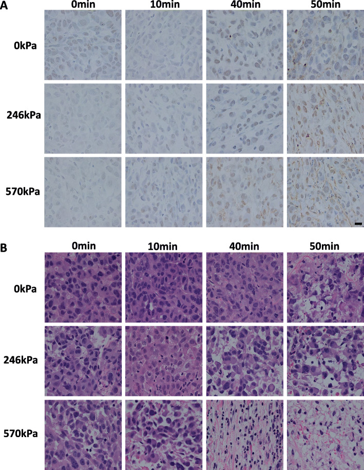
The study here investigated quantitative ultrasound (QUS) parameters to assess tumour response to ultrasound-stimulated microbubbles (USMB) and hyperthermia (HT) treatment in vivo. Mice bearing prostate cancer xenografts were exposed to various treatment conditions including 1% (v/v) Definity microbubbles stimulated at ultrasound pressures 246 kPa and 570 kPa and HT duration of 0, 10, 40, and 50 min. Ultrasound radiofrequency (RF) data were collected using an ultrasound transducer with a central frequency of 25 MHz. QUS parameters based on form factor models were used as potential biomarkers of cell death in prostate cancer xenografts.

RESULTS

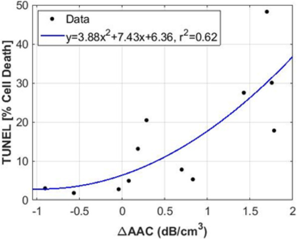
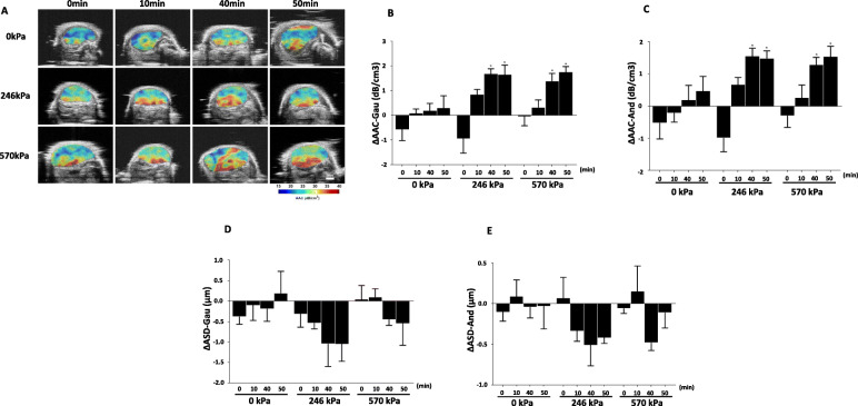
The average acoustic concentration (AAC) parameter from spherical gaussian and the fluid-filled spherical models were the most efficient imaging biomarker of cell death. Statistical significant increases of AAC were found in the combined treatment groups: 246 kPa + 40 min, 246 kPa + 50 min, and 570 kPa + 50 min, in comparison with control tumours (0 kPa + 0 min). Changes in AAC correlates strongly (r = 0.62) with cell death fraction quantified from the histopathological analysis.

CONCLUSION

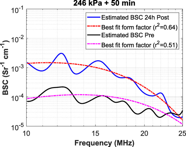
Scattering property estimates from spherical gaussian and fluid-filled spherical models are useful imaging biomarkers for assessing tumour response to treatment. Our observation of changes in AAC from high ultrasound frequencies was consistent with previous findings where parameters related to the backscatter intensity (AAC) increased with cell death.

摘要

背景

本研究通过定量超声(QUS)参数来评估超声刺激微泡(USMB)和热疗(HT)联合治疗体内肿瘤的反应。研究中使用携带前列腺癌异种移植的小鼠作为研究对象,对其施加不同的治疗条件,包括在超声压力 246 kPa 和 570 kPa 下分别使用 1%(v/v)浓度的 Definity 微泡,以及 0、10、40 和 50 min 的 HT 处理时长。使用中心频率为 25 MHz 的超声换能器采集超声射频(RF)数据。基于形态因子模型的 QUS 参数被用作前列腺癌异种移植细胞死亡的潜在生物标志物。

结果

基于球形高斯和充满液体的球形模型的平均声强(AAC)参数是最有效的细胞死亡成像生物标志物。在联合治疗组中,246 kPa+40 min、246 kPa+50 min 和 570 kPa+50 min,与对照组肿瘤(0 kPa+0 min)相比,AAC 参数均有统计学显著增加。AAC 的变化与组织病理学分析量化的细胞死亡分数密切相关(r=0.62)。

结论

球形高斯和充满液体的球形模型的散射特性估计值是评估肿瘤对治疗反应的有用成像生物标志物。我们观察到高频超声下 AAC 的变化与之前的发现一致,即与反向散射强度(AAC)相关的参数随着细胞死亡而增加。

https://cdn.ncbi.nlm.nih.gov/pmc/blobs/0500/8417963/51c2a0fce888/12885_2021_8706_Fig4_HTML.jpg
https://cdn.ncbi.nlm.nih.gov/pmc/blobs/0500/8417963/44d0086b706a/12885_2021_8706_Fig1_HTML.jpg
https://cdn.ncbi.nlm.nih.gov/pmc/blobs/0500/8417963/bdc7694c49e6/12885_2021_8706_Fig2_HTML.jpg
https://cdn.ncbi.nlm.nih.gov/pmc/blobs/0500/8417963/d3d1e7ac3fcf/12885_2021_8706_Fig3_HTML.jpg
https://cdn.ncbi.nlm.nih.gov/pmc/blobs/0500/8417963/51c2a0fce888/12885_2021_8706_Fig4_HTML.jpg

相似文献

1
In vivo assessment of prostate cancer response using quantitative ultrasound characterization of ultrasonic scattering properties.利用超声散射特性定量超声特征评估前列腺癌的体内反应。
BMC Cancer. 2021 Sep 3;21(1):991. doi: 10.1186/s12885-021-08706-7.
2
Noninvasive Evaluation of Breast Tumor Response to Combined Ultrasound-Stimulated Microbubbles and Hyperthermia Therapy Using Quantitative Ultrasound-Based Texture Analysis Method.应用基于定量超声的纹理分析方法无创评估联合超声刺激微泡和热疗治疗乳腺癌肿瘤的疗效。
J Ultrasound Med. 2024 Jan;43(1):137-150. doi: 10.1002/jum.16347. Epub 2023 Oct 24.
3
Ultrasound microbubble potentiated enhancement of hyperthermia-effect in tumours.超声微泡增强肿瘤热疗效果。
PLoS One. 2019 Dec 18;14(12):e0226475. doi: 10.1371/journal.pone.0226475. eCollection 2019.
4
Quantitative ultrasound characterization of therapy response in prostate cancer in vivo.前列腺癌体内治疗反应的定量超声特征分析
Am J Transl Res. 2021 May 15;13(5):4437-4449. eCollection 2021.
5
Quantitative Ultrasound for Evaluation of Tumour Response to Ultrasound-Microbubbles and Hyperthermia.超声微泡与超声热疗治疗肿瘤疗效的定量超声评价
Technol Cancer Res Treat. 2023 Jan-Dec;22:15330338231200993. doi: 10.1177/15330338231200993.
6
Quantitative ultrasound characterization of tumor cell death: ultrasound-stimulated microbubbles for radiation enhancement.肿瘤细胞死亡的定量超声表征:用于增强放疗的超声激发微泡
PLoS One. 2014 Jul 22;9(7):e102343. doi: 10.1371/journal.pone.0102343. eCollection 2014.
7
Quantification of Ultrasonic Scattering Properties of In Vivo Tumor Cell Death in Mouse Models of Breast Cancer.乳腺癌小鼠模型中体内肿瘤细胞死亡的超声散射特性定量分析
Transl Oncol. 2015 Dec;8(6):463-73. doi: 10.1016/j.tranon.2015.11.001.
8
Optimization of microbubble enhancement of hyperthermia for cancer therapy in an in vivo breast tumour model.优化微泡增强在体内乳腺癌模型中的热疗治疗癌症。
PLoS One. 2020 Aug 14;15(8):e0237372. doi: 10.1371/journal.pone.0237372. eCollection 2020.
9
Tumour Vascular Shutdown and Cell Death Following Ultrasound-Microbubble Enhanced Radiation Therapy.超声微泡增强放射治疗后肿瘤血管关闭和细胞死亡。
Theranostics. 2018 Jan 1;8(2):314-327. doi: 10.7150/thno.19010. eCollection 2018.
10
Cellular characterization of ultrasound-stimulated microbubble radiation enhancement in a prostate cancer xenograft model.前列腺癌异种移植模型中超声刺激微泡辐射增强的细胞特征
Dis Model Mech. 2014 Mar;7(3):363-72. doi: 10.1242/dmm.012922. Epub 2014 Jan 30.

引用本文的文献

1
Tumor monitoring and detection of lymph node metastasis using quantitative ultrasound and immune cytokine profiling in dogs undergoing radiation therapy: a pilot study.使用定量超声和免疫细胞因子分析对接受放射治疗的犬进行肿瘤监测及淋巴结转移检测:一项初步研究。
ArXiv. 2025 Mar 25:arXiv:2503.19243v1.
2
A priori prediction of breast cancer response to neoadjuvant chemotherapy using quantitative ultrasound, texture derivative and molecular subtype.基于定量超声、纹理特征和分子亚型预测乳腺癌新辅助化疗反应。
Sci Rep. 2023 Dec 19;13(1):22687. doi: 10.1038/s41598-023-49478-3.
3
Quantitative Ultrasound for Evaluation of Tumour Response to Ultrasound-Microbubbles and Hyperthermia.

本文引用的文献

1
Optimization of microbubble enhancement of hyperthermia for cancer therapy in an in vivo breast tumour model.优化微泡增强在体内乳腺癌模型中的热疗治疗癌症。
PLoS One. 2020 Aug 14;15(8):e0237372. doi: 10.1371/journal.pone.0237372. eCollection 2020.
2
Ultrasound microbubble potentiated enhancement of hyperthermia-effect in tumours.超声微泡增强肿瘤热疗效果。
PLoS One. 2019 Dec 18;14(12):e0226475. doi: 10.1371/journal.pone.0226475. eCollection 2019.
3
Probing the Cellular Size Distribution in Cell Samples Undergoing Cell Death.
超声微泡与超声热疗治疗肿瘤疗效的定量超声评价
Technol Cancer Res Treat. 2023 Jan-Dec;22:15330338231200993. doi: 10.1177/15330338231200993.
探测发生细胞死亡的细胞样本中的细胞大小分布。
Ultrasound Med Biol. 2019 Jul;45(7):1787-1798. doi: 10.1016/j.ultrasmedbio.2019.01.006. Epub 2019 Apr 22.
4
Tumour Vascular Shutdown and Cell Death Following Ultrasound-Microbubble Enhanced Radiation Therapy.超声微泡增强放射治疗后肿瘤血管关闭和细胞死亡。
Theranostics. 2018 Jan 1;8(2):314-327. doi: 10.7150/thno.19010. eCollection 2018.
5
Quantitative ultrasound imaging of therapy response in bladder cancer in vivo.体内膀胱癌治疗反应的定量超声成像
Oncoscience. 2016 Apr 18;3(3-4):122-33. doi: 10.18632/oncoscience.302. eCollection 2016.
6
Review of Quantitative Ultrasound: Envelope Statistics and Backscatter Coefficient Imaging and Contributions to Diagnostic Ultrasound.定量超声综述:包络统计与背向散射系数成像及其对诊断超声的贡献
IEEE Trans Ultrason Ferroelectr Freq Control. 2016 Feb;63(2):336-51. doi: 10.1109/TUFFC.2015.2513958. Epub 2016 Jan 8.
7
Quantification of Ultrasonic Scattering Properties of In Vivo Tumor Cell Death in Mouse Models of Breast Cancer.乳腺癌小鼠模型中体内肿瘤细胞死亡的超声散射特性定量分析
Transl Oncol. 2015 Dec;8(6):463-73. doi: 10.1016/j.tranon.2015.11.001.
8
Non-invasive evaluation of breast cancer response to chemotherapy using quantitative ultrasonic backscatter parameters.使用定量超声背散射参数评估乳腺癌对化疗的反应。
Med Image Anal. 2015 Feb;20(1):224-36. doi: 10.1016/j.media.2014.11.009. Epub 2014 Nov 25.
9
Quantitative ultrasound characterization of tumor cell death: ultrasound-stimulated microbubbles for radiation enhancement.肿瘤细胞死亡的定量超声表征:用于增强放疗的超声激发微泡
PLoS One. 2014 Jul 22;9(7):e102343. doi: 10.1371/journal.pone.0102343. eCollection 2014.
10
Low-frequency quantitative ultrasound imaging of cell death in vivo.低频定量超声成像技术在体检测细胞死亡。
Med Phys. 2013 Aug;40(8):082901. doi: 10.1118/1.4812683.